SALL2 / Sal-like protein 2 · IHC design guide

Design Immunohistochemistry for SALL2

Plan chromogenic SALL2 IHC in paraffin sections using the nuclear tissue pattern as a scoring guide (HPA tissue IHC). The catalog antibody has documented staining in mouse and rat brain sections (datasheet A05853-1); compare signal in the cell populations reported by the HPA (HPA tissue IHC).

Evidence assembled Oct 2026 · For research use; verify linked source records and product datasheet before use
Immunohistochemistry protocol sheet for SALL2 (IHC for SALL2): expected localisation Nuclear staining in several tissues (HPA tissue IHC), antibody A05853-1, validated IHC image, and IHC protocol steps
Printable SALL2 IHC protocol sheet — expected localisation Nuclear staining in several tissues (HPA tissue IHC), antibody A05853-1, controls and protocol steps. Open the full SALL2 IHC guide →

SALL2 Immunohistochemistry Experimental Design Guide

Expected localisation, validated protocols, controls and antibodies — the at-a-glance facts below, then the full design guide.

Must know before staining
Expected localisation Nuclear staining in several tissues (HPA tissue IHC)
Staining pattern Nuclei in glia, cerebellar granular layer and glands (HPA tissue IHC)
Antigen retrieval EDTA pH 8.0 HIER, heat-mediated (datasheet A05853-1)
Positive control ⓘ Caudate+3 more · see all
Negative control ⓘ Adipose tissue+4 more · see all
Important caveats
Reasons your staining may differ from the expected pattern.
Fixation Keep fixation conditions consistent across sections. (standard IHC practice; not target-specific)
Caveat Glial staining is lower in cortex than caudate (HPA tissue IHC)
Regulation Highest expression in adult brain (UniProt)
Isoform / epitope 2 isoforms; epitope coverage is unspecified (UniProt)
Section 1

Recommended SALL2 IHC & IF Protocols

The catalog antibody’s IHC-P protocol is accompanied by published SALL2 staining protocols for colon tissue microarrays (PMC12387037) and breast cancer tissues (PMC6895605).

Recommended immunohistochemistry (IHC-P) protocol parameters
SampleParaffin-embedded mouse brain tissue; fixative not specified (datasheet A05853-1)
FixationImage fixative and duration unreported (datasheet A05853-1); verify before use.
Sectioning4–5 µm sections on charged slides (standard)
DeparaffinisationXylene, graded ethanol series to water (standard)
Antigen retrievalHeat retrieval: EDTA pH 8.0 (datasheet A05853-1); 20 min, 95–100 °C (standard)
Peroxidase block3% H2O2, 10 min, room temperature (standard)
Blocking10% goat serum (datasheet A05853-1)
Primary antibodyRabbit anti-SALL2, 2-5 μg/ml (datasheet A05853-1)
Primary incubationOvernight at 4 °C (datasheet A05853-1)
DetectionHRP-conjugated secondary, DAB chromogen (datasheet A05853-1)
CounterstainHematoxylin, blue, dehydrate and mount (standard)
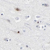
Expected resultSALL2-positive staining in glial cells of caudate (HPA tissue IHC: Medium). HPA tissue profile: Nuclear expression in several tissues. No signal in the no-primary control.
💡Decision noteStart with heat-mediated EDTA pH 8.0 retrieval (datasheet A05853-1). The colon tissue microarray study reports a high-pH automated IHC platform (PMC12387037).
Section 2

What Is the Expected SALL2 Staining Pattern?

SALL2 is annotated as nuclear and has no transmembrane segment (UniProt Q9Y467). In tissue IHC, expect nuclear staining in selected cells, including glial cells in caudate and hippocampus, cerebellar granular-layer cells, and thyroid glandular cells (HPA tissue IHC). HPA rates the tissue staining Approved, supported by external characterization (HPA tissue IHC).

What am I looking at on my slide?
Discrete nuclear staining in caudate or hippocampal glial cells.This fits the reported Medium staining in those cells and the broader nuclear tissue profile (HPA tissue IHC). Assess the named cells rather than treating every nucleus in the section as an expected positive.
Predominantly cytoplasmic staining in tissue IHC, with little nuclear signal.This departs from the reported nuclear tissue pattern (HPA tissue IHC; UniProt Q9Y467). Review morphology, background, and controls before calling it SALL2. Vesicular ICC-IF staining is reported separately and does not establish a cytoplasmic tissue IHC pattern (HPA subcellular ICC-IF).
Staining in adipocytes or bronchial respiratory epithelial cells.HPA reports SALL2 as Not detected in these named cell populations (HPA tissue IHC). Check whether staining follows cell boundaries, pigment, or endogenous chromogenic activity before interpreting it as target signal (general IHC practice).
Diffuse colour across tissue, including spaces between cells.An even deposit without recognizable nuclear enrichment is difficult to reconcile with the tissue profile (HPA tissue IHC). Review the negative control and detection background; diffuse colour alone cannot identify its cause (general IHC practice).
No nuclear signal in caudate glial cells or thyroid glandular cells.Both are reported at Medium staining, making them useful reference populations (HPA tissue IHC). Confirm their presence in the section, then review the IHC-validated antibody and detection workflow before concluding that SALL2 is absent (general IHC practice).
💡Expected SALL2 appearanceCall positive when defined glial, granular-layer, or thyroid glandular cells show predominantly nuclear Medium staining (HPA tissue IHC); diffuse colour or staining confined to unrelated cells is suspect (general IHC practice).
How each factor affects the staining
Choice of reference cellsCaudate and hippocampal glia, cerebellar granular-layer cells, and thyroid glandular cells have reported Medium staining (HPA tissue IHC). Cerebral-cortex glia are reported Low, so a weak cortical result alone is a less clear check of assay performance (HPA tissue IHC).
Antibody evidenceThe reported tissue pattern is Approved, with external characterization supporting staining (HPA tissue IHC). Antibody HPA004162 is IHC Approved; the supplied record does not assign it an IHC Enhanced status (HPA antibodies). Treat the listed pattern as a reference, not proof that every stained cell is specific.
Cell and compartment selectionSALL2 is nuclear and lacks a transmembrane segment (UniProt Q9Y467). Score nuclear signal in the specified cell populations against tissue morphology and controls (HPA tissue IHC; general IHC practice). Its topology does not, by itself, predict sensitivity to fixation or antigen retrieval.
Isoforms and processingUniProt lists 2 isoforms and one full-length chain, with no signal peptide or propeptide annotated (UniProt Q9Y467). The supplied evidence gives no antibody epitope or isoform-specific tissue pattern, so staining cannot be assigned to either isoform from localization alone.
IF/ICC: should vesicles count as expected?Yes, within the separate ICC-IF context: HPA reports vesicles as the approved main location and nucleoplasm as an approved additional location (HPA subcellular ICC-IF). Tissue IHC instead reports nuclear expression (HPA tissue IHC). Compare each assay with its own reference pattern.
Why is my staining missing, weak or wrong?
SituationLikely causeNext action
Known-positive tissue has no nuclear staining.The expected cells may be absent from the section, or an IHC workflow step may have failed (HPA tissue IHC; general IHC practice).Identify caudate glia or thyroid glandular cells on the section, then check antibody, retrieval, detection, and control results against the established IHC workflow (HPA tissue IHC; general IHC practice). No SALL2-specific retrieval condition is supplied.
Nuclear signal is very faint in cerebral cortex.Cerebral-cortex glia are reported Low, whereas caudate and hippocampal glia are Medium (HPA tissue IHC).Assess a reported Medium population alongside the cortex and keep exposure or chromogen development comparable (HPA tissue IHC; general IHC practice). Avoid calling assay failure from weak cortical staining alone.
Broad cytoplasmic colour obscures nuclei.Nonspecific antibody binding or detection background may obscure compartment assessment (general IHC practice); nuclear tissue staining is the reference pattern (HPA tissue IHC).Compare with an appropriate negative control, review blocking and antibody concentration, and score only cells whose nuclear boundaries remain interpretable (general IHC practice). Do not import the ICC-IF vesicle pattern into tissue IHC scoring (HPA subcellular ICC-IF).
Colour appears in HPA Not detected cell types.Endogenous detection activity or nonspecific binding can mimic a positive chromogenic result (general IHC practice); HPA reports adipocytes and bronchial respiratory epithelial cells as Not detected (HPA tissue IHC).Inspect a negative control and tissue morphology, then review the detection system and its endogenous-activity blocking step (general IHC practice). Report the unexpected cell type explicitly rather than extending HPA's positive-cell list.
Background is strong throughout the section.Inadequate blocking, excess antibody, or prolonged chromogen development can raise background (general IHC practice). The HPA reference is a cell-resolved nuclear pattern (HPA tissue IHC).Check the negative control and adjust blocking, antibody concentration, washing, or development within the validated IHC workflow (general IHC practice). Judge improvement by clearer nuclear contrast in reported positive cells (HPA tissue IHC).
ICC-IF shows puncta but tissue IHC is nuclear.The assay references differ: vesicles are the approved main ICC-IF location, with nucleoplasm additional; tissue IHC reports nuclear expression (HPA subcellular ICC-IF; HPA tissue IHC).Interpret each preparation against its own HPA assay record and matched controls (HPA tissue IHC; HPA subcellular ICC-IF; general IHC practice). Do not use ICC-IF puncta as a tissue IHC positive criterion.

Sample controls for SALL2 IHC & IF

🧪Run cerebellum first and look for staining in cells of the granular layer (HPA: Medium in cerebellar granular-layer cells). Use appendix glandular cells as the negative tissue (HPA: Not detected); cells without staining on the positive slide should show only background, although the supplied HPA rows do not establish a specific internal negative cell type.
Positive control tissue: Caudate (Glial cells, HPA Medium)
Negative control tissue: Adipose tissue (HPA Not detected)
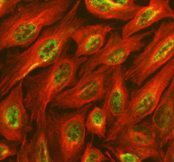
ICC-IF cell lines (HPA subcellular resource): HPA ICC-IF images show SALL2 in HEK293, RT-4, U2OS, with annotated localisation: Vesicles (approved) (HPA subcellular).
Technical controls: Include a no-primary (secondary-only) control, a host- and class-matched rabbit IgG control with clonality matched where known, and SALL2 knockout tissue if available (selected caption: rabbit anti-SALL2, A05853-1). For brain sections developed with HRP-DAB, quench endogenous peroxidase and assess background staining (selected caption: mouse brain, HRP-DAB).
⚠️Feasibility: A target-specific fixation window or fixation effect is unreported, and the selected tissue-IHC caption does not state the fixative (selected caption: fixative unreported). Heat-mediated EDTA retrieval at pH 8.0 is a practical starting condition, but retrieval dependency is unreported (selected caption: EDTA retrieval). The supplied evidence does not establish that frozen sections or IF are easier; brain autofluorescence would need assessment if using IF (HPA: Medium in cerebellar granular-layer cells).

HPA tissue IHC evidence for SALL2

Comprehensive Human Protein Atlas IHC scoring per tissue (reliability: Approved — External characterization data supports antibody staining.). Rows are taken directly from the HPA tissue chart — click any row's HPA link to view the source.

Positive expression · recommended positive controls

TissueCell typeLevelEvidenceSource
Caudate Glial cells Medium Protein (IHC) HPA →
Cerebellum Cells in granular layer Medium Protein (IHC) HPA →
Hippocampus Glial cells Medium Protein (IHC) HPA →
Thyroid gland Glandular cells Medium Protein (IHC) HPA →

Undetected expression · recommended negative controls

TissueCell typeLevelEvidenceSource
Adipose tissue Adipocytes Not detected Protein (IHC) HPA →
Appendix Glandular cells Not detected Protein (IHC) HPA →
Breast Adipocytes Not detected Protein (IHC) HPA →
Bronchus Respiratory epithelial cells Not detected Protein (IHC) HPA →
Cervix Glandular cells Not detected Protein (IHC) HPA →
Section 3

Advanced SALL2 IHC Tips

Troubleshoot SALL2 staining in paraffin section IHC using the catalog antibody’s mouse brain protocol and the reported tissue and subcellular patterns.

What retrieval should I use when nuclear SALL2 staining is weak?
Start with heat mediated retrieval in EDTA at pH 8.0 for paraffin sections (datasheet A05853-1). The catalog antibody detected SALL2 in mouse brain after this retrieval, followed by 2 μg/ml primary antibody overnight at 4°C (datasheet A05853-1). If nuclei remain weak, check heating consistency, section adhesion and reagent coverage before changing retrieval conditions (standard IHC practice). Run any adjusted condition beside the original EDTA condition on matched sections, because stronger DAB staining alone does not establish improved specificity (standard IHC practice). Score nuclear signal separately from diffuse staining, using the reported nuclear tissue pattern as the reference (HPA tissue IHC).
How should I troubleshoot fixation related loss of SALL2 staining?
The mouse brain caption identifies a paraffin embedded section but does not state its fixative, so target specific fixation sensitivity is unknown (datasheet A05853-1). Record the fixative, fixation duration and processing history for each section before comparing staining intensity (standard IHC practice). With matched sections, examine whether staining changes track processing batches while retrieval remains EDTA at pH 8.0 and primary incubation remains 2 μg/ml overnight at 4°C (datasheet A05853-1; standard IHC practice). Include a brain section processed alongside the test specimen to distinguish a batch problem from a tissue difference (UniProt Q9Y467 tissue specificity; standard IHC practice).
Should I score SALL2 in nuclei, cytoplasm or vesicles?
For chromogenic tissue IHC, score nuclear staining as the primary SALL2 readout because the tissue profile reports nuclear expression in several tissues (HPA tissue IHC). The protein is annotated as nuclear and lacks a transmembrane segment (UniProt Q9Y467 subcellular location and topology). HPA cell imaging reports vesicles as the main location and nucleoplasm as an additional location, so document convincing extranuclear signal separately (HPA subcellular). Compare compartment patterns with a matched negative control and with surrounding cells before calling diffuse cytoplasmic DAB positive (standard IHC practice). A visible counterstain helps assign DAB to individual nuclei, especially in densely cellular brain regions (standard IHC practice).
Can this antibody distinguish the two SALL2 isoforms in tissue?
SALL2 has 2 annotated isoforms, but the supplied antibody caption gives no epitope coordinates or isoform selectivity (UniProt Q9Y467 isoforms; datasheet A05853-1). Treat a positive DAB nucleus as SALL2 immunoreactivity rather than proof of a particular isoform (HPA tissue IHC; standard IHC interpretation). If isoform resolution matters, obtain the antibody’s epitope information and compare it with isoform sequences before interpreting staining (standard antibody validation practice). Phosphoserines at positions 243, 797, 802 and 806 are annotated, but their effect on this antibody’s staining is unreported (UniProt Q9Y467 modified residues; datasheet A05853-1). Keep retrieval and detection conditions matched across specimens when comparing intensity (standard IHC practice).
How can I investigate SALL2 with IF after seeing nuclear IHC staining?
Use a separately validated IF antibody and multiplex it with a validated marker for the expected glial population, since glial cells show SALL2 staining in caudate and hippocampus (HPA tissue IHC; standard IF practice). Select a fluorophore whose emission avoids the specimen’s strongest autofluorescence, and assess each channel with single stain controls (standard IF practice). SALL2 is annotated as nuclear with no transmembrane segment, while cell imaging also reports vesicles and nucleoplasm (UniProt Q9Y467 location and topology; HPA subcellular). Permeabilise sufficiently for antibody access to these intracellular epitopes, then check whether stronger treatment alters morphology or background (standard IF practice). Do not transfer the paraffin IHC antibody concentration directly to IF (datasheet A05853-1; standard antibody validation practice).
What should I check when DAB staining is widespread?
First compare the stained section with a no primary control to separate primary dependent staining from detection background (standard IHC practice). The documented workflow uses 10% goat serum blocking, a peroxidase conjugated secondary and DAB development (datasheet A05853-1). Check the peroxidase block, secondary antibody specificity, wash quality and DAB development time when the control also darkens (standard chromogenic IHC practice). If background appears only with primary antibody, reassess its 2 μg/ml concentration and overnight incubation at 4°C on matched sections (datasheet A05853-1; standard IHC practice). Require a discernible nuclear pattern before treating diffuse colour as SALL2 expression (HPA tissue IHC).
How should I quantify SALL2 across brain sections? ⚠ ANSWER MARKED FOR VERIFICATION
Define the cell population and anatomical region before scoring, because reported brain staining includes glial cells in caudate and hippocampus and cells in the cerebellar granular layer (HPA tissue IHC). For nuclear DAB, report the percentage of positive cells or an H-score based on intensity and positive fraction, with the scoring thresholds held constant (standard IHC scoring practice). Normalise positive counts to the number of evaluable cells in that population; if using density, report positive nuclei per mm² of viable tissue (standard IHC quantification practice). Exclude folds, torn areas and poorly preserved regions by a prespecified rule (standard IHC practice). Analyse nuclear and extranuclear signal separately (HPA tissue IHC; HPA subcellular).
How do I distinguish credible SALL2 positivity from artefact?
Prioritise reproducible nuclear staining in intact cells, since nuclear expression is reported for tissue IHC and SALL2 is annotated as nuclear (HPA tissue IHC; UniProt Q9Y467 subcellular location). Glial cells in caudate and hippocampus and cells of the cerebellar granular layer provide reported positive patterns for comparison (HPA tissue IHC). Vesicular staining has been reported in cell imaging, so record a convincing extranuclear pattern without assuming it proves tissue expression (HPA subcellular; standard IHC interpretation). Discount colour concentrated at section edges, folds or necrotic areas, and compare it with a no primary control for endogenous enzyme signal (standard chromogenic IHC practice). Interpret absent staining against the expected cell population and processing controls (standard IHC practice).
Boster reagents

Best SALL2 / Sal-like protein 2 IHC Antibodies

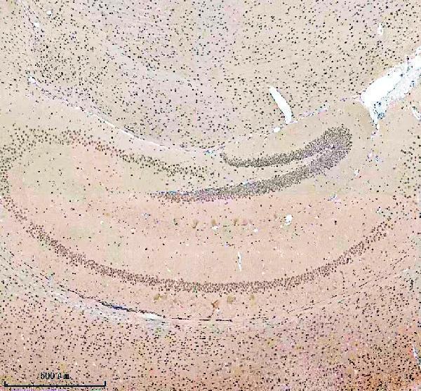
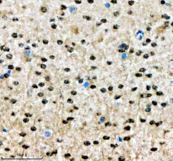
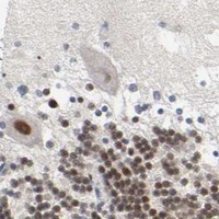
A05853-1 has SALL2 IHC images from paraffin-embedded mouse and rat brain sections, plus IF images from those sections and U2OS cells (catalog image captions).

Real IHC data IHC analysis of SALL2 using anti-SALL2 antibody (A05853-1). SALL2 was detected in a paraffin-embedded section of mouse brain tissue. Heat mediated antigen retrieval was performed in EDTA buffer (pH 8.0, epitope retrieval solution). The tissue section was blocked with 10% goat serum. The tissue section was then incubated with 2 μg/ml rabbit anti-SALL2 Antibody (A05853-1) overnight at 4°C. Peroxidase Conjugated Goat Anti-rabbit IgG was used as secondary antibody and incubated for 30 minutes at 37°C. The tissue section was developed using HRP Conjugated Rabbit IgG Super Vision Assay Kit (Catalog # SV0002) with DAB as the chromogen.
Anti-SALL2 Antibody ®
Cat # A05853-1

A05853-1 is listed for IHC, IF and ICC, with Human, Mouse and Rat reactivity (catalog applications and reactivity). Its IHC images show paraffin-embedded mouse and rat brain sections; its IF images show those sections and U2OS cells (catalog image captions).

Which to pick: For tissue IHC, choose A05853-1: its paraffin-section captions document EDTA retrieval at pH 8.0 and 2 μg/ml primary antibody (A05853-1 IHC image captions). For IF/ICC, the same SKU has a U2OS cell IF image and mouse and rat brain-section IF images (A05853-1 IF image captions). For cross-species work, its listed reactivity covers Human, Mouse and Rat, while the pictured tissue IHC evidence covers mouse and rat; the section fixative is unreported (catalog reactivity; A05853-1 IHC image captions).

Each figure is that product's own IHC / IF validation image from its datasheet.

References

  1. UniProt Consortium. UniProt entry Q9Y467 (SALL2_HUMAN, Sal-like protein 2).
  2. Human Protein Atlas. SALL2 tissue IHC expression (reliability: Approved).
  3. Human Protein Atlas. SALL2 subcellular location (ICC-IF): Mainly localized to vesicles. In addition localized to the nucleoplasm..
  4. Human Protein Atlas. SALL2 antibody validation summary (1 antibodies).
  5. NSUN2 mediates SALL2 m5C methylation to inhibit ferroptosis and promote breast cancer progression. Cancer biology & therapy 2026 — PMC13327353.
  6. SALL2-Mediated Suppression of WNT Signaling Through Transcriptional Control of AXIN2 in Colorectal Cancer Cells. International journal of molecular sciences 2025 — PMC12387037.
  7. Mutation of SALL2 causes recessive ocular coloboma in humans and mice. Human molecular genetics 2014 — PMC3990155.
  8. Epigenetic silencing of SALL2 confers tamoxifen resistance in breast cancer. EMBO molecular medicine 2019 — PMC6895605.
  9. PubMed PMID:8975705 — UniProt-cited evidence.
  10. PubMed PMID:9205841 — UniProt-cited evidence.
  11. PubMed PMID:12168954 — UniProt-cited evidence.