This website uses cookies to ensure you get the best experience on our website.


- Table of Contents
SART1 is a nuclear spliceosome component (UniProt), with nuclear staining in most cell types by tissue IHC (HPA tissue IHC). This guide helps plan paraffin-section staining using catalog antibody A05414-1 at 2–5 μg/ml (datasheet A05414-1) and interpret differences between cell populations (HPA tissue IHC).

Expected localisation, validated protocols, controls and antibodies — the at-a-glance facts below, then the full design guide.
| Expected localisation | Nuclear staining in most cell types (HPA tissue IHC) | |
| Staining pattern | Nuclear signal across most cell types (HPA tissue IHC) | |
| Antigen retrieval | EDTA pH 8.0 HIER, heat-mediated (datasheet A05414-1) | |
| Positive control | Bronchus+4 more · see all | |
| Negative control | Adipose tissue+4 more · see all |
| Fixation | Keep tissue fixation consistent. (standard IHC practice; not target-specific) | |
| Caveat | Some cell populations show no detectable staining (HPA tissue IHC) | |
| Regulation | Preferential in proliferating cells (UniProt) | |
| Isoform / epitope | One isoform; a single 1–800 chain (UniProt) |
Compare the catalog antibody’s IHC-P protocol with the published SART1 protocol for paraffin liver sections (PMC5203921).
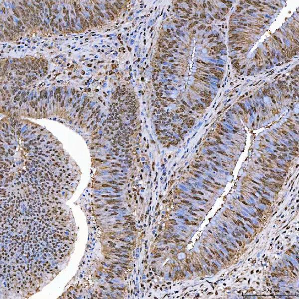
| Sample | Paraffin-embedded human colorectal adenocarcinoma tissue; fixative not specified (datasheet A05414-1) |
| Fixation | Image fixative and duration unreported (datasheet A05414-1); verify before use. |
| Sectioning | 4–5 µm sections on charged slides (standard) |
| Deparaffinisation | Xylene, graded ethanol series to water (standard) |
| Antigen retrieval | Heat retrieval: EDTA pH 8.0 (datasheet A05414-1); 20 min, 95–100 °C (standard) |
| Peroxidase block | 3% H2O2, 10 min, room temperature (standard) |
| Blocking | 10% goat serum (datasheet A05414-1) |
| Primary antibody | Rabbit anti-SART1, 2-5 μg/ml (datasheet A05414-1) |
| Primary incubation | Overnight at 4 °C (datasheet A05414-1) |
| Detection | HRP-conjugated secondary, DAB chromogen (datasheet A05414-1) |
| Counterstain | Hematoxylin, blue, dehydrate and mount (standard) |
| Expected result | SART1-positive staining in respiratory epithelial cells of bronchus (HPA tissue IHC: High). HPA tissue profile: Nuclear expression in most cell types. No signal in the no-primary control. |
SART1 should appear predominantly in nuclei across many cell types in paraffin-section IHC (HPA: nuclear expression in most cell types; reliability Supported, with medium consistency and pending external verification). Strong examples include bronchial respiratory epithelium, esophageal squamous epithelium, placental trophoblasts and stomach glands (HPA: High in these cells). Its spliceosomal role and lack of a transmembrane segment support a nuclear expectation (UniProt O43290: function and topology).
| Distinct nuclear staining in respiratory epithelial cells of bronchus, with interpretable nuclei and little background. | This matches a strong positive reference pattern (HPA: High in bronchial respiratory epithelial cells; nuclear expression in most cell types). Compare intensity within the same run rather than treating every nucleus as equally positive; the HPA tissue profile varies by cell type (HPA: High, Medium, Low and Not detected entries). |
| Predominantly membranous staining, or broad cytoplasmic staining without a convincing nuclear component. | Treat this as a compartment mismatch and investigate nonspecific signal or detection artefact (HPA: nuclear tissue profile; UniProt O43290: nucleus, no transmembrane segment). An additional Golgi location was observed by ICC-IF, so a limited extranuclear signal alone does not establish that an IHC slide is specific (HPA: supported additional Golgi localization in ICC-IF). |
| Strong staining in adipocytes, alveolar cells or cardiomyocytes while the intended positive cells are weak. | Question specificity or endogenous chromogen-generating activity before scoring this as SART1 (HPA: Not detected in those named cell types; general chromogenic IHC practice). “Not detected” describes HPA observations, not an absolute biological absence; the antibody, detection system and tissue processing still need appropriate controls (HPA: tissue IHC profile; general IHC practice). |
| Uniform haze across tissue, including areas between cells, obscures nuclear boundaries. | This is uninterpretable background rather than evidence of broad SART1 expression (HPA: expected nuclear tissue profile; general IHC practice). Review blocking, antibody concentration, washes and chromogen development with a no-primary control; these are general assay checks, not documented SART1-specific sensitivities (general chromogenic IHC practice). |
| No visible nuclear signal in bronchial respiratory epithelium or placental trophoblasts. | A failed run, weak detection or an unsuitable antibody condition is possible because both are reported High (HPA: High in bronchial respiratory epithelial cells and placental trophoblasts). Check section quality and a same-run positive control before calling the sample negative; HPA reliability is Supported and pending external verification (HPA: reliability description; general IHC practice). |
| Which tissue and cells provide a useful comparison? | Use an HPA High population, such as esophageal squamous epithelial cells, alongside the study tissue; appendix glandular cells are Medium, while adipocytes are Not detected (HPA: tissue IHC profile). These are cell-specific observations, so whole-section staining should not be reduced to a single tissue-wide score (HPA: named cell-type levels). |
| How strong is the IHC localization evidence? | The tissue pattern has Supported reliability, described as medium consistency with RNA and pending external verification (HPA: tissue IHC reliability). HPA031188 and HPA031189 each have IHC Supported status; HPA031190 has no supplied IHC status (HPA: antibody validation entries). Interpret a discrepant slide with controls rather than treating the reference pattern as infallible. |
| Could processing or topology change the expected compartment? | UniProt lists one isoform, a chain spanning residues 1–800, no signal peptide or propeptide, and no transmembrane segment (UniProt O43290: isoform, processing and topology). The supplied record gives no cleavage, shedding or tissue-specific epitope-access result; none should be invoked to explain an absent nuclear stain (UniProt O43290: processing record). |
| Does antigen retrieval have a SART1-specific setting? | No target-specific fixation or retrieval effect is established by these sources (HPA: tissue staining and antibody status; UniProt O43290: protein annotations). For paraffin IHC, assess retrieval against a same-run positive section and follow the IHC-validated antibody’s documented conditions where available (general IHC practice). Do not infer an optimal retrieval condition from HPA intensity or UniProt phosphorylation sites. |
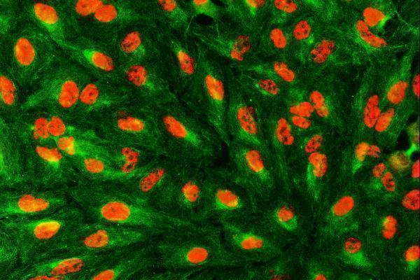
| What should IF/ICC show? | In its separate IF/ICC assay, expect mainly nuclear speckles, with an additional supported Golgi location (HPA: enhanced nuclear-speckle and supported Golgi ICC-IF localizations). This answers the localization question only; the ICC-IF observation does not supply an IHC protocol or prove that chromogenic paraffin sections will resolve speckles (HPA: subcellular ICC-IF profile). |
| Situation | Likely cause | Next action |
|---|---|---|
| Known-positive epithelium is blank in the same run. | Primary-antibody or detection conditions may have failed; the named epithelium is an HPA High reference (HPA: High in bronchial respiratory or esophageal squamous epithelial cells). | Verify control-section integrity, antibody use and detection reagents; then assess the documented paraffin-IHC retrieval conditions (general IHC practice). The sources do not establish a SART1-specific fixation sensitivity. |
| Signal is mostly at cell borders. | That distribution conflicts with the nuclear expectation and lacks support from the annotated topology (HPA: nuclear tissue profile; UniProt O43290: no transmembrane segment). | Review the no-primary control, chromogen deposition and primary-antibody dilution; repeat with an IHC Supported antibody if needed (general IHC practice; HPA: HPA031188 and HPA031189 IHC Supported). |
| Unexpected cells stain more strongly than the positive reference. | Cross-reactivity or endogenous detection activity is possible when staining appears in cells HPA lists as Not detected (HPA: adipocytes, alveolar cells and cardiomyocytes Not detected; general chromogenic IHC practice). | Compare matched sections with no-primary and detection controls, then score named cell populations separately (general IHC practice; HPA: cell-specific tissue profile). |
| Brown haze prevents nuclear scoring. | Background from concentrated primary antibody, incomplete washes or overdeveloped chromogen can obscure compartment assignment (general chromogenic IHC practice). | Adjust the general assay conditions against a same-run positive control and confirm that nuclear boundaries become readable (general IHC practice; HPA: expected nuclear profile). |
| A specimen is called negative because one cell type is unstained. | HPA reports differing levels within its tissue survey, including High, Low and Not detected cell populations (HPA: tissue IHC profile). | Record the cell population and nuclear intensity separately, and compare with a named HPA reference population; do not extend a negative call to the entire section (HPA: cell-specific tissue profile; general IHC scoring practice). |
| A punctate or Golgi-like signal is used to validate the paraffin IHC result. | Those finer locations come from ICC-IF, whereas HPA summarizes tissue IHC as nuclear (HPA: subcellular ICC-IF profile and tissue IHC profile). | Judge chromogenic IHC by interpretable nuclear staining in the expected cells and assay controls; consult the separate IF/ICC guide for that application (HPA: tissue IHC profile; general IHC practice). |


Comprehensive Human Protein Atlas IHC scoring per tissue (reliability: Supported — Medium consistency between antibody staining and RNA expression data. Pending external verification.). Rows are taken directly from the HPA tissue chart — click any row's HPA link to view the source.
| Tissue | Cell type | Level | Evidence | Source |
|---|---|---|---|---|
| Bronchus | Respiratory epithelial cells | High | Protein (IHC) | HPA → |
| Esophagus | Squamous epithelial cells | High | Protein (IHC) | HPA → |
| Nasopharynx | Respiratory epithelial cells | High | Protein (IHC) | HPA → |
| Oral mucosa | Squamous epithelial cells | High | Protein (IHC) | HPA → |
| Placenta | Trophoblastic cells | High | Protein (IHC) | HPA → |
| Tissue | Cell type | Level | Evidence | Source |
|---|---|---|---|---|
| Adipose tissue | Adipocytes | Not detected | Protein (IHC) | HPA → |
| Bone marrow | Hematopoietic cells | Not detected | Protein (IHC) | HPA → |
| Endometrium | Cells in endometrial stroma | Not detected | Protein (IHC) | HPA → |
| Fallopian tube | Glandular cells | Not detected | Protein (IHC) | HPA → |
| Heart muscle | Cardiomyocytes | Not detected | Protein (IHC) | HPA → |
Troubleshoot SART1 chromogenic IHC by checking retrieval, nuclear staining, controls, and scoring; apply separate validation criteria when using IF.
A05414-1 has human paraffin-section IHC images and human cell and tissue IF images (catalog image captions); Human, Mouse and Rat reactivity is listed (catalog reactivity).
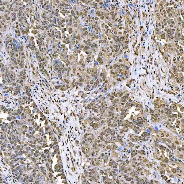
A05414-1 has IHC images from paraffin sections of human colorectal adenocarcinoma, ovarian cancer, breast cancer and laryngeal squamous cell carcinoma (catalog IHC image captions). The same SKU has IF images from A549 cells and a paraffin section of human breast cancer (catalog IF image captions).
Which to pick: Choose A05414-1 for chromogenic IHC on human paraffin sections: its colorectal adenocarcinoma image used EDTA retrieval at pH 8.0, 2 μg/ml primary antibody and DAB detection (A05414-1 IHC image caption); the fixative is unreported (A05414-1 IHC image caption). Choose A05414-1 for IF/ICC because those applications are listed and its IF images show A549 cells and human breast cancer tissue (catalog applications; catalog IF image captions). For cross-species work, A05414-1 lists Human, Mouse and Rat reactivity, while the supplied IHC images show human tissue (catalog reactivity; catalog IHC image captions); it is rabbit-hosted, with no clone specified (catalog host and clone fields).