This website uses cookies to ensure you get the best experience on our website.


- Table of Contents
Plan chromogenic SART3 IHC in paraffin sections using mainly nuclear staining as the expected tissue pattern (HPA tissue IHC). Breast glandular cells and bone marrow hematopoietic cells provide high-staining references, while low consistency between staining and RNA expression warrants caution when scoring (HPA tissue IHC).

Expected localisation, validated protocols, controls and antibodies — the at-a-glance facts below, then the full design guide.
| Expected localisation | Mainly nuclear tissue staining (HPA tissue IHC) | |
| Staining pattern | Ubiquitous, mainly nuclear staining (HPA tissue IHC) | |
| Antigen retrieval | EDTA pH 8.0 HIER, heat-mediated (datasheet A03973-2) | |
| Positive control | Adrenal gland+4 more · see all | |
| Negative control | None in HPA (detected in all 45 tissues); use no-primary + isotype controls |
| Fixation | Keep fixation conditions consistent across sections. (standard IHC practice; not target-specific) | |
| Caveat | Staining and RNA expression show low consistency (HPA tissue IHC) | |
| Regulation | Ubiquitously expressed (UniProt) | |
| Isoform / epitope | 4 isoforms; epitope coverage is unspecified (UniProt) |
The catalog antibody’s IHC-P protocol (datasheet A03973-2) is accompanied by published SART3 staining conditions for human HCC tissue (PMC5256867; PMC10496991).
| Sample | Paraffin-embedded human breast cancer tissue; fixative not specified (datasheet A03973-2) |
| Fixation | Image fixative and duration unreported (datasheet A03973-2); verify before use. |
| Sectioning | 4–5 µm sections on charged slides (standard) |
| Deparaffinisation | Xylene, graded ethanol series to water (standard) |
| Antigen retrieval | Heat retrieval: EDTA pH 8.0 (datasheet A03973-2); 20 min, 95–100 °C (standard) |
| Peroxidase block | 3% H2O2, 10 min, room temperature (standard) |
| Blocking | 10% goat serum (datasheet A03973-2) |
| Primary antibody | Rabbit anti-SART3, 2-5 μg/ml (datasheet A03973-2) |
| Primary incubation | Overnight at 4 °C (datasheet A03973-2) |
| Detection | HRP-conjugated secondary, DAB chromogen (datasheet A03973-2) |
| Counterstain | Hematoxylin, blue, dehydrate and mount (standard) |
| Expected result | SART3-positive staining in glandular cells of adrenal gland (HPA tissue IHC: High). HPA tissue profile: Ubiquitous mainly nuclear expression. No signal in the no-primary control. |
SART3 is expected mainly in nuclei, consistent with UniProt nucleoplasm, Cajal body and nuclear speckle annotations; UniProt also lists cytoplasm and no transmembrane segment (UniProt Q15020). HPA describes ubiquitous, mainly nuclear tissue staining, with high staining in several glandular, hematopoietic, epithelial and neuronal cell populations (HPA tissue IHC). Its tissue IHC reliability is Approved, with low consistency between staining and RNA expression (HPA tissue IHC).
| Predominantly nuclear staining in an HPA high-staining population, such as bone marrow hematopoietic cells or breast glandular cells (HPA tissue IHC). | This fits the reported mainly nuclear tissue pattern (HPA tissue IHC) and UniProt nuclear locations (UniProt Q15020). Compare cell identity and nuclear localisation together; a positive-looking region alone does not establish antibody specificity (standard IHC interpretation). |
| Predominantly cytoplasmic staining with weak or absent nuclear staining. | This departs from HPA's mainly nuclear tissue pattern (HPA tissue IHC). UniProt also lists cytoplasm, so some cytoplasmic signal cannot be dismissed categorically (UniProt Q15020). Check morphology and controls before treating a cytoplasm-dominant result as SART3-specific (standard IHC interpretation). |
| Strong staining in cells other than the annotated high-staining population within a sampled tissue (HPA tissue IHC). | Consider cross-reactivity or endogenous detection activity, especially if the signal also appears in a reagent control (standard IHC practice). SART3 is ubiquitously expressed, so an unlisted cell type is not, by itself, a valid negative control (UniProt Q15020; HPA tissue IHC). |
| Diffuse colour over nuclei, cytoplasm and extracellular areas, without clear cellular boundaries. | This is harder to reconcile with HPA's mainly nuclear pattern (HPA tissue IHC). Non-specific reagent binding, excess detection signal or endogenous enzyme activity can create broad chromogenic background; inspect controls before scoring individual nuclei (standard IHC practice). |
| No convincing nuclear signal in an HPA high-staining population, such as bronchial respiratory epithelial cells (HPA tissue IHC). | First assess section quality, cell identification, controls and assay conditions (standard IHC practice). HPA reports high staining in that population, but its Approved tissue profile has low consistency with RNA expression, so a single negative section needs cautious interpretation (HPA tissue IHC). |
| Subcellular reference | HPA reports ubiquitous mainly nuclear tissue staining, while UniProt lists nucleoplasm, Cajal bodies, nuclear speckles and cytoplasm (HPA tissue IHC; UniProt Q15020). Nuclear predominance is the practical IHC expectation; cytoplasmic signal needs context. |
| Tissue benchmarks | HPA calls adrenal and breast glandular cells, bone marrow hematopoietic cells, and several neuronal populations High; lung alveolar cells, oral squamous epithelial cells and liver cholangiocytes are Low (HPA tissue IHC). Low means weaker reported staining, not a validated absence. |
| Antibody evidence | The HPA tissue profile is Approved but has low agreement between antibody staining and RNA expression (HPA tissue IHC). Among listed antibodies, HPA044322 is IHC Approved; HPA028840 has ICC Supported status with no listed IHC status (HPA antibodies). |
| Molecular context | SART3 has four reported isoforms and no transmembrane segment or signal peptide (UniProt Q15020). The supplied record gives no antibody epitope, so isoform recognition cannot be inferred; topology supports evaluating intracellular compartments rather than a membrane rim (UniProt Q15020). |
| Preparation and detection | The supplied UniProt and HPA records give no SART3-specific fixation sensitivity or antigen retrieval condition. If adjusting retrieval or detection, treat those changes as general IHC optimisation and judge them against morphology and controls (standard IHC practice). |
| Situation | Likely cause | Next action |
|---|---|---|
| Known high-staining cells show no nuclear signal (HPA tissue IHC). | The cells may be misidentified, the section may be compromised, or the staining workflow may have failed (standard IHC practice). | Confirm tissue morphology and run a suitable assay control; review retrieval, antibody dilution and detection conditions as general IHC checks (standard IHC practice). Do not infer SART3-specific fixation sensitivity from HPA staining levels. |
| Colour is mainly cytoplasmic or outlines membranes. | A membrane rim conflicts with the reported mainly nuclear IHC pattern and UniProt's lack of a transmembrane segment (HPA tissue IHC; UniProt Q15020). UniProt does list cytoplasm (UniProt Q15020). | Recheck nuclear counterstain, cellular boundaries and reagent controls; score any cytoplasmic signal separately from nuclear signal (standard IHC practice). |
| Unexpected cells stain more strongly than the annotated population (HPA tissue IHC). | Cross-reactivity or endogenous enzyme activity is possible, but ubiquitous expression limits claims that a cell type must be negative (UniProt Q15020; standard IHC practice). | Compare the annotated cells in the same section and inspect reagent controls; use an independent specificity check before assigning the unexpected signal to SART3 (standard IHC practice). |
| Broad background obscures nuclei. | Non-specific binding, excessive detection signal or endogenous peroxidase can produce diffuse chromogenic colour (standard IHC practice). | Review blocking, washing, antibody concentration and endogenous-enzyme controls; retain conditions that preserve interpretable nuclear staining and morphology (standard IHC practice). |
| A low-staining population appears negative. | HPA labels lung alveolar cells, oral squamous epithelial cells and liver cholangiocytes Low, rather than absent (HPA tissue IHC). | Interpret alongside a high-staining population and assay controls; avoid calling a Low population a confirmed negative or using it alone to reject the assay (HPA tissue IHC; standard IHC practice). |
| What should IF/ICC show? | HPA supports nucleoplasmic localisation in ICC-IF and lists HAP1 and HEL image sets; UniProt additionally lists other nuclear sites and cytoplasm (HPA subcellular; UniProt Q15020). | Use the separate IF/ICC guide for that application. Compare any IF image with the supported nucleoplasmic reference; do not transfer an IF pattern or protocol setting directly into IHC-P scoring (HPA subcellular; standard assay interpretation). |
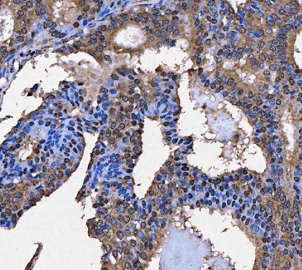
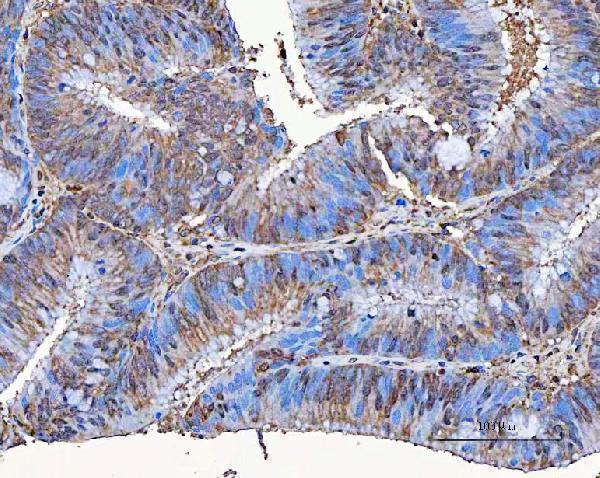
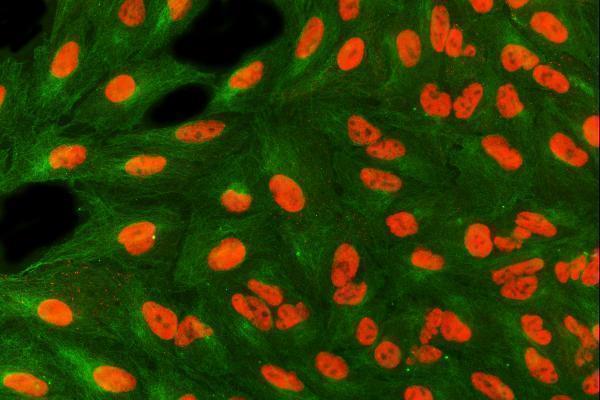


Comprehensive Human Protein Atlas IHC scoring per tissue (reliability: Approved — Low consistency between antibody staining and RNA expression data.). Rows are taken directly from the HPA tissue chart — click any row's HPA link to view the source.
| Tissue | Cell type | Level | Evidence | Source |
|---|---|---|---|---|
| Adrenal gland | Glandular cells | High | Protein (IHC) | HPA → |
| Bone marrow | Hematopoietic cells | High | Protein (IHC) | HPA → |
| Breast | Glandular cells | High | Protein (IHC) | HPA → |
| Bronchus | Respiratory epithelial cells | High | Protein (IHC) | HPA → |
| Caudate | Neuronal cells | High | Protein (IHC) | HPA → |
| Tissue | Cell type | Level | Evidence | Source |
|---|---|---|---|---|
| None in HPA: SART3 is detected in all 45 scored tissues. Use a no-primary (secondary-only) and an isotype control instead. | ||||
Troubleshoot SART3 staining by checking retrieval, nuclear localisation, and background before comparing signal across paraffin sections.
The IHC-validated anti-SART3 antibody has image data from human paraffin tissue sections and IF data from A549 cells and human paraffin tissue sections (catalog: A03973-2 image captions).
A03973-2 has IHC image data from paraffin sections of human breast cancer, colorectal adenocarcinoma and laryngeal squamous cell carcinoma (catalog: A03973-2 IHC image captions). It also has IF image data from A549 cells and a paraffin section of human esophageal squamous carcinoma (catalog: A03973-2 IF image captions).
Which to pick: For tissue IHC, choose A03973-2: its paraffin-section image captions document EDTA pH 8.0 retrieval, 2 μg/ml primary antibody and DAB detection; the fixative is unreported (catalog: A03973-2 IHC image captions). For IF/ICC, A03973-2 lists both applications and has corresponding image data (catalog: A03973-2 applications and IF image captions). No cross-species option is documented here; A03973-2 lists human reactivity only, and its clonality is unspecified (catalog: A03973-2 reactivity and clone fields).