This website uses cookies to ensure you get the best experience on our website.


- Table of Contents
Plan paraffin-section SASS6 IHC using the IHC-validated antibody at 2–5 μg/ml (datasheet A05762-1). Compare medium staining in endometrial glandular cells with undetected staining in adrenal glandular cells, while accounting for uncertain tissue staining reliability (HPA tissue IHC).

Expected localisation, validated protocols, controls and antibodies — the at-a-glance facts below, then the full design guide.
| Expected localisation | Cytoplasmic tissue staining (HPA tissue IHC) | |
| Staining pattern | Cytoplasmic staining in most tissues and some immune cells (HPA tissue IHC) | |
| Antigen retrieval | EDTA pH 8.0 HIER, heat-mediated (datasheet A05762-1) | |
| Positive control | Adipose tissue+4 more · see all | |
| Negative control | Adrenal gland+4 more · see all |
| Fixation | Keep paraffin-section fixation consistent across samples. (standard IHC practice; not target-specific) | |
| Caveat | Tissue staining is uncertain; external verification is pending (HPA tissue IHC) | |
| Regulation | Expression regulation is not annotated (UniProt) | |
| Isoform / epitope | No annotated isoforms or processing; one 1–657 chain (UniProt) |
Compare the catalog antibody's IHC-P protocol (datasheet A05762-1) with the published paraffin-section protocol (PMC11499408).
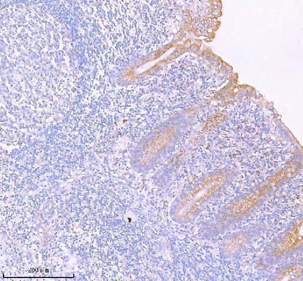
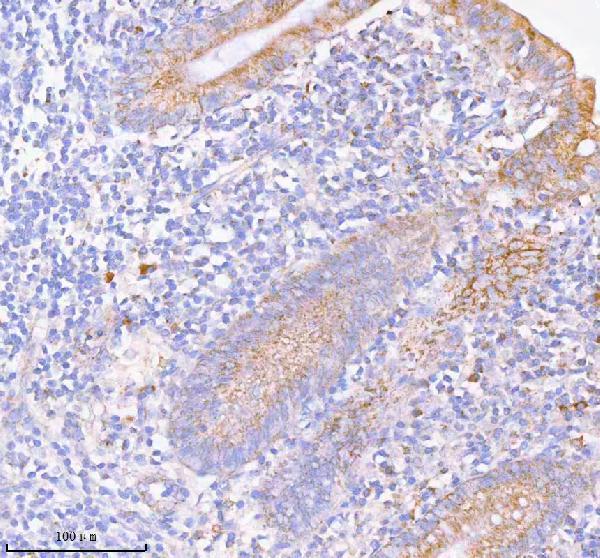
| Sample | Paraffin-embedded human appendiceal carcinoid tissue; fixative not specified (datasheet A05762-1) |
| Fixation | Image fixative and duration unreported (datasheet A05762-1); verify before use. |
| Sectioning | 4–5 µm sections on charged slides (standard) |
| Deparaffinisation | Xylene, graded ethanol series to water (standard) |
| Antigen retrieval | Heat retrieval: EDTA pH 8.0 (datasheet A05762-1); 20 min, 95–100 °C (standard) |
| Peroxidase block | 3% H2O2, 10 min, room temperature (standard) |
| Blocking | 10% goat serum (datasheet A05762-1) |
| Primary antibody | Rabbit anti-SASS6, 2-5 μg/ml (datasheet A05762-1) |
| Primary incubation | Overnight at 4 °C (datasheet A05762-1) |
| Detection | HRP-conjugated secondary, DAB chromogen (datasheet A05762-1) |
| Counterstain | Hematoxylin, blue, dehydrate and mount (standard) |
| Expected result | SASS6-positive staining in adipocytes of adipose tissue (HPA tissue IHC: Medium). HPA tissue profile: Cytoplasmic expression in most tissues and in a subset of immune cells. No signal in the no-primary control. |
SASS6 is associated with centrosomes and nascent procentrioles, with no transmembrane segment (UniProt Q6UVJ0). In paraffin section IHC, assess cytoplasmic staining in the cell populations reported by HPA, such as endometrial glandular cells or kidney glomerular cells, both stained at medium intensity (HPA: tissue IHC). Treat that pattern as provisional: HPA rates the tissue staining and its listed antibody HPA028187 as uncertain (HPA: tissue IHC; HPA: antibody validation).
| Cytoplasmic staining in endometrial glandular cells or kidney glomerular cells, with limited staining in adjacent cells. | This is compatible with HPA's medium staining observations in those cell populations (HPA: tissue IHC). Score the relevant cells and compartment; the uncertain tissue and antibody ratings mean the pattern alone does not establish specificity (HPA: tissue IHC; HPA: antibody validation). |
| Small cytoplasmic foci are visible, but many otherwise stained cells lack a resolvable punctum. | A focal component is plausible because SASS6 associates transiently with nascent procentrioles (UniProt Q6UVJ0). Chromogenic section IHC may not resolve every centriole; interpret the overall cellular pattern alongside controls rather than requiring a punctum in each positive cell (standard IHC practice). |
| Predominantly nuclear or membrane rim staining appears without a convincing cytoplasmic component. | That compartment pattern conflicts with UniProt's centrosome and centriole annotation and its lack of a transmembrane segment (UniProt Q6UVJ0). Consider nonspecific staining or a detection artefact, and check controls before assigning it to SASS6 (standard IHC practice). |
| Strong staining appears in alveolar cells or cardiomyocytes while the selected comparison tissue remains faint. | HPA reports SASS6 as not detected in those respective cell populations (HPA: lung alveolar cells; HPA: heart cardiomyocytes). This mismatch raises concern for cross-reactivity or endogenous detection activity; it is a warning, not proof, given HPA's uncertain IHC reliability (HPA: tissue IHC). |
| The whole section has a diffuse tint, or no cells stain in a selected HPA medium-staining tissue. | Diffuse colour obscures compartment scoring; a blank comparison tissue makes this run uninformative (standard IHC practice). Repeat with suitable detection controls, and remember that HPA's medium observations are provisional rather than guaranteed positives (HPA: tissue IHC, uncertain). |
| Cell population used for interpretation | HPA reports medium staining in adipocytes, caudate glia, selected endothelial and glandular cells, and kidney glomerular cells (HPA: tissue IHC). It also reports low or undetected staining in specified other populations; score the annotated cell type, not a whole-organ average (HPA: tissue IHC). |
| Intracellular location and topology | SASS6 is a centrosome and centriole component associated transiently with nascent procentrioles; it has no transmembrane segment (UniProt Q6UVJ0). Cytoplasmic staining can be assessed in section IHC, while a sharply resolved centriole is not required for a compatible chromogenic result (standard IHC practice). |
| Strength of IHC evidence | HPA describes cytoplasmic expression in most tissues and a subset of immune cells, but labels tissue staining uncertain and HPA028187 IHC uncertain (HPA: tissue IHC; HPA: antibody validation). Use tissue comparisons and controls to qualify an interpretation; HPA's reported intensity is not a validated threshold. |
| IF/ICC Q: Should the IHC result reproduce ciliary IF staining? | No direct match is established. HPA approves primary cilium localization in ICC-IF but marks cytosol and vesicles uncertain (HPA: subcellular). UniProt places SASS6 at centrosomes and centrioles (UniProt Q6UVJ0). Interpret each application using its own imaging context. |
| Situation | Likely cause | Next action |
|---|---|---|
| No staining in the selected endometrial glandular or kidney glomerular cells. | The run may have failed, or the selected section may differ from HPA's medium-staining observation; HPA rates its IHC evidence uncertain (HPA: tissue IHC). | Confirm that the intended cells are present, inspect the run controls, and review the catalog antibody's IHC-P instructions before repeating (standard IHC practice). Do not infer absence of SASS6 from a single blank section. |
| Diffuse brown colour makes cytoplasmic staining hard to distinguish. | Nonspecific background or detection activity can obscure cellular localization (standard chromogenic IHC practice). | Compare with a no-primary control; review blocking, washing, and detection exposure. If using peroxidase detection, check the endogenous enzyme quench (standard IHC practice). Reassess only after background permits compartment scoring. |
| Alveolar cells or cardiomyocytes stain strongly. | Those cells are reported as not detected by HPA, so cross-reactivity or endogenous detection activity is possible (HPA: lung alveolar cells; HPA: heart cardiomyocytes). | Compare the same run with an HPA medium-staining cell population and a no-primary control; inspect whether colour tracks cellular compartments or tissue-wide background (HPA: tissue IHC; standard IHC practice). |
| Signal is mainly nuclear or outlines cell membranes. | That distribution conflicts with the annotated centrosome and centriole location and absence of a transmembrane segment (UniProt Q6UVJ0). | Check the no-primary control and tissue morphology, then reassess antibody specificity with an independent control if available (standard IHC practice). Do not score the misplaced signal as a confident SASS6 positive. |
| Only weak cytoplasmic staining is visible in a tissue expected to stain. | HPA reports low staining in some cell populations and medium staining in others; its overall IHC reliability is uncertain (HPA: tissue IHC). The sampled cells may affect the comparison. | Identify and score the exact cell population, compare sections processed in the same run, and confirm that the signal exceeds background (standard IHC practice). Avoid applying a single intensity cutoff across tissues. |
| A ciliary IF image seems inconsistent with the section IHC pattern. | HPA's approved primary cilium location comes from ICC-IF, while its tissue IHC describes cytoplasmic expression and is uncertain (HPA: subcellular; HPA: tissue IHC). | Keep the applications' observations distinct: score the cell type and chromogenic compartment on the section, and consult the separate IF/ICC guide for fluorescence interpretation (standard IHC/IF practice). |
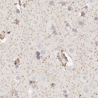


Comprehensive Human Protein Atlas IHC scoring per tissue (reliability: Uncertain — Antibody staining in cells/structures not annotated, view images. Pending external verification.). Rows are taken directly from the HPA tissue chart — click any row's HPA link to view the source.
| Tissue | Cell type | Level | Evidence | Source |
|---|---|---|---|---|
| Adipose tissue | Adipocytes | Medium | Protein (IHC) | HPA → |
| Breast | Adipocytes | Medium | Protein (IHC) | HPA → |
| Caudate | Glial cells | Medium | Protein (IHC) | HPA → |
| Cerebral cortex | Endothelial cells | Medium | Protein (IHC) | HPA → |
| Colon | Endothelial cells | Medium | Protein (IHC) | HPA → |
| Tissue | Cell type | Level | Evidence | Source |
|---|---|---|---|---|
| Adrenal gland | Glandular cells | Not detected | Protein (IHC) | HPA → |
| Appendix | Glandular cells | Not detected | Protein (IHC) | HPA → |
| Cerebellum | Cells in granular layer | Not detected | Protein (IHC) | HPA → |
| Fallopian tube | Glandular cells | Not detected | Protein (IHC) | HPA → |
| Heart muscle | Cardiomyocytes | Not detected | Protein (IHC) | HPA → |
Troubleshoot chromogenic SASS6 IHC in paraffin sections using the catalog antibody’s tissue image, UniProt localisation, and cautiously interpreted HPA observations.
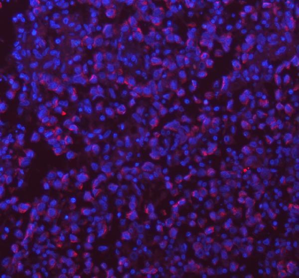
A05762-1 has IHC data from human paraffin sections of appendiceal carcinoid and breast cancer, plus IF data from a human paraffin section of testis cancer (catalog image captions).
A05762-1 has IHC images from human paraffin sections of appendiceal carcinoid and breast cancer (catalog IHC captions). Its IF image shows a human paraffin section of testis cancer; the catalog lists Human, Mouse and Rat reactivity (catalog IF caption; catalog reactivity).
Which to pick: Choose A05762-1 for tissue IHC: its IHC captions document staining of human paraffin sections with the rabbit antibody, while the fixative is unreported (catalog IHC captions). For IF, A05762-1 has a human tissue section image, but the supplied data do not establish ICC validation (catalog IF caption; catalog applications). For work across species, A05762-1 lists Human, Mouse and Rat reactivity; its IHC dilution listing specifies Human and Mouse, and the supplied IHC images show human tissue (catalog reactivity; catalog dilution listing; catalog IHC captions).