SAV1 / Protein salvador homolog 1 · IHC design guide

Design Immunohistochemistry for SAV1

Plan chromogenic SAV1 IHC in paraffin sections with the catalog antibody at 2–5 μg/mL (datasheet A04183-2). Use the reported cytoplasmic tissue pattern and its low consistency with RNA expression to guide interpretation (HPA tissue IHC).

Evidence assembled Oct 2026 · For research use; verify linked source records and product datasheet before use
Immunohistochemistry protocol sheet for SAV1 (IHC for SAV1): expected localisation Cytoplasm in tissue (HPA tissue IHC); nucleus annotated (UniProt), antibody A04183-2, validated IHC image, and IHC protocol steps
Printable SAV1 IHC protocol sheet — expected localisation Cytoplasm in tissue (HPA tissue IHC); nucleus annotated (UniProt), antibody A04183-2, controls and protocol steps. Open the full SAV1 IHC guide →

SAV1 Immunohistochemistry Experimental Design Guide

Expected localisation, validated protocols, controls and antibodies — the at-a-glance facts below, then the full design guide.

Must know before staining
Expected localisation Cytoplasm in tissue (HPA tissue IHC); nucleus annotated (UniProt)
Staining pattern Cytoplasmic staining in most tissues; glandular cells positive (HPA tissue IHC)
Antigen retrieval EDTA pH 8.0 HIER, heat-mediated (datasheet A04183-2)
Positive control ⓘ Adrenal gland+4 more · see all
Negative control ⓘ Adipose tissue+4 more · see all
Important caveats
Reasons your staining may differ from the expected pattern.
Fixation Matched tissue-IHC evidence does not establish the fixation claim. Validate the specimen-specific method before use. (selected-SKU IHC image A04183-2)
Caveat Antibody staining has low consistency with RNA expression (HPA tissue IHC)
Regulation Higher expression in fetal than adult heart (UniProt)
Isoform / epitope No listed isoforms; full-length chain 1–383 (UniProt)
Section 1

Recommended SAV1 IHC & IF Protocols

Compare the catalog antibody’s IHC-P protocol (datasheet A04183-2) with three published SAV1 tissue protocols (PMC10742029; PMC4978403; PMC11083677).

Recommended immunohistochemistry (IHC-P) protocol parameters
SampleParaffin-embedded human lung cancer tissue; fixative not specified (datasheet A04183-2)
FixationImage fixative and duration unreported (datasheet A04183-2); verify before use.
Sectioning4–5 µm sections on charged slides (standard)
DeparaffinisationXylene, graded ethanol series to water (standard)
Antigen retrievalHeat retrieval: EDTA pH 8.0 (datasheet A04183-2); 20 min, 95–100 °C (standard)
Peroxidase block3% H2O2, 10 min, room temperature (standard)
Blocking10% goat serum (datasheet A04183-2)
Primary antibodyRabbit anti-SAV1, 2-5 μg/ml (datasheet A04183-2)
Primary incubationOvernight at 4 °C (datasheet A04183-2)
DetectionHRP-conjugated secondary, DAB chromogen (datasheet A04183-2)
CounterstainHematoxylin, blue, dehydrate and mount (standard)
Expected resultSAV1-positive staining in glandular cells of adrenal gland (HPA tissue IHC: Medium). HPA tissue profile: Cytoplasmic expression in most tissues. No signal in the no-primary control.
💡Decision noteStart with heat-mediated EDTA retrieval at pH 8.0 (datasheet A04183-2); the three emitted articles do not specify retrieval conditions (PMC10742029; PMC4978403; PMC11083677).
Section 2

What Is the Expected SAV1 Staining Pattern?

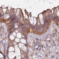
SAV1 is primarily cytoplasmic in tissue IHC (HPA: cytoplasmic expression in most tissues); UniProt also lists nuclear localization (UniProt Q9H4B6: nucleus, cytoplasm). Expect staining in glandular cells of colon, breast and other listed positive tissues, and in bronchial respiratory epithelial cells (HPA: medium staining). SAV1 has no transmembrane segment (UniProt Q9H4B6: topology). Interpret tissue results cautiously: HPA rates the IHC antibody Approved but reports low consistency between staining and RNA expression (HPA: reliability description).

What am I looking at on my slide?
Cytoplasmic staining in colon glandular cells or bronchial respiratory epithelial cells.This fits the reported compartment and cell types (HPA: cytoplasmic expression in most tissues; medium in these cells). Compare staining within the specified cells, since a positive tissue can contain cells with different staining patterns (general IHC practice).
Predominantly nuclear staining, with little cytoplasmic signal.Review cautiously: UniProt lists the nucleus as a possible SAV1 location, while HPA reports cytoplasmic tissue staining and supported cytosolic ICC-IF localization (UniProt Q9H4B6: subcellular location; HPA: tissue IHC, ICC-IF). A nuclear-only result does not match HPA's principal observed pattern; check controls and detection artefacts before assigning it to SAV1 (general IHC practice).
Strong staining confined to adipocytes or esophageal squamous epithelial cells.These cell types were not detected in the supplied tissue IHC record (HPA: adipose tissue, esophagus). Check for cross-reactivity or endogenous detection activity with suitable controls (general IHC practice). An unexpected stain alone cannot establish which explanation applies.
Diffuse color across several compartments, including cell-free areas.This does not provide a clear cellular localization; HPA describes cytoplasmic expression in most tissues (HPA: tissue IHC profile). Review background in the no-primary control, blocking, washes and chromogen development before scoring cells as positive (general IHC practice).
No visible signal in colon glandular cells despite preserved tissue morphology.Colon glandular cells are reported at medium staining (HPA: colon), so inspect the IHC workflow and a concurrent positive control (general IHC practice). The HPA antibody is Approved, but staining has low consistency with RNA expression; absence in one section is not proof that SAV1 is absent (HPA: reliability description).
💡Expected SAV1 appearanceCall a section positive when its specified cells show discernible, mainly cytoplasmic staining—for example, medium staining in colon glandular cells (HPA: tissue IHC); broad acellular color or isolated staining of HPA-undetected cell types is suspect (HPA: tissue IHC; general IHC practice).
How each factor affects the staining
Compartment used for scoringPrioritize interpretable cytoplasmic staining in tissue IHC (HPA: tissue IHC profile). UniProt also lists the nucleus, so record a nuclear component separately and assess its controls (UniProt Q9H4B6: subcellular location; general IHC practice).
Choice of comparison cellsColon glandular and bronchial respiratory epithelial cells are medium-staining examples; adipocytes and esophageal squamous epithelial cells are reported as not detected (HPA: tissue IHC). Use the named cell types when comparing sections.
Strength of validation evidenceThe listed antibody, HPA001808, has Approved IHC status, alongside a warning of low consistency between staining and RNA expression (HPA: antibody status, tissue reliability). Treat discordant staining as a finding to verify.
Intracellular protein featuresSAV1 has no transmembrane segment or signal peptide and is annotated in the cytoplasm and nucleus (UniProt Q9H4B6: topology, processing, subcellular location). The supplied sources do not establish a SAV1-specific antigen retrieval or fixation response.
IF/ICC: what localization should I expect?HPA reports supported cytosolic localization in ICC-IF, with images from A-431, SiHa and U2OS (HPA: subcellular). Interpret IF localization against that observation; this tissue IHC section supplies no IF protocol.
Why is my staining missing, weak or wrong?
SituationLikely causeNext action
Cytoplasmic stain is faint in a selected positive cell type.The observed level may vary by cell type; some listed cells stain low rather than medium (HPA: tissue IHC). Weak detection can also arise during a general IHC workflow.Confirm the exact HPA cell type and its reported level, then review the concurrent positive control, retrieval and detection steps (HPA: tissue IHC; general IHC practice). No SAV1-specific retrieval condition is established here.
Nuclei dominate the staining.A nuclear location is annotated, but HPA's tissue profile is cytoplasmic and its ICC-IF localization is cytosolic (UniProt Q9H4B6: subcellular location; HPA: tissue IHC, ICC-IF).Score nuclear and cytoplasmic compartments separately; compare no-primary and positive controls before interpreting nuclear-only staining (general IHC practice).
Color appears in an HPA-undetected cell type.Possible cross-reactivity or endogenous detection activity (general IHC practice); HPA reports adipocytes and esophageal squamous epithelial cells as not detected (HPA: tissue IHC).Check a no-primary control and the detection system, then compare staining with a named HPA-positive cell type on a suitable section (general IHC practice; HPA: tissue IHC).
The entire section has diffuse background.Nonspecific reagent binding, inadequate washing or excess chromogen development can obscure cellular staining (general IHC practice).Inspect the no-primary control, optimize blocking and washes, and shorten detection development if background persists (general IHC practice). Reassess whether cytoplasmic cells remain distinguishable (HPA: tissue IHC profile).
A positive comparison section shows no signal.A failed staining run is possible, while HPA also cautions that staining has low consistency with RNA expression (general IHC practice; HPA: reliability description).Verify control performance and review the IHC workflow before calling SAV1 absent; record the cell type and section examined (general IHC practice).
Tissue IHC and ICC-IF seem to disagree.HPA describes cytoplasmic staining in most tissues and supported cytosolic localization in ICC-IF (HPA: tissue IHC, subcellular). Differences in the cells examined may complicate comparison.Compare compartment calls in the specific cells tested and document controls for each assay (general IHC practice); do not infer an IF staining procedure from the tissue IHC pattern.

Sample controls for SAV1 IHC & IF

🧪Run breast first: its glandular cells should show SAV1 staining (HPA: Medium in breast glandular cells). Use adipose tissue as the negative tissue (HPA: Not detected in adipocytes); adipocytes, if present within the breast section, should remain unstained (HPA: Not detected in adipocytes).
Positive control tissue: Adrenal gland (Glandular cells, HPA Medium)
Negative control tissue: Adipose tissue (HPA Not detected)
ICC-IF cell lines (HPA subcellular resource): HPA ICC-IF images show SAV1 in A-431, SiHa, U2OS, with annotated localisation: Cytosol (supported) (HPA subcellular).
Technical controls: Include no-primary (secondary-only), a concentration-matched rabbit isotype control for the rabbit primary, and a SAV1 knockout or validated peptide-block control (selected-SKU caption: rabbit anti-SAV1 antibody; standard IHC controls). Quench endogenous peroxidase and check for residual signal in the breast section before interpreting DAB staining (selected-SKU caption: peroxidase/DAB detection; standard IHC practice).
⚠️Feasibility: A paraffin-section example uses heat-mediated EDTA retrieval at pH 8.0, but it does not establish that retrieval is required; the selected-SKU caption does not report a fixative, and a target-specific fixation window or fixation effect is unreported (selected-SKU tissue-IHC caption). Frozen-section performance and whether IF is easier than paraffin IHC are unreported; ICC-IF images in A-431, SiHa and U2OS support cytosolic localisation (HPA: Cytosol supported; ICC-IF images). In breast sections, score glandular cells separately from adipocytes so unstained fat does not dilute the assessment (HPA: Medium in breast glandular cells; Not detected in adipocytes).

HPA tissue IHC evidence for SAV1

Comprehensive Human Protein Atlas IHC scoring per tissue (reliability: Approved — Low consistency between antibody staining and RNA expression data.). Rows are taken directly from the HPA tissue chart — click any row's HPA link to view the source.

Positive expression · recommended positive controls

TissueCell typeLevelEvidenceSource
Adrenal gland Glandular cells Medium Protein (IHC) HPA →
Appendix Glandular cells Medium Protein (IHC) HPA →
Breast Glandular cells Medium Protein (IHC) HPA →
Bronchus Respiratory epithelial cells Medium Protein (IHC) HPA →
Colon Glandular cells Medium Protein (IHC) HPA →

Undetected expression · recommended negative controls

TissueCell typeLevelEvidenceSource
Adipose tissue Adipocytes Not detected Protein (IHC) HPA →
Bone marrow Hematopoietic cells Not detected Protein (IHC) HPA →
Esophagus Squamous epithelial cells Not detected Protein (IHC) HPA →
Liver Cholangiocytes Not detected Protein (IHC) HPA →
Oral mucosa Squamous epithelial cells Not detected Protein (IHC) HPA →
Section 3

Advanced SAV1 IHC Tips

Troubleshoot SAV1 staining in paraffin sections by checking retrieval, compartment, cell type, and controls before comparing chromogenic signal across samples.

What should I change when SAV1 staining is weak after antigen retrieval?
Start with heat-mediated retrieval in EDTA pH 8.0 for paraffin SAV1 IHC (datasheet A04183-2). The catalog antibody stained a paraffin section of human lung cancer tissue after this retrieval, followed by 2 μg/ml primary antibody overnight at 4°C (datasheet A04183-2). Check that sections remain attached and compare retrieved sections with an otherwise identical unretrieved control before increasing retrieval exposure (standard IHC practice). Keep antibody concentration, incubation, and DAB development matched while testing retrieval, so a signal change has an interpretable cause (standard IHC practice). Record the heating and cooling conditions used, because the caption does not specify their duration (datasheet A04183-2).
Could fixation explain inconsistent SAV1 staining between paraffin blocks?
The selected image documents a paraffin section but does not report its fixative, so SAV1 sensitivity to a particular fixative or fixation time is unknown (datasheet A04183-2). Record fixative, delay before fixation, duration, and specimen thickness for each block before attributing a difference to SAV1 expression (standard IHC practice). Compare blocks using the same EDTA pH 8.0 retrieval and 2 μg/ml primary conditions where appropriate (datasheet A04183-2). Include a consistently processed control section in each staining run to reveal processing or staining variation (standard IHC practice). Neither tissue staining patterns nor SAV1 topology establishes a target-specific fixation effect (HPA: tissue IHC; UniProt Q9H4B6 topology).
Should I score nuclear or cytoplasmic SAV1 staining in tissue sections?
Score cytoplasmic staining separately from nuclear staining: tissue IHC describes cytoplasmic expression in most tissues, while supported subcellular localization is cytosolic (HPA: tissue IHC; HPA: subcellular). Nuclear localization is also recorded, so a nuclear signal merits documentation and validation instead of automatic exclusion (UniProt Q9H4B6 subcellular location). Use the counterstain to mark nuclear boundaries, then compare compartments within the same defined cell population (standard IHC practice). SAV1 has no transmembrane segment, so a sharp membrane-only pattern needs scrutiny as a possible artefact (UniProt Q9H4B6 topology). Report compartment-specific scores rather than combining unlike patterns into one intensity value (standard IHC practice).
How can I assess whether epitope accessibility is changing SAV1 staining?
The record lists 0 alternative isoforms and does not locate this antibody’s epitope, so do not assign staining differences to isoform switching (UniProt Q9H4B6 isoforms; datasheet A04183-2). SAV1 contains WW domains at residues 199–232 and 234–267, and a SARAH domain at 321–368 (UniProt Q9H4B6 domains). Reported phosphorylation sites include 94, 136, and 210; their effect on this antibody’s binding is unestablished without epitope information (UniProt Q9H4B6 modified residues; datasheet A04183-2). When staining varies, compare identically processed sections and a second independently characterized antibody if available (standard IHC practice). Document the tested retrieval and antibody conditions before interpreting a compartment or intensity shift as biological (standard IHC practice).
How should I check a SAV1 IHC pattern by multiplex immunofluorescence?
Use IF as a separate validation experiment and mark the cell population being compared; glandular cells show medium tissue IHC staining in several listed tissues (HPA: tissue IHC). Pair SAV1 with an appropriate cell-type marker, then inspect each channel and its single-label control before interpreting overlap (standard IF practice). Choose a fluorophore away from a strongly autofluorescent tissue channel, using a far-red channel when green background dominates (standard IF practice). SAV1 is supported in cytosol and has no transmembrane segment, so test permeabilisation for intracellular access while recognizing that this antibody’s epitope is unspecified (HPA: subcellular; UniProt Q9H4B6 topology; datasheet A04183-2). Do not transfer a fixation condition from the paraffin caption to IF; none is reported there (datasheet A04183-2).
How can I separate SAV1 signal from diffuse brown background?
Run a no-primary section alongside the test section to assess secondary-reagent, endogenous-enzyme, and chromogen background (standard IHC practice). The selected IHC image used 10% goat serum block, a goat anti-rabbit peroxidase secondary for 30 minutes at 37°C, and DAB development (datasheet A04183-2). Include an endogenous peroxidase block when using HRP and DAB, and compare background before lengthening primary incubation or chromogen development (standard IHC practice). Check whether brown pigment persists in the no-primary section or concentrates at folds and section edges (standard IHC practice). Keep exposure and wash conditions consistent across controls so diffuse staining can be traced to a workflow step (standard IHC practice).
What is a defensible way to quantify SAV1 DAB staining? ⚠ ANSWER MARKED FOR VERIFICATION
Define the cell population and compartment before scoring, because tissue IHC describes largely cytoplasmic staining whereas nuclear localization is also recorded (HPA: tissue IHC; UniProt Q9H4B6 subcellular location). An H-score combines the percentage of viable target cells at intensity grades 0–3, yielding 0–300; report the grading rules with each comparison (standard IHC practice). Alternatively, report percent positive cells using one threshold established from controls and held constant across sections (standard IHC practice). Normalize cell counts to viable cells of the same type, or report positive-cell density per mm² of viable tissue (standard IHC practice). Exclude folds, necrosis, and section edges using prespecified rules, and record nuclear and cytoplasmic results separately (standard IHC practice).
Which staining patterns support genuine SAV1 detection rather than artefact?
Look for reproducible staining within defined viable cells and a plausible compartment: cytoplasmic tissue expression is reported, with cytosol supported in subcellular imaging (HPA: tissue IHC; HPA: subcellular). Bronchial respiratory epithelial cells have medium reported staining, while adipocytes are reported as not detected; these are comparative references, not absolute validation controls (HPA: tissue IHC). Treat membrane-only staining cautiously because SAV1 has no transmembrane segment, while nuclear staining requires its own validation (UniProt Q9H4B6 topology; UniProt Q9H4B6 subcellular location). Reject apparent signal confined to edges, folds, or necrotic regions, and check a no-primary section for endogenous peroxidase or DAB background (standard IHC practice). Interpret weak differences cautiously because antibody staining and RNA expression have low consistency in the tissue dataset (HPA: reliability description).
Boster reagents

Best SAV1 / Protein salvador homolog 1 IHC Antibodies

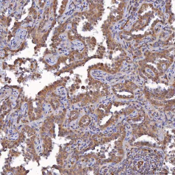
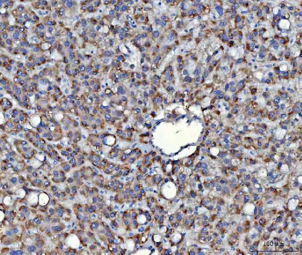
A04183-2 has IHC data from human paraffin sections of lung cancer, liver cancer and spleen (catalog IHC image captions). No IF images are provided (catalog: IF image list empty).

Real IHC data IHC analysis of SAV1 using anti-SAV1 antibody (A04183-2). SAV1 was detected in a paraffin-embedded section of human lung cancer tissue. Heat mediated antigen retrieval was performed in EDTA buffer (pH 8.0, epitope retrieval solution). The tissue section was blocked with 10% goat serum. The tissue section was then incubated with 2 μg/ml rabbit anti-SAV1 Antibody (A04183-2) overnight at 4°C. Peroxidase Conjugated Goat Anti-rabbit IgG was used as secondary antibody and incubated for 30 minutes at 37°C. The tissue section was developed using HRP Conjugated Rabbit IgG Super Vision Assay Kit (Catalog # SV0002) with DAB as the chromogen.
Anti-SAV1 Antibody ®
Cat # A04183-2

A04183-2 is listed for human IHC (catalog: applications and reactivity). Its IHC images show staining in paraffin sections of human lung cancer, liver cancer and spleen (catalog IHC image captions).

Which to pick: Choose A04183-2 for human paraffin-section IHC; its captions document EDTA retrieval at pH 8.0 and staining at 2 μg/ml, but do not report the fixative (catalog IHC image captions). IF/ICC is not listed as a validated application for A04183-2, so there is no supported IF/ICC choice here (catalog: applications and IF image list). Cross-species use is likewise unsupported by this catalog entry, which lists human reactivity only (catalog: reactivity).

Each figure is that product's own IHC / IF validation image from its datasheet.

References

  1. UniProt Consortium. UniProt entry Q9H4B6 (SAV1_HUMAN, Protein salvador homolog 1).
  2. Human Protein Atlas. SAV1 tissue IHC expression (reliability: Approved).
  3. Human Protein Atlas. SAV1 subcellular location (ICC-IF): Localized to the cytosol..
  4. Human Protein Atlas. SAV1 antibody validation summary (1 antibodies).
  5. Impaired Expression of the Salvador Homolog-1 Gene Is Associated with the Development and Progression of Colorectal Cancer. Cancers 2023 — PMC10742029.
  6. Downregulation of SAV1 plays a role in pathogenesis of high-grade clear cell renal cell carcinoma. BMC cancer 2011 — PMC3292516.
  7. The Hippo/MST Pathway Member SAV1 Plays a Suppressive Role in Development of the Prehierarchical Follicles in Hen Ovary. PloS one 2016 — PMC4978403.
  8. Loss of SAV1 in Kidney Proximal Tubule Induces Maladaptive Repair after Ischemia and Reperfusion Injury. International journal of molecular sciences 2024 — PMC11083677.
  9. PubMed PMID:11027580 — UniProt-cited evidence.
  10. PubMed PMID:14702039 — UniProt-cited evidence.
  11. PubMed PMID:15489334 — UniProt-cited evidence.