This website uses cookies to ensure you get the best experience on our website.


- Table of Contents
Plan SBNO1 IHC-P around nuclear staining in most tissues (HPA tissue IHC). Use appendix glandular cells, which show high staining (HPA tissue IHC), as a positive tissue reference when interpreting chromogenic signal.

Expected localisation, validated protocols, controls and antibodies — the at-a-glance facts below, then the full design guide.
| Expected localisation | Nuclear staining in most tissues (HPA tissue IHC) | |
| Staining pattern | Nuclear staining across most tissues (HPA tissue IHC) | |
| Antigen retrieval | EDTA pH 8.0 HIER, heat-mediated (datasheet A13130) | |
| Positive control | Adrenal gland+4 more · see all | |
| Negative control | None in HPA (detected in all 45 tissues); use no-primary + isotype controls |
| Fixation | Keep paraffin-section fixation consistent (standard IHC practice; not target-specific); Matched tissue-IHC evidence does not establish the fixation claim. Validate the specimen-specific method before use. (selected-SKU IHC image A13130) | |
| Caveat | Staining and RNA show medium consistency (HPA tissue IHC) | |
| Regulation | RNA is tissue enhanced in testis (HPA tissue RNA) | |
| Isoform / epitope | Four isoforms; epitope coverage is unreported (UniProt) |
Compare the catalog antibody’s IHC-P protocol with the published human CCA tissue protocol (PMC13067845).
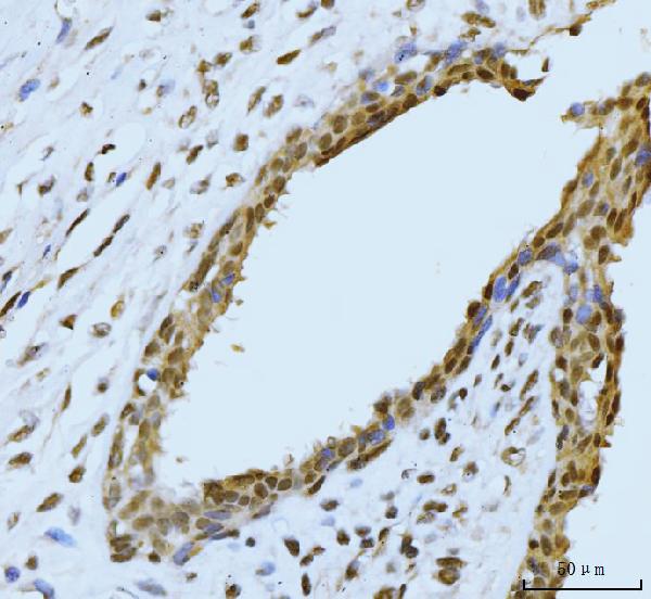
| Sample | Paraffin-embedded human appendiceal adenocarcinoma tissue; fixative not specified (datasheet A13130) |
| Fixation | Image fixative and duration unreported (datasheet A13130); verify before use. |
| Sectioning | 4–5 µm sections on charged slides (standard) |
| Deparaffinisation | Xylene, graded ethanol series to water (standard) |
| Antigen retrieval | Heat retrieval: EDTA pH 8.0 (datasheet A13130); 20 min, 95–100 °C (standard) |
| Peroxidase block | 3% H2O2, 10 min, room temperature (standard) |
| Blocking | 10% goat serum (datasheet A13130) |
| Primary antibody | Rabbit anti-SBNO1, 2-5 μg/ml (datasheet A13130) |
| Primary incubation | Overnight at 4 °C (datasheet A13130) |
| Detection | Streptavidin-biotin complex (SABC), DAB chromogen (datasheet A13130) |
| Counterstain | Hematoxylin, blue, dehydrate and mount (standard) |
| Expected result | SBNO1-positive staining in glandular cells of adrenal gland (HPA tissue IHC: High). HPA tissue profile: Nuclear expression in most tissues. No signal in the no-primary control. |
SBNO1 should appear predominantly in nuclei across most tissues (HPA tissue IHC: nuclear expression in most tissues; UniProt A3KN83: nucleus). High staining is reported in several glandular, respiratory epithelial, hematopoietic, and endothelial cell populations (HPA tissue IHC: High in the listed populations). UniProt annotates no transmembrane segment (UniProt A3KN83 topology). Treat the tissue pattern as a guide, with medium consistency between staining and RNA data and external verification pending (HPA tissue IHC: Approved).
| Distinct nuclear chromogen in adrenal or breast glandular cells, bronchial respiratory epithelial cells, or bone marrow hematopoietic cells. | This fits the reported compartment and high-staining populations (HPA tissue IHC: nuclear expression in most tissues; High in those cells). Compare cells within the section before scoring: the HPA level describes those populations, not every cell in the tissue (HPA tissue IHC). |
| Chromogen is mainly cytoplasmic or outlines cell membranes while nuclei remain pale. | That distribution conflicts with the reported nuclear pattern (HPA tissue IHC: nuclear expression; UniProt A3KN83: nucleus, no transmembrane segment). Review counterstain alignment and detection controls before assigning a cellular compartment (general IHC practice). |
| The strongest staining is in a cell population other than the documented high-staining population within the same tissue. | Check cell identity and possible cross-reactivity or endogenous detection activity (general IHC practice). This is a discrepancy, not proof of a false positive: HPA reports nuclear expression in most tissues and lists selected high-staining populations (HPA tissue IHC). |
| Weak, diffuse chromogen covers tissue structures and obscures nuclear boundaries. | Interpretation is unreliable when background prevents nuclear scoring (general IHC practice). Compare a no-primary control and reassess blocking, antibody concentration, washing, and chromogen development (general IHC practice). |
| No nuclear signal appears in a section expected to contain a listed high-staining population. | First confirm that the relevant cells are present; examples include appendix glandular cells or colon endothelial cells (HPA tissue IHC: High). If present, investigate assay performance with a suitable positive section and detection controls (general IHC practice). |
| Compartment and topology | Nuclear staining is the primary localisation criterion (UniProt A3KN83: nucleus; HPA tissue IHC: nuclear expression in most tissues). No transmembrane segment is annotated, so a dominant membrane outline needs investigation (UniProt A3KN83 topology). |
| Choice of comparison tissue | Adrenal, appendix, breast, cervix, and duodenal glandular cells; bronchial respiratory epithelial cells; bone marrow hematopoietic cells; and colon endothelial cells are listed as High (HPA tissue IHC). Glial cells in hippocampus and caudate are listed as Low, making them poor choices as sole positive controls (HPA tissue IHC). |
| Evidence strength | The tissue profile is Approved, with medium consistency between antibody staining and RNA expression and external verification pending (HPA tissue IHC). HPA042388 is IHC Approved; the supplied antibody record does not report Enhanced IHC validation (HPA antibodies). Interpret unexpected patterns with appropriate controls (general IHC practice). |
| Isoforms and antibody coverage | Four SBNO1 isoforms are listed (UniProt A3KN83: isoforms 1–4). The supplied record does not map the IHC antibody epitope or establish detection of each isoform (HPA antibodies; UniProt A3KN83). Do not attribute staining differences to a particular isoform from these data alone. |
| IF/ICC question: where should signal appear? | In ICC-IF, nucleoplasmic signal is the approved HPA localisation; image lines listed are A-431, A-549, U2OS, and NIH 3T3 (HPA subcellular; HPA antibodies: HPA042388 ICC Approved). This supports a localisation check, not an IF protocol or a tissue IHC intensity prediction. |
| Situation | Likely cause | Next action |
|---|---|---|
| A listed high-staining population shows no nuclear chromogen. | The relevant cells may be absent from the section, or an assay step may have failed (general IHC practice); the population is reported High, not guaranteed positive in every specimen (HPA tissue IHC). | Confirm cell identity and preservation, then run a suitable positive section and detection controls; review the antibody's documented IHC-P conditions (general IHC practice). |
| Signal appears mainly in cytoplasm or along cell borders. | The observed compartment disagrees with nuclear SBNO1 localisation (UniProt A3KN83: nucleus; HPA tissue IHC: nuclear expression). Background or misassigned cell boundaries may complicate scoring (general IHC practice). | Check the nuclear counterstain and no-primary control, then score only signal confidently assigned to nuclei (general IHC practice). |
| Brown deposit is diffuse across the section. | Excess background, including insufficient blocking or washing, can obscure cellular localisation (general IHC practice). The supplied sources do not establish SBNO1-specific background behaviour (HPA tissue IHC; UniProt A3KN83). | Compare a no-primary control; review blocking, wash steps, antibody concentration, and development time under the assay's standard workflow (general IHC practice). |
| Unexpected cells stain strongly within a tissue. | Cross-reactivity or endogenous detection activity is possible (general IHC practice). The HPA list names selected High populations and does not define every other population as negative (HPA tissue IHC). | Confirm cell identity and nuclear localisation; use no-primary and detection-system controls before calling the staining target specific (general IHC practice). |
| A hippocampal or caudate glial section gives little signal. | Glial cells in those regions are listed as Low (HPA tissue IHC); weak staining there alone does not demonstrate assay failure. | Assess a section containing an HPA-listed High population and verify the expected nuclear pattern before adjusting the assay (HPA tissue IHC; general IHC practice). |
| An IF/ICC image shows a bright nonnuclear pattern. | That pattern conflicts with the approved nucleoplasmic ICC-IF localisation (HPA subcellular). Autofluorescence or nonspecific signal can also affect IF interpretation (general IF practice). | Check nuclear counterstain registration and appropriate fluorescence controls, then compare localisation with the HPA nucleoplasmic reference (general IF practice; HPA subcellular). |



Comprehensive Human Protein Atlas IHC scoring per tissue (reliability: Approved — Medium consistency between antibody staining and RNA expression data. Pending external verification.). Rows are taken directly from the HPA tissue chart — click any row's HPA link to view the source.
| Tissue | Cell type | Level | Evidence | Source |
|---|---|---|---|---|
| Adrenal gland | Glandular cells | High | Protein (IHC) | HPA → |
| Appendix | Glandular cells | High | Protein (IHC) | HPA → |
| Bone marrow | Hematopoietic cells | High | Protein (IHC) | HPA → |
| Breast | Glandular cells | High | Protein (IHC) | HPA → |
| Bronchus | Respiratory epithelial cells | High | Protein (IHC) | HPA → |
| Tissue | Cell type | Level | Evidence | Source |
|---|---|---|---|---|
| None in HPA: SBNO1 is detected in all 45 scored tissues. Use a no-primary (secondary-only) and an isotype control instead. | ||||
Troubleshoot SBNO1 staining by checking nuclear localisation, the validated retrieval conditions, and matched controls before comparing tissue scores.
The catalog lists one anti-SBNO1 antibody with paraffin-section IHC images from human tumors and an IF image from A549 cells (catalog: A13130 image captions).
A13130 has IHC images from human appendiceal adenocarcinoma, breast cancer, esophageal squamous carcinoma, and gall bladder adenosquamous carcinoma paraffin sections (catalog: A13130 IHC captions). A13130 also has an IF image from A549 cells and lists Human, Mouse, and Rat reactivity (catalog: A13130 IF caption and reactivity).
Which to pick: For tissue IHC, choose A13130: its paraffin-section captions document EDTA retrieval at pH 8.0 and primary antibody at 2 μg/ml; the fixative is unreported (catalog: A13130 IHC captions). For IF/ICC, A13130 has an A549 IF image using 5 μg/ml primary antibody (catalog: A13130 IF caption). A13130 is the only listed cross-species candidate, with Human, Mouse, and Rat reactivity; its host is Rabbit, while clonality and mouse or rat IHC image evidence are unreported (catalog: A13130 reactivity, host, clone, and IHC captions).