This website uses cookies to ensure you get the best experience on our website.


- Table of Contents
Plan chromogenic IHC on paraffin sections using the general cytoplasmic tissue pattern (HPA tissue IHC) and ER membrane location (UniProt). The catalog antibody has an IHC dilution of 2–5 μg/ml (datasheet A00588-1); interpret staining with HPA’s low staining–RNA consistency in mind (HPA tissue IHC).

Expected localisation, validated protocols, controls and antibodies — the at-a-glance facts below, then the full design guide.
| Expected localisation | General cytoplasmic staining (HPA tissue IHC) | |
| Staining pattern | Cytoplasmic staining in adipocytes and glandular cells (HPA tissue IHC) | |
| Antigen retrieval | EDTA pH 8.0 HIER, heat-mediated (datasheet A00588-1) | |
| Positive control | Adipose tissue+4 more · see all | |
| Negative control | None in HPA (detected in all 45 tissues); use no-primary + isotype controls |
| Fixation | Keep fixation consistent across sections (standard IHC practice; not target-specific); Matched tissue-IHC evidence does not establish the fixation claim. Validate the specimen-specific method before use. (selected-SKU IHC image A00588-1) | |
| Caveat | Staining has low consistency with RNA expression (HPA tissue IHC) | |
| Regulation | Higher expression in adult adipose tissue (UniProt) | |
| Isoform / epitope | No annotated isoforms; cytoplasmic vs ER-lumenal epitopes may matter (UniProt) |
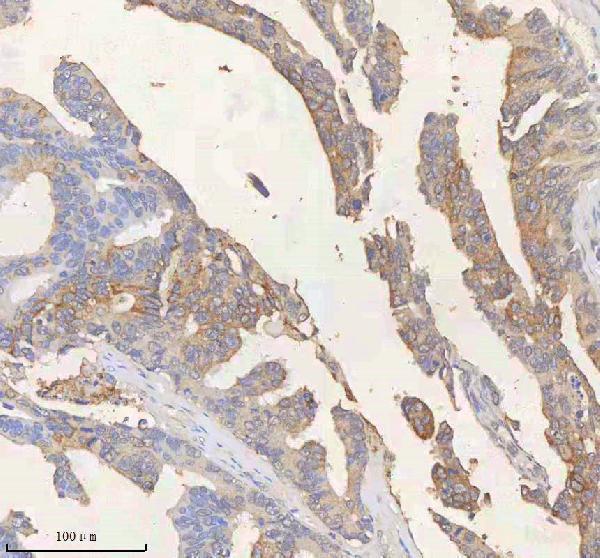
Compare the catalog antibody’s IHC-P protocol (datasheet: A00588-1) with published colon and ovarian carcinoma IHC protocols (PMC13566249; PMC8469522).
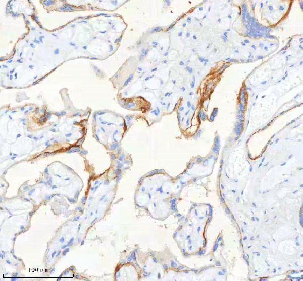
| Sample | Paraffin-embedded human placenta tissue; fixative not specified (datasheet A00588-1) |
| Fixation | Image fixative and duration unreported (datasheet A00588-1); verify before use. |
| Sectioning | 4–5 µm sections on charged slides (standard) |
| Deparaffinisation | Xylene, graded ethanol series to water (standard) |
| Antigen retrieval | Heat retrieval: EDTA pH 8.0 (datasheet A00588-1); 20 min, 95–100 °C (standard) |
| Peroxidase block | 3% H2O2, 10 min, room temperature (standard) |
| Blocking | 10% goat serum (datasheet A00588-1) |
| Primary antibody | Rabbit anti-SCD, 2-5 μg/ml (datasheet A00588-1) |
| Primary incubation | Overnight at 4 °C (datasheet A00588-1) |
| Detection | HRP-conjugated secondary, DAB chromogen (datasheet A00588-1) |
| Counterstain | Hematoxylin, blue, dehydrate and mount (standard) |
| Expected result | SCD-positive staining in adipocytes of adipose tissue (HPA tissue IHC: High). HPA tissue profile: General cytoplasmic expression. No signal in the no-primary control. |
SCD is a four-pass endoplasmic reticulum membrane protein, so positive cells should show cytoplasmic rather than nuclear staining (UniProt O00767 topology; HPA: general cytoplasmic IHC expression). HPA reports high staining in adipocytes, selected glandular and respiratory epithelial cells, and selected brain cells (HPA: tissue IHC). Its tissue IHC profile is Approved, with low consistency between antibody staining and RNA expression (HPA: tissue IHC reliability).
| Cytoplasmic staining in adipocytes or other HPA-listed positive cell types, with discernible cell boundaries. | This fits the reported IHC pattern; the ER membrane location supports a cytoplasmic distribution (HPA: tissue IHC; UniProt O00767 subcellular location). Judge intensity within the relevant cell population: HPA reports High adipocyte staining, but its Approved profile has low staining–RNA consistency (HPA: tissue IHC). |
| Predominantly nuclear staining, or a pattern confined to an unrelated compartment. | That distribution conflicts with the annotated ER membrane location and HPA’s general cytoplasmic IHC pattern (UniProt O00767 subcellular location; HPA: tissue IHC). Treat it as possible nonspecific staining or a staining artefact; review controls before scoring those cells as SCD positive (general IHC practice). |
| Strong staining in a cell population outside the expected population for the tissue being assessed. | Check the tissue’s HPA cell-level pattern before assigning SCD: staining in an unexpected cell type can reflect cross-reactivity or endogenous detection activity (HPA: tissue IHC; general chromogenic IHC practice). HPA’s supplied list is limited to selected positive tissues, so an unlisted cell type alone does not establish a false positive (HPA: supplied tissue IHC profile). |
| Diffuse color across cells and extracellular areas, with little separation between positive and background regions. | This is difficult to interpret as the reported cytoplasmic pattern (HPA: tissue IHC). General chromogenic IHC causes include inadequate blocking, excess antibody or detection reagent, and incomplete washing; compare a no-primary control before assigning cellular positivity (general IHC practice). |
| No cellular signal in an adipose section expected to contain positive adipocytes. | HPA reports High staining in adipocytes, making them a useful positive reference, although its tissue IHC reliability notes low staining–RNA consistency (HPA: adipose tissue IHC; HPA: reliability). First assess tissue preservation, positive-control performance, retrieval, and detection as workflow checks; no SCD-specific retrieval or fixation response is supplied (general IHC practice; supplied UniProt/HPA record). |
| Membrane topology and unknown antibody epitope (UniProt O00767 topology; supplied record). | SCD has four membrane spans and cytoplasmic and short lumenal regions (UniProt O00767 topology). The supplied data do not map an antibody epitope, so topology alone cannot predict which retrieval or permeabilization condition will expose it (supplied UniProt/HPA record). |
| Tissue pattern and validation strength (HPA: tissue IHC; HPA: antibodies). | HPA lists High staining in adipocytes, adrenal and other glandular cells, respiratory epithelium, and selected brain cells (HPA: tissue IHC). The tissue profile is Approved with low staining–RNA consistency; HPA012107 is IHC Approved, while no IHC status is supplied for HPA063921 (HPA: reliability; HPA: antibodies). |
| Processing and isoforms (UniProt O00767 processing; supplied record). | UniProt lists one chain spanning residues 1–359, no signal peptide or propeptide, and no annotated isoforms (UniProt O00767 processing; supplied record). These annotations provide no basis for expecting a cleaved or shed staining product; they do not identify the antibody epitope (UniProt O00767 processing; supplied record). |
| Chromogenic detection controls (general IHC practice). | Endogenous enzyme activity can resemble specific color when the detection system uses an enzyme chromogen (general chromogenic IHC practice). Interpret unexpected staining alongside a no-primary control and the relevant detection-system control; this is a general workflow issue, not an SCD-specific activity claim (general IHC practice). |
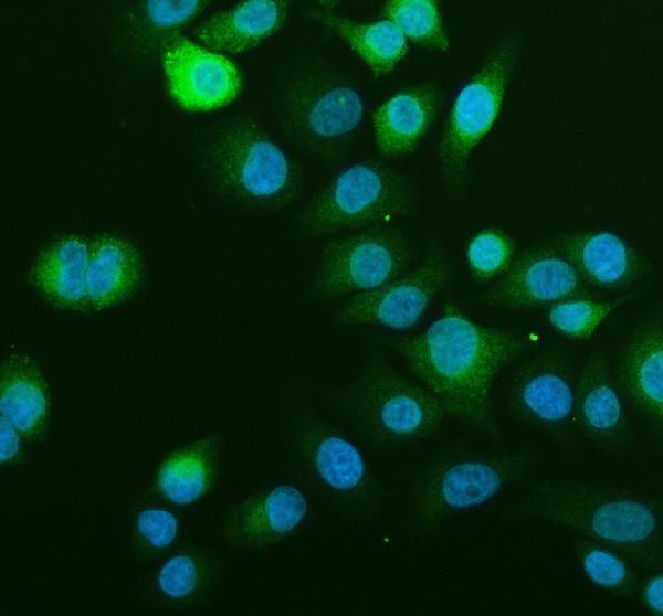
| IF/ICC Q: What localization should be expected? (HPA: subcellular ICC-IF). | A: HPA reports SCD mainly in the endoplasmic reticulum, with ER as its enhanced main location (HPA: subcellular ICC-IF). That supports an ER-associated cytoplasmic fluorescence pattern; it does not supply an IF protocol for paraffin-section IHC (HPA: subcellular ICC-IF; supplied record). |
| Situation | Likely cause | Next action |
|---|---|---|
| No signal in adipocytes expected to stain strongly (HPA: adipose tissue IHC). | A failed staining run, ineffective retrieval, or detection failure is possible; the supplied sources do not establish SCD-specific fixation or retrieval sensitivity (general IHC practice; supplied UniProt/HPA record). | Check the run’s positive control, antibody and detection steps, and the retrieval condition used for that assay before interpreting the section as negative (general IHC practice). |
| Nuclear staining dominates the slide (UniProt O00767 ER location; HPA: cytoplasmic IHC profile). | The pattern is inconsistent with the annotated location and may reflect nonspecific binding or an artefact (UniProt O00767 subcellular location; HPA: tissue IHC; general IHC practice). | Review the no-primary control and antibody concentration, then score only staining with credible cellular localization (general IHC practice). |
| Unexpected cells stain more strongly than the relevant HPA-listed cells (HPA: tissue IHC). | Cross-reactivity or endogenous chromogenic activity is possible; the supplied HPA list does not establish every cell type that can express SCD (general IHC practice; HPA: supplied tissue IHC profile). | Confirm cell identity on the counterstained section, compare HPA’s cell-level tissue pattern, and inspect detection controls (HPA: tissue IHC; general IHC practice). |
| Color is diffuse across the section (HPA: general cytoplasmic IHC profile). | High background can obscure the reported cellular pattern; excess reagent, insufficient blocking, or incomplete washes are general possibilities (HPA: tissue IHC; general IHC practice). | Compare the no-primary control and adjust blocking, reagent concentration, or washes according to the assay’s validated workflow (general IHC practice). |
| Adipocyte staining is hard to distinguish from surrounding color (HPA: High in adipocytes). | Adipocytes have limited visible cytoplasm around lipid spaces, so weak cellular color can be difficult to separate from background (general histology practice). | Inspect the thin cytoplasmic rim and neighboring cells at appropriate magnification, compare the no-primary control, and avoid scoring color in empty lipid spaces as SCD (general histology and IHC practice). |


Comprehensive Human Protein Atlas IHC scoring per tissue (reliability: Approved — Low consistency between antibody staining and RNA expression data.). Rows are taken directly from the HPA tissue chart — click any row's HPA link to view the source.
| Tissue | Cell type | Level | Evidence | Source |
|---|---|---|---|---|
| Adipose tissue | Adipocytes | High | Protein (IHC) | HPA → |
| Adrenal gland | Glandular cells | High | Protein (IHC) | HPA → |
| Appendix | Glandular cells | High | Protein (IHC) | HPA → |
| Breast | Adipocytes | High | Protein (IHC) | HPA → |
| Bronchus | Respiratory epithelial cells | High | Protein (IHC) | HPA → |
| Tissue | Cell type | Level | Evidence | Source |
|---|---|---|---|---|
| None in HPA: SCD is detected in all 45 scored tissues. Use a no-primary (secondary-only) and an isotype control instead. | ||||
Troubleshoot SCD staining in paraffin sections by checking retrieval, cell identity and the expected endoplasmic reticulum pattern (datasheet A00588-1; UniProt O00767 topology).
Anti-SCD antibodies have real images for human paraffin-section IHC and A549 cell IF/ICC (catalog: A00588-1 IHC captions; A00588-2 IF caption).
A00588-1 is listed for human IHC, with paraffin-section images from placenta, rectum adenocarcinoma, and liver cancer (catalog: applications, reactivity, IHC captions). A00588-2 is listed for human IF/ICC, with an image from A549 cells (catalog: applications, reactivity, IF caption).
Which to pick: Choose A00588-1 for human paraffin-section IHC because its own images document that preparation; the fixative is unreported (catalog: A00588-1 IHC captions). Choose A00588-2 for human IF/ICC because its application list and A549 image support it (catalog: A00588-2 applications and IF caption). Neither SKU has documented cross-species reactivity, and clonality is unreported for both (catalog: reactivity and clone fields).