This website uses cookies to ensure you get the best experience on our website.


- Table of Contents
Plan chromogenic SCN4B IHC in paraffin sections using the variable cytoplasmic tissue pattern as a reference (HPA tissue IHC). Start with the catalog antibody's 0.5–1 μg/mL IHC-P range (datasheet A06453-1), and assess membrane staining in light of the annotated cell-membrane location (UniProt).

Expected localisation, validated protocols, controls and antibodies — the at-a-glance facts below, then the full design guide.
| Expected localisation | Cytoplasm observed (HPA tissue IHC); membrane annotated (UniProt) | |
| Staining pattern | Variable cytoplasmic staining across tissue cell types (HPA tissue IHC) | |
| Antigen retrieval | Citrate pH 6 HIER, heat-mediated (datasheet A06453-1) | |
| Positive control | Adrenal gland+4 more · see all | |
| Negative control | Adipose tissue+2 more · see all |
| Fixation | Keep fixation consistent across paraffin sections (standard IHC practice; not target-specific); Matched tissue-IHC evidence does not establish the fixation claim. Validate the specimen-specific method before use. (selected-SKU IHC image A06453-1) | |
| Caveat | IHC staining has low consistency with RNA expression (HPA tissue IHC) | |
| Regulation | Higher expression in dorsal root ganglia (UniProt) | |
| Isoform / epitope | 3 isoforms; map the epitope to the extracellular or cytoplasmic region (UniProt) |
The catalog antibody’s IHC-P protocol is paired with one published SCN4B IHC protocol covering human tissue arrays and mouse tumors (PMC5150224 Methods).
| Sample | Paraffin-embedded human renal cancer tissue; fixative not specified (datasheet A06453-1) |
| Fixation | Image fixative and duration unreported (datasheet A06453-1); verify before use. |
| Sectioning | 4–5 µm sections on charged slides (standard) |
| Deparaffinisation | Xylene, graded ethanol series to water (standard) |
| Antigen retrieval | Heat retrieval: Citrate pH 6, 20 min (datasheet A06453-1) |
| Peroxidase block | 3% H2O2, 10 min, room temperature (standard) |
| Blocking | 10% goat serum (datasheet A06453-1) |
| Primary antibody | Rabbit anti-SCN4B, 0.5-1μg/ml (datasheet A06453-1) |
| Primary incubation | Overnight at 4 °C (datasheet A06453-1) |
| Detection | Streptavidin-biotin complex (SABC), DAB chromogen (datasheet A06453-1) |
| Counterstain | Hematoxylin, blue, dehydrate and mount (standard) |
| Expected result | SCN4B-positive staining in glandular cells of adrenal gland (HPA tissue IHC: Medium). HPA tissue profile: Cytoplasmic expression in all tissues at variable levels. No signal in the no-primary control. |
SCN4B is a cell-membrane protein with an extracellular region at residues 31–162 and one transmembrane segment at 163–183 (UniProt Q8IWT1 topology). In tissue IHC, HPA reports variable cytoplasmic staining, including medium staining in cerebral cortical neuronal cells (HPA: tissue IHC). Treat compartment calls cautiously: HPA rates its IHC evidence Approved but reports low consistency between antibody staining and RNA expression (HPA: reliability).
| Medium, cell-associated staining in cerebral cortical neuronal cells (HPA: cerebral cortex, Medium). | This matches an observed HPA IHC pattern. Membrane-associated staining is biologically plausible (UniProt Q8IWT1: cell membrane), but HPA describes its tissue IHC pattern as cytoplasmic; membrane enrichment is therefore not a required pass criterion (HPA: tissue IHC). |
| Predominantly nuclear staining, or staining confined to an unrelated subcellular compartment. | That compartment conflicts with SCN4B's annotated cell-membrane location (UniProt Q8IWT1: subcellular location). Check counterstain alignment and detection controls before treating it as specific; misplaced chromogen or nonspecific signal can mislead compartment scoring (general IHC practice). |
| Strong staining in adipocytes or bone-marrow hematopoietic cells. | HPA reports SCN4B as not detected in those cell populations (HPA: adipose tissue; bone marrow). Unexpected signal raises a specificity or endogenous-detection concern; compare a no-primary control and the same staining run's positive tissue before assigning a biological interpretation (general IHC practice). |
| Diffuse color across tissue and empty areas, with little distinction between cells. | A field-wide deposit is difficult to reconcile with HPA's cell-specific observations (HPA: tissue IHC). Review the no-primary control, blocking, washes and chromogen development for background (general IHC practice); HPA's reported cytoplasmic staining alone does not establish an artefact (HPA: tissue IHC). |
| No discernible staining in cerebral cortical neuronal cells. | That misses one HPA-observed medium-staining population (HPA: cerebral cortex, Medium). First verify tissue identity and a working detection control; then review the catalog antibody's IHC-P instructions and the run conditions (general IHC practice). One negative section cannot establish absence of SCN4B. |
| Compartment evidence | UniProt places SCN4B at the cell membrane, whereas HPA describes variable cytoplasmic tissue IHC staining (UniProt Q8IWT1: subcellular location; HPA: tissue IHC). Record the observed compartment without forcing agreement between these sources. |
| Choice of comparison cells | Cerebral cortical neuronal cells and cerebellar granular-layer cells have medium HPA IHC staining; adipocytes and bone-marrow hematopoietic cells are not detected (HPA: tissue IHC). Compare named cell populations rather than treating an entire section as uniformly positive or negative. |
| Antibody evidence | HPA017293 is IHC Approved, while HPA reports low consistency between antibody staining and RNA expression (HPA: antibody record; reliability). Interpret unexpected cell types conservatively; the supplied record does not establish an IHC Enhanced validation claim. |
| Antigen features | The mature chain begins at residue 31; three glycosylation sites lie in the extracellular region, and UniProt lists three isoforms (UniProt Q8IWT1: processing, topology, glycosylation, isoforms). Without an antibody epitope map, these facts cannot predict retrieval needs or isoform-specific staining. |
| Situation | Likely cause | Next action |
|---|---|---|
| No signal in an expected positive cell population. | Cerebral cortical neuronal cells show medium staining in HPA, so a blank run needs investigation (HPA: cerebral cortex). | Confirm the cell population and positive detection control; check the catalog antibody's IHC-P instructions, reagent preparation and development time (general IHC practice). |
| Broad, weak haze obscures cell boundaries. | Background from detection or incomplete washing may obscure a cell-associated pattern (general IHC practice). | Compare the no-primary control, then review blocking, washes and chromogen development (general IHC practice); retain HPA's cytoplasmic tissue pattern as a possible specific appearance (HPA: tissue IHC). |
| Strong color appears in adipocytes. | HPA lists adipocytes as not detected; strong signal there is unexpected (HPA: adipose tissue). | Compare a no-primary control and a positive tissue from the same run; assess endogenous detection activity and nonspecific antibody staining before scoring adipocytes as positive (general IHC practice). |
| The apparent signal is mainly nuclear. | Nuclear restriction conflicts with the annotated cell-membrane location (UniProt Q8IWT1: subcellular location). | Check nuclear counterstain alignment, focus and detection controls, then rescore only clearly localized signal (general IHC practice). |
| Cytoplasmic staining appears without a clear membrane outline. | HPA reports cytoplasmic tissue IHC despite UniProt's membrane annotation (HPA: tissue IHC; UniProt Q8IWT1: subcellular location). | Report the observed compartment and cell type; use negative cells and controls to judge specificity. Do not reject the slide solely because membrane enrichment is unresolved (HPA: tissue IHC; general IHC practice). |
| Can an IF/ICC result be judged by this IHC pattern? | HPA summarizes SCN4B as membrane-associated in its subcellular record but supplies no ICC-IF image-bearing cell lines here (HPA: subcellular record). | Treat membrane labeling as a localization expectation (UniProt Q8IWT1: subcellular location; HPA: subcellular summary). Use the separate IF/ICC guide for assay design; this IHC tissue record does not validate an IF staining pattern. |


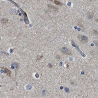
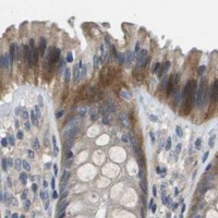
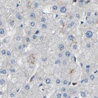

Comprehensive Human Protein Atlas IHC scoring per tissue (reliability: Approved — Low consistency between antibody staining and RNA expression data.). Rows are taken directly from the HPA tissue chart — click any row's HPA link to view the source.
| Tissue | Cell type | Level | Evidence | Source |
|---|---|---|---|---|
| Adrenal gland | Glandular cells | Medium | Protein (IHC) | HPA → |
| Appendix | Glandular cells | Medium | Protein (IHC) | HPA → |
| Breast | Glandular cells | Medium | Protein (IHC) | HPA → |
| Bronchus | Ciliated cells (cell body) | Medium | Protein (IHC) | HPA → |
| Caudate | Glial cells | Medium | Protein (IHC) | HPA → |
Troubleshoot SCN4B staining in paraffin sections by checking retrieval, compartment, cell identity, and controls before interpreting chromogenic signal.
A06453-1 has real SCN4B IHC images from human renal cancer and rat spleen paraffin sections; ICC and IHC-F are listed applications (catalog: IHC image captions, applications).
A06453-1 will render with its human renal cancer paraffin-section IHC figure (catalog: card image caption). An additional caption documents rat spleen paraffin-section IHC; Human, Mouse and Rat reactivity is listed, but no IF image is supplied (catalog: IHC image captions, reactivity, IF image alts).
Which to pick: For tissue IHC, pick A06453-1: its figure documents 1 μg/ml on a human renal cancer paraffin section after citrate pH 6 retrieval; the fixative is unreported (catalog: card image caption). For IF/ICC, A06453-1 lists ICC and IHC-F applications and an IF dilution of 0.5–1 μg/ml, but supplies no IF image (catalog: applications, IF dilution, IF image alts). For cross-species work, A06453-1 lists Human, Mouse and Rat reactivity, with tissue IHC captions for human and rat only; it is rabbit-hosted, and clonality is unreported (catalog: reactivity, IHC image captions, host, clone).