This website uses cookies to ensure you get the best experience on our website.


- Table of Contents
Plan SCNN1B staining in paraffin sections using reported cytoplasmic tissue staining and the expected apical membrane location (HPA tissue IHC; UniProt). Compare appropriate tissue controls and interpret staining cautiously because antibody staining and RNA expression show low consistency (HPA tissue IHC).

Expected localisation, validated protocols, controls and antibodies — the at-a-glance facts below, then the full design guide.
| Expected localisation | Cytoplasmic in tissue (HPA tissue IHC); apical membrane expected (UniProt) | |
| Staining pattern | Cytoplasmic staining in several tissues (HPA tissue IHC) | |
| Antigen retrieval | Tris-EDTA pH 8.0 HIER, heat-mediated (datasheet A01670) | |
| Positive control | Stomach+4 more · see all | |
| Negative control | Bone marrow+4 more · see all |
| Fixation | Keep fixation consistent across compared paraffin sections (selected-SKU IHC image A01670) | |
| Caveat | Tissue staining has low concordance with RNA (HPA tissue IHC) | |
| Regulation | pro-PCSK9 promotes ENaC degradation (UniProt) | |
| Isoform / epitope | 2 isoforms; check extracellular versus cytoplasmic epitope (UniProt) |
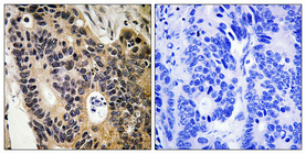
The catalog antibody protocol uses heat-mediated Tris-EDTA retrieval at pH 8.0 (datasheet A01670). The published colorectal tissue protocol below provides IHC staining details (PMC11484387).
| Sample | Paraffin-embedded Human colon cancer tissue; fixative not specified (datasheet A01670) |
| Fixation | Image fixative and duration unreported (datasheet A01670); verify before use. |
| Sectioning | 4–5 µm sections on charged slides (standard) |
| Deparaffinisation | Xylene, graded ethanol series to water (standard) |
| Antigen retrieval | Heat retrieval: Tris-EDTA pH 8.0 (datasheet A01670); 20 min, 95–100 °C (standard) |
| Peroxidase block | 3% H2O2, 10 min, room temperature (standard) |
| Blocking | 10% normal serum of the secondary host, 30 min, room temperature (standard) |
| Primary antibody | Rabbit anti-SCNN1B, 1:100-1:300 (datasheet A01670) |
| Primary incubation | Overnight at 4 °C (standard) |
| Detection | HRP-polymer secondary, DAB chromogen 5–10 min (standard) |
| Counterstain | Hematoxylin, blue, dehydrate and mount (standard) |
| Expected result | SCNN1B-positive staining in glandular cells of stomach (HPA tissue IHC: High). HPA tissue profile: Cytoplasmic expression in several tissues. No signal in the no-primary control. |
SCNN1B is an apical cell membrane protein that can also occur on cytoplasmic vesicle membranes (UniProt P51168 localization; topology: 2 transmembrane segments). In paraffin sections, examine epithelial cells, especially kidney tubules and bronchial respiratory epithelium, and stomach glandular cells (HPA: medium, medium and high staining, respectively). HPA describes cytoplasmic staining across several tissues, but rates its tissue IHC profile Uncertain because staining and RNA expression have low consistency (HPA: tissue IHC).
| Apical-edge staining in kidney tubular cells, with or without adjacent cytoplasmic staining. | This fits the expected membrane compartment (UniProt P51168: apical cell membrane) and an observed positive cell population (HPA: medium in kidney tubular cells). Cytoplasmic staining can be compatible with vesicle localization (UniProt P51168: cytoplasmic vesicle membrane), but HPA's uncertain tissue profile means the pattern alone does not verify antibody specificity (HPA: tissue IHC, Uncertain). |
| Predominantly cytoplasmic staining in stomach glandular cells, without a clearly resolved apical rim. | HPA reports high staining in stomach glandular cells and describes cytoplasmic expression in several tissues (HPA: tissue IHC). Interpret this as an observed IHC pattern, while noting that UniProt places SCNN1B at the apical membrane and on cytoplasmic vesicles (UniProt P51168 localization). Cytoplasmic signal alone cannot establish which structure contains the antigen (general IHC interpretation). |
| Strong, isolated nuclear staining or staining concentrated in an unrelated compartment. | That distribution does not match the annotated apical membrane or cytoplasmic vesicle membrane locations (UniProt P51168 localization). Treat it as a possible artefact and compare with tissue morphology and controls before assigning SCNN1B identity (general IHC practice); HPA's Uncertain reliability makes compartment agreement especially useful, though it is not proof (HPA: tissue IHC). |
| Staining in cells reported as negative, such as bone marrow hematopoietic cells or colon endothelial cells. | These populations are reported as not detected in HPA tissue IHC (HPA: bone marrow hematopoietic cells; colon endothelial cells). Unexpected chromogen may reflect cross-reactivity or endogenous detection activity (general IHC practice). Check a no-primary control and the detection workflow; because HPA rates its profile Uncertain, do not treat its negative calls as absolute biological exclusions (HPA: tissue IHC). |
| Uniform haze across epithelium and surrounding tissue, or no signal in a kidney section. | Uniform haze without cell or compartment structure is difficult to assign to SCNN1B (general IHC interpretation). Kidney tubular cells are an observed medium positive population, and kidney protein expression is reported independently by UniProt (HPA: kidney tubular cells; UniProt P51168: kidney protein expression). A blank kidney run warrants a technical check, but cannot by itself prove absence of SCNN1B. |
| Membrane topology and epitope position. | SCNN1B has cytoplasmic residues 1–50 and 554–640, an extracellular region at 72–532, and 2 transmembrane segments (UniProt P51168 topology). An antibody's epitope position would affect how staining is interpreted, but no epitope is supplied here; do not infer its accessibility or prescribe antigen retrieval from topology alone. |
| Observed tissue pattern and reliability. | HPA reports high stomach glandular staining and medium staining in kidney tubular and bronchial respiratory epithelial cells, yet labels the overall tissue IHC profile Uncertain because of low staining–RNA consistency (HPA: tissue IHC). Use these populations as comparison tissues, with morphology and controls, rather than as definitive specificity standards (general IHC practice). |
| Cytoplasmic signal alongside expected membrane localization. | UniProt lists both apical cell membrane and cytoplasmic vesicle membrane locations (UniProt P51168 localization); HPA describes cytoplasmic expression in several tissues (HPA: tissue IHC). Record membrane and cytoplasmic compartments separately when scoring (general IHC practice). The supplied evidence does not establish that every cytoplasmic deposit represents vesicular SCNN1B. |
| Isoforms and antibody validation. | UniProt lists 2 isoforms (UniProt P51168 isoforms). The supplied records do not identify the catalog antibody's epitope or isoform coverage, so staining cannot be assigned to a particular isoform. The listed HPA antibody, HPA015612, has an Uncertain IHC status (HPA: antibody validation); compare results with independent evidence before making a strong specificity claim (general IHC practice). |
| IF/ICC comparison. | HPA summarizes the subcellular location as membrane but provides no main location or cell lines with ICC-IF images (HPA: subcellular record). This offers limited support for a membrane expectation; it does not validate an IF/ICC image pattern. Address IF/ICC setup and interpretation in its separate guide. |
| Situation | Likely cause | Next action |
|---|---|---|
| Kidney tubules show no staining. | A weak run is possible because HPA reports medium staining in tubular cells; tissue variation also remains possible under HPA's Uncertain profile (HPA: kidney and tissue IHC). | Confirm that tubules are present, inspect the run's positive and negative controls, and review antibody dilution, retrieval and chromogen steps as general IHC checks (general IHC practice). Do not infer a SCNN1B-specific retrieval requirement from these sources. |
| Stomach glandular cells are blank in an intended positive comparison. | HPA reports high staining in this cell population, so a blank result raises a run or specimen question; HPA's uncertain reliability prevents treating one section as a guaranteed positive (HPA: stomach; tissue IHC). | Verify glandular morphology and control performance, then review primary incubation and detection reagents (general IHC practice). Compare another documented positive population, such as kidney tubules, if available (HPA: kidney tubular cells, medium). |
| Signal appears chiefly in nuclei. | A nuclear-only pattern conflicts with the annotated apical and vesicle membrane locations (UniProt P51168 localization); nonspecific binding or chromogen artefact is possible (general IHC practice). | Compare the nuclear signal with a no-primary control, check counterstain and precipitate, and score membrane and cytoplasmic staining separately (general IHC practice). Do not call nuclear staining a confirmed SCNN1B pattern from the supplied evidence. |
| Bone marrow hematopoietic cells stain strongly. | HPA reports these cells as not detected (HPA: bone marrow hematopoietic cells). Cross-reactivity or endogenous peroxidase activity could account for unexpected chromogen in an IHC run (general IHC practice). | Inspect a no-primary control and the peroxidase-blocking step, then compare the distribution with a reported positive epithelial population (general IHC practice; HPA: kidney tubular cells, medium). Treat the HPA negative call as provisional because tissue reliability is Uncertain (HPA: tissue IHC). |
| Diffuse background obscures cell borders. | Indistinct staining prevents reliable compartment scoring (general IHC interpretation); the supplied records do not identify a SCNN1B-specific cause. | Review blocking, washing, primary antibody dilution and chromogen development against the run controls (general IHC practice). Reassess apical and cytoplasmic distribution only after tissue structures are readable (UniProt P51168 localization; general IHC practice). |
| Can IF/ICC confirm an apical SCNN1B pattern? | HPA provides a membrane summary but no ICC-IF image-bearing cell lines or specified main location (HPA: subcellular record). | Use the separate IF/ICC guide for that application. Interpret any IF/ICC image with compartment markers and controls as general imaging practice; the supplied HPA entry alone cannot confirm an apical IF/ICC pattern. |


Comprehensive Human Protein Atlas IHC scoring per tissue (reliability: Uncertain — Low consistency between antibody staining and RNA expression data. Pending external verification.). Rows are taken directly from the HPA tissue chart — click any row's HPA link to view the source.
| Tissue | Cell type | Level | Evidence | Source |
|---|---|---|---|---|
| Stomach | Glandular cells | High | Protein (IHC) | HPA → |
| Bronchus | Respiratory epithelial cells | Medium | Protein (IHC) | HPA → |
| Cervix | Squamous epithelial cells | Medium | Protein (IHC) | HPA → |
| Hippocampus | Neuronal cells | Medium | Protein (IHC) | HPA → |
| Kidney | Cells in tubules | Medium | Protein (IHC) | HPA → |
| Tissue | Cell type | Level | Evidence | Source |
|---|---|---|---|---|
| Bone marrow | Hematopoietic cells | Not detected | Protein (IHC) | HPA → |
| Colon | Endothelial cells | Not detected | Protein (IHC) | HPA → |
| Fallopian tube | Glandular cells | Not detected | Protein (IHC) | HPA → |
| Ovary | Ovarian stroma cells | Not detected | Protein (IHC) | HPA → |
| Rectum | Glandular cells | Not detected | Protein (IHC) | HPA → |
Use compartment aware controls and matched processing to troubleshoot SCNN1B staining in paraffin sections (UniProt P51168 topology; datasheet A01670).
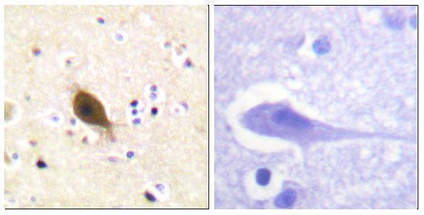
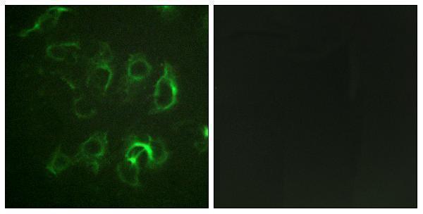
A01670 has IHC images of paraffin-embedded human colon cancer and brain tissue, plus an IF image of HUVEC cells (catalog image captions). Listed reactivity is human, mouse, and rat (catalog).
A01670 is listed for IHC and IF, with IHC images of paraffin-embedded human colon cancer and brain tissue (catalog applications; IHC image captions). Its IF image shows HUVEC cells; the catalog lists human, mouse, and rat reactivity (IF image caption; catalog reactivity).
Which to pick: For tissue IHC, choose A01670 based on its paraffin-section images; the fixative is unreported (IHC image captions). For IF/ICC work, A01670 has an IF image in HUVEC cells, but ICC validation and clonality are unreported (IF image caption; catalog). For cross-species work, A01670 lists human, mouse, and rat reactivity, while the supplied IHC and IF images show human samples only (catalog reactivity; image captions).