This website uses cookies to ensure you get the best experience on our website.


- Table of Contents
Plan SDHB chromogenic IHC around granular cytoplasmic staining across cell types (HPA tissue IHC). Use the IHC-validated antibody with tissue controls and score cytoplasmic signal, bearing in mind SDHB’s inner mitochondrial membrane location (UniProt).

Expected localisation, validated protocols, controls and antibodies — the at-a-glance facts below, then the full design guide.
| Expected localisation | Granular cytoplasm (HPA tissue IHC) | |
| Staining pattern | Granular cytoplasmic staining across cell types (HPA tissue IHC) | |
| Antigen retrieval | Citrate pH 6 HIER, heat-mediated (datasheet M01090-1) | |
| Positive control | Adrenal gland+4 more · see all | |
| Negative control | None in HPA (detected in all 45 tissues); use no-primary + isotype controls |
| Fixation | Keep fixation conditions consistent across sections (standard IHC practice; not target-specific) | |
| Caveat | Low staining in adipocytes and chondrocytes (HPA tissue IHC) | |
| Regulation | RNA enhanced in skeletal muscle, tongue (HPA tissue RNA) | |
| Isoform / epitope | No isoforms listed; mature chain spans residues 29–280 (UniProt) |
The catalog antibody protocol uses citrate pH 6 heat retrieval (datasheet M01090-1). Three published SDHB IHC protocols provide additional sample, staining and detection details (PMC3415983; PMC4718191; PMC8418866).
| Sample | Paraffin-embedded human lung cancer tissues; fixative not specified (datasheet M01090-1) |
| Fixation | Image fixative and duration unreported (datasheet M01090-1); verify before use. |
| Sectioning | 4–5 µm sections on charged slides (standard) |
| Deparaffinisation | Xylene, graded ethanol series to water (standard) |
| Antigen retrieval | Heat retrieval: Citrate pH 6, 20 min (datasheet M01090-1) |
| Peroxidase block | 3% H2O2, 10 min, room temperature (standard) |
| Blocking | 10% goat serum (datasheet M01090-1) |
| Primary antibody | Mouse monoclonal (clone 2I3) anti-SDHB, 0.5-1μg/ml (datasheet M01090-1) |
| Primary incubation | Overnight at 4 °C (datasheet M01090-1) |
| Detection | Streptavidin-biotin complex (SABC), DAB chromogen (datasheet M01090-1) |
| Counterstain | Hematoxylin, blue, dehydrate and mount (standard) |
| Expected result | SDHB-positive staining in glandular cells of adrenal gland (HPA tissue IHC: High). HPA tissue profile: Granular cytoplasmic expression in all cell types. No signal in the no-primary control. |
SDHB is an inner mitochondrial membrane protein without a transmembrane segment (UniProt P21912 topology). In paraffin-section IHC, expect granular cytoplasmic staining across cell types (HPA: granular cytoplasmic expression in all cell types). HPA rates the tissue IHC profile Enhanced, reflecting high consistency between antibody staining and RNA expression data (HPA: Enhanced reliability). Interpret intensity in the tissue and cell type examined rather than requiring every cell to stain equally.
| Granular cytoplasmic staining in colon glandular cells or bronchial respiratory epithelial cells. | This matches the reported compartment and cells; both examples have High staining (HPA: granular cytoplasmic expression; High in colon glandular and bronchial respiratory epithelial cells). Assess the granules against tissue morphology and nearby background. |
| Predominantly nuclear or cell-edge staining, with little granular cytoplasmic signal. | This differs from the tissue IHC pattern (HPA: granular cytoplasmic expression). Review controls and detection before calling it SDHB. Additional nucleoplasm and plasma membrane locations are uncertain in ICC-IF (HPA: subcellular location); they do not establish an expected paraffin IHC pattern. |
| Strong staining confined to an unexpected cell population while expected cells lack granules. | Treat the distribution as suspicious, not as proof of a new SDHB-positive cell type: HPA describes granular expression in all cell types and gives cell-specific intensity examples (HPA: tissue IHC). Check cross-reactivity and endogenous detection activity using appropriate controls (general IHC practice). |
| Diffuse, uniform color over tissue or blank areas instead of discrete cytoplasmic granules. | Background can obscure the reported pattern (HPA: granular cytoplasmic expression). Compare a no-primary control, inspect wash and blocking steps, and assess whether the detection system contributes color (general IHC practice). Diffuse color alone does not establish SDHB localization. |
| No convincing signal in colon glandular cells despite preserved tissue morphology. | Colon glandular cells are reported High (HPA: colon glandular cells, High). First evaluate the positive control and assay steps; absent staining in one section cannot by itself establish loss of SDHB expression (general IHC practice). |
| Compartment and topology | SDHB is assigned to the mitochondrial inner membrane and has no transmembrane segment (UniProt P21912 topology). The tissue readout is granular cytoplasmic staining (HPA: tissue IHC); score that visible pattern rather than inferring membrane-surface staining from the protein name. |
| Tissue and cell-type intensity | HPA reports granular cytoplasmic expression in all cell types, with High staining in its listed colon glandular and bronchial respiratory epithelial cells (HPA: tissue IHC). Match an intensity judgment to the cell type on the slide; the examples are observations, not a universal intensity cutoff. |
| Low-staining cell examples | Adipocytes and chondrocytes are listed as Low (HPA: adipocytes, Low; chondrocytes, Low). Weak staining there is less informative as a sole assay control than a documented High cell population (general IHC practice). Low is an intensity category, not evidence of complete absence. |
| Protein processing | UniProt assigns SDHB a mature chain spanning residues 29–280 and lists no isoforms (UniProt P21912 processing and isoforms). These annotations do not specify the antibody epitope or predict whether a particular antigen-retrieval condition will reveal it. |
| Antibody-level validation | HPA rates IHC staining Enhanced for HPA002868, CAB009822 and CAB068235, and Supported for CAB068233 and CAB068234 (HPA: antibody IHC validation). These ratings support review of staining patterns; they do not supply a dilution, retrieval setting or slide-specific performance guarantee. |
| Antigen retrieval | Retrieval is a general paraffin IHC workflow variable (general IHC practice). The supplied HPA and UniProt records give no SDHB-specific retrieval condition or fixation-sensitivity result; optimize with controls before interpreting a weak slide. |
| Situation | Likely cause | Next action |
|---|---|---|
| Known High colon glandular cells show no granular staining. | An assay-step failure is possible; the expected cell-level observation is High (HPA: colon glandular cells, High). The records do not identify a target-specific fixation cause. | Check a documented positive section, reagent sequence, detection and counterstain; review the chosen retrieval condition as general IHC practice. Do not score biological loss until assay performance is established. |
| The section has widespread flat color that hides cell detail. | Excess background or endogenous detection activity may mask the granular cytoplasmic pattern (HPA: tissue IHC; general IHC practice). The appearance alone does not identify which step failed. | Run a no-primary control and review blocking, washes and chromogen development (general IHC practice). Read SDHB only where granules remain distinguishable from background (HPA: granular cytoplasmic expression). |
| A nuclear rim or plasma-membrane outline dominates. | This is discordant with tissue IHC granules (HPA: tissue IHC). HPA labels additional nucleoplasm and plasma membrane ICC-IF locations uncertain (HPA: subcellular location). | Recheck cell morphology, control staining and detection specificity (general IHC practice); avoid treating an uncertain ICC-IF location as confirmation of the chromogenic IHC result. |
| Adipocytes or chondrocytes stain faintly while other cells stain strongly. | Those cell types have Low reported staining (HPA: adipocytes, Low; chondrocytes, Low). Unequal intensity by itself does not imply a failed stain. | Judge the section against its cell types and include a documented High population for comparison (HPA: tissue IHC; general IHC practice). Do not use Low as a required negative result. |
| Only one unexpected cell population is intensely colored. | A restricted pattern conflicts with HPA's granular expression across cell types if expected cells are unstained (HPA: tissue IHC). Cross-reactivity or endogenous detection activity are possible assay explanations (general IHC practice). | Compare morphology and a no-primary control; repeat with another appropriately validated antibody if needed (general IHC practice; HPA: antibody IHC validation). Do not assign a new cell-specific expression pattern from this slide alone. |
| IF/ICC: should mitochondrial puncta be expected? | HPA supports mitochondria as the main ICC-IF location; additional nucleoplasm and plasma membrane locations are uncertain (HPA: subcellular location). This evidence describes localization, not an IF protocol. | Expect a mitochondrial distribution when interpreting IF/ICC (HPA: mitochondria, supported; UniProt P21912 localization). Use the separate IF/ICC guide for experimental setup. |


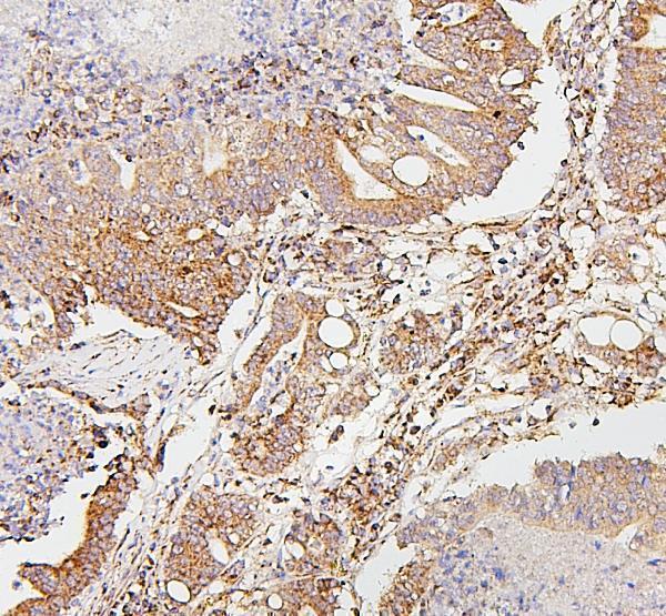
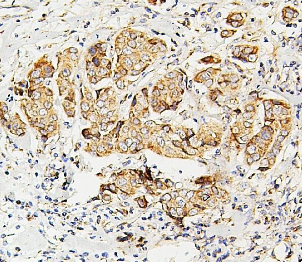


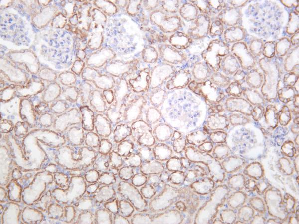
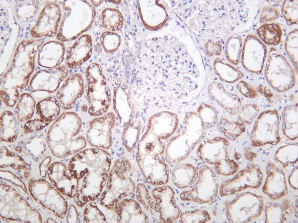


Comprehensive Human Protein Atlas IHC scoring per tissue (reliability: Enhanced — High consistency between antibody staining and RNA expression data.). Rows are taken directly from the HPA tissue chart — click any row's HPA link to view the source.
| Tissue | Cell type | Level | Evidence | Source |
|---|---|---|---|---|
| Adrenal gland | Glandular cells | High | Protein (IHC) | HPA → |
| Appendix | Glandular cells | High | Protein (IHC) | HPA → |
| Breast | Glandular cells | High | Protein (IHC) | HPA → |
| Bronchus | Respiratory epithelial cells | High | Protein (IHC) | HPA → |
| Caudate | Neuronal cells | High | Protein (IHC) | HPA → |
| Tissue | Cell type | Level | Evidence | Source |
|---|---|---|---|---|
| None in HPA: SDHB is detected in all 45 scored tissues. Use a no-primary (secondary-only) and an isotype control instead. | ||||
Use the paraffin-section IHC example and the reported mitochondrial staining pattern to troubleshoot SDHB while keeping unreported processing details explicit.
Anti-SDHB IHC images cover human cancer tissues and rat kidney (catalog IHC captions: M01090-1, M01090-2, PA1718, M01090-5); an IF/ICC image shows U20S cells (A01090 IF caption).
M01090-1 shows human lung cancer, and M01090-2 shows human intestinal cancer in paraffin sections (catalog IHC captions: M01090-1, M01090-2). PA1718 shows paraffin-section human liver cancer, while M01090-5 shows rat kidney (catalog IHC captions: PA1718, M01090-5).
Which to pick: For tissue IHC, choose mouse monoclonal M01090-1 for human lung cancer with citrate retrieval, M01090-2 for human intestinal cancer with citrate retrieval, or PA1718 for human liver cancer with EDTA retrieval (catalog IHC captions: M01090-1, M01090-2, PA1718; catalog clones: M01090-1, M01090-2). For IF/ICC, consider rabbit monoclonal M01090-5 because IF is listed at 1:200–1:1000; its supplied image captions document IHC rather than IF (M01090-5 application list, dilution and image captions). For cross-species tissue IHC, M01090-5 has human, mouse and rat kidney images; the four rendered IHC captions do not report the fixative (M01090-5 image captions; catalog IHC captions: M01090-1, M01090-2, PA1718, M01090-5).