SEC31A / Protein transport protein Sec31A · IHC design guide

Design Immunohistochemistry for SEC31A

Plan SEC31A chromogenic IHC in paraffin sections using the reported cytoplasmic tissue pattern (HPA tissue IHC). Compare strongly stained adipocytes or adrenal glandular cells with cardiomyocytes, where staining was not detected (HPA tissue IHC).

Evidence assembled Oct 2026 · For research use; verify linked source records and product datasheet before use
Immunohistochemistry protocol sheet for SEC31A (IHC for SEC31A): expected localisation General cytoplasmic tissue staining (HPA tissue IHC), antibody A04582, validated IHC image, and IHC protocol steps
Printable SEC31A IHC protocol sheet — expected localisation General cytoplasmic tissue staining (HPA tissue IHC), antibody A04582, controls and protocol steps. Open the full SEC31A IHC guide →

SEC31A Immunohistochemistry Experimental Design Guide

Expected localisation, validated protocols, controls and antibodies — the at-a-glance facts below, then the full design guide.

Must know before staining
Expected localisation General cytoplasmic tissue staining (HPA tissue IHC)
Staining pattern Cytoplasmic staining in multiple tissue cell types (HPA tissue IHC)
Antigen retrieval EDTA pH 8.0 HIER, heat-mediated (datasheet A04582)
Positive control ⓘ Adipose tissue+4 more · see all
Negative control ⓘ Heart muscle
Important caveats
Reasons your staining may differ from the expected pattern.
Fixation Keep fixation consistent across sections (standard IHC practice; not target-specific)
Caveat Cardiomyocytes have no detectable staining (HPA tissue IHC)
Regulation Broad expression; no regulator specified (UniProt)
Isoform / epitope 10 isoforms; epitope coverage is unverified (UniProt)
Section 1

Recommended SEC31A IHC & IF Protocols

The catalog antibody’s IHC-P protocol is accompanied by two published chromogenic SEC31A IHC protocols (PMC11600210; PMC12502155).

Recommended immunohistochemistry (IHC-P) protocol parameters
SampleParaffin-embedded human liver cancer tissue; fixative not specified (datasheet A04582)
FixationImage fixative and duration unreported (datasheet A04582); verify before use.
Sectioning4–5 µm sections on charged slides (standard)
DeparaffinisationXylene, graded ethanol series to water (standard)
Antigen retrievalHeat retrieval: EDTA pH 8.0 (datasheet A04582); 20 min, 95–100 °C (standard)
Peroxidase block3% H2O2, 10 min, room temperature (standard)
Blocking10% goat serum (datasheet A04582)
Primary antibodyRabbit anti-SEC31A, 2-5 μg/ml (datasheet A04582)
Primary incubationOvernight at 4 °C (datasheet A04582)
DetectionHRP-conjugated secondary, DAB chromogen (datasheet A04582)
CounterstainHematoxylin, blue, dehydrate and mount (standard)
Expected resultSEC31A-positive staining in adipocytes of adipose tissue (HPA tissue IHC: High). HPA tissue profile: General cytoplasmic expression. No signal in the no-primary control.
💡Decision noteStart with heat-mediated EDTA pH 8.0 retrieval for the catalog antibody (datasheet: A04582); the published protocols used enzyme retrieval or citrate buffer (PMC11600210; PMC12502155).
Section 2

What Is the Expected SEC31A Staining Pattern?

SEC31A should show predominantly cytoplasmic staining across many cell types, consistent with its abundant, ubiquitous expression (HPA: general cytoplasmic expression; UniProt O94979: tissue specificity). Its association with COPII vesicles and endoplasmic reticulum exit sites may produce cytoplasmic puncta when resolution permits (UniProt O94979: subcellular location). It has no transmembrane segment (UniProt O94979: topology). HPA rates its tissue IHC profile Approved, with medium consistency between staining and RNA data (HPA: tissue IHC).

What am I looking at on my slide?
Cytoplasmic staining appears in many cells, with strong signal in adipocytes or cerebellar Purkinje cells.This matches the reported general cytoplasmic profile and High staining in those cells (HPA: tissue IHC). Score the relevant cell population and compartment together; broad expression alone does not establish antibody specificity (UniProt O94979: tissue specificity; HPA: Approved, medium consistency).
A crisp nuclear-only or cell-surface rim pattern dominates while cytoplasm is unstained.Treat the compartment mismatch as suspect: SEC31A is reported in cytosol, COPII vesicles and at endoplasmic reticulum exit sites (UniProt O94979: subcellular location). Check morphology and staining controls before interpreting it as a new location; the supplied sources do not support a dominant nuclear or surface pattern.
Strong chromogen appears in cardiomyocytes, especially if it persists on a no-primary control.HPA reports SEC31A as Not detected in cardiomyocytes (HPA: heart muscle). Recheck cell identification and assess cross-reactivity or endogenous detection activity. A positive reaction here warrants investigation, although HPA's Approved profile has only medium staining–RNA consistency (HPA: tissue IHC).
A diffuse haze covers tissue and background structures without a clear cellular boundary.The result is difficult to score against HPA's cellular cytoplasmic profile (HPA: tissue IHC). In routine IHC, inadequate blocking, insufficient washing or excess detection reagent can raise background; compare a no-primary control and repeat the stain after correcting the implicated step.
No convincing cytoplasmic signal appears in an otherwise intact, expected-positive specimen.High staining is reported in adipocytes, adrenal glandular cells, cortical neuronal cells and several other populations (HPA: tissue IHC). Check that the section actually contains the chosen population, then review the IHC-validated antibody, retrieval, detection and counterstain controls. A negative result alone cannot establish absent SEC31A.
💡Expected SEC31A appearanceCall a section positive when the relevant cells show discernible cytoplasmic signal, potentially punctate, with stronger staining in HPA High populations such as adipocytes or Purkinje cells; isolated nuclear staining or strong cardiomyocyte staining is suspect (HPA: tissue IHC; UniProt O94979: subcellular location).
How each factor affects the staining
Expected cell populations and scoring referenceHPA reports Low tissue specificity and general cytoplasmic expression, with High staining in specified cells but Not detected in cardiomyocytes (HPA: tissue IHC). Identify the cell type before comparing intensity; these observations are references, not universal thresholds.
Compartment and topologySEC31A occurs in cytosol, COPII-coated vesicle membranes and endoplasmic reticulum exit sites, without a transmembrane segment (UniProt O94979: subcellular location and topology). Interpret cytoplasmic signal in that context; a fine punctate component may be hard to resolve in chromogenic sections.
Antibody validationThe listed rabbit polyclonal antibody HPA005457 is Approved for IHC and Supported for ICC (HPA: antibodies). The tissue profile has medium staining–RNA consistency (HPA: tissue IHC), so validation status supports use but does not settle an unexpected compartment or cell-type result.
Isoforms and modified residuesUniProt lists 10 isoforms and five modified residues, including phosphoserines and phosphothreonines (UniProt O94979: isoforms and modified residues). Without an epitope map, these annotations do not predict which forms an antibody detects or explain a tissue-specific staining difference.
IF/ICC Q&A: What localisation should be checked?Look for vesicular and cytosolic signal: HPA labels vesicles Supported and cytosol Approved in ICC-IF, with images from A-431, U-251MG and U2OS (HPA: subcellular). This is an interpretation reference for IF/ICC, not an IHC protocol option.
Why is my staining missing, weak or wrong?
SituationLikely causeNext action
Expected-positive cells have no chromogenic signal.The target population may be absent from the section, or a routine IHC step may have failed; HPA reports High staining in adipocytes and adrenal glandular cells (HPA: tissue IHC).Verify cell identity and section integrity, then check retrieval, primary-antibody application and detection with an appropriate positive control. Do not infer target absence from one failed stain.
Only nuclei or a sharp surface rim stain.That pattern conflicts with the reported cytosolic, vesicular and endoplasmic reticulum exit-site localisation (UniProt O94979: subcellular location).Review cellular morphology and no-primary controls; repeat with the IHC-validated antibody and inspect cytoplasmic signal before accepting the result.
Cardiomyocytes stain strongly.HPA reports cardiomyocytes as Not detected (HPA: heart muscle); cross-reactivity or endogenous detection activity is possible in routine IHC.Confirm the cell type, run a no-primary control and review the detection chemistry. Treat persistent signal as unresolved rather than assigning it to SEC31A.
Background obscures cytoplasmic staining.Diffuse deposition can arise from routine IHC blocking, washing or reagent-concentration problems; it cannot be assigned to SEC31A from these sources.Compare background with a no-primary control, then optimize blocking and washes and review reagent concentrations. Score only signal distinguishable from background.
Positive cells show inconsistent intensity across a section.Cell populations may differ: HPA reports High staining in some cells and Low staining in smooth muscle cells and skeletal myocytes (HPA: tissue IHC). Technical unevenness is also possible.Compare like cell types in well-preserved areas and inspect section-wide staining uniformity before assigning a biological intensity difference.
IF/ICC shows diffuse cytosol but no obvious vesicular puncta.HPA supports vesicular localisation and approves cytosolic localisation (HPA: subcellular); puncta may be difficult to distinguish from diffuse signal under the imaging conditions.Assess focus and resolution, then compare the observed compartments with HPA's vesicle-and-cytosol reference. Do not use the absence of visible puncta alone to reject cytosolic staining.

Sample controls for SEC31A IHC & IF

🧪Run adipose tissue first and score adipocytes for SEC31A staining (HPA: High in adipocytes); run heart muscle as the negative tissue and score cardiomyocytes (HPA: Not detected in cardiomyocytes). On the adipose slide, cells without specific cytoplasmic staining should show counterstain only, but the supplied HPA rows do not identify a validated internal negative cell type.
Positive control tissue: Adipose tissue (Adipocytes, HPA High)
Negative control tissue: Heart muscle (HPA Not detected)
ICC-IF cell lines (HPA subcellular resource): HPA ICC-IF images show SEC31A in A-431, U-251MG, U2OS, with annotated localisation: Vesicles (supported), Cytosol (approved) (HPA subcellular).
Technical controls: Include a no-primary (secondary-only) control, a concentration-matched rabbit IgG isotype control (selected-SKU caption: rabbit primary), and a SEC31A knockout control or validated peptide competition control. Quench endogenous peroxidase before HRP/DAB detection (selected-SKU caption: peroxidase secondary and DAB).
⚠️Feasibility: A target-specific fixation window or fixation effect is unreported, and the selected-SKU tissue-IHC caption does not state the fixative. The caption documents heat retrieval in EDTA at pH 8.0 for a paraffin section, but does not establish that retrieval is required (selected-SKU tissue-IHC caption). The supplied evidence does not establish whether frozen sections or IF are easier; adipose lipid vacuoles can complicate interpretation of staining around adipocytes (standard IHC practice).

HPA tissue IHC evidence for SEC31A

Comprehensive Human Protein Atlas IHC scoring per tissue (reliability: Approved — Medium consistency between antibody staining and RNA expression data.). Rows are taken directly from the HPA tissue chart — click any row's HPA link to view the source.

Positive expression · recommended positive controls

TissueCell typeLevelEvidenceSource
Adipose tissue Adipocytes High Protein (IHC) HPA →
Adrenal gland Glandular cells High Protein (IHC) HPA →
Breast Adipocytes High Protein (IHC) HPA →
Caudate Neuronal cells High Protein (IHC) HPA →
Cerebellum Purkinje cells High Protein (IHC) HPA →

Undetected expression · recommended negative controls

TissueCell typeLevelEvidenceSource
Heart muscle Cardiomyocytes Not detected Protein (IHC) HPA →
Section 3

Advanced SEC31A IHC Tips

Troubleshoot SEC31A staining in paraffin sections by checking retrieval, cytoplasmic localisation, controls and scoring; use the IF guidance for complementary localisation studies.

What retrieval should I try first when SEC31A staining is weak?
Use heat-mediated antigen retrieval in EDTA at pH 8.0 first (datasheet A04582). That condition preceded SEC31A detection in a paraffin-embedded human liver cancer section incubated with 2 μg/ml primary antibody overnight at 4°C (datasheet A04582). If staining remains weak, compare retrieval duration on adjacent sections while keeping antibody concentration, detection and exposure to DAB consistent (standard IHC practice). Include a known staining control and a no-primary control in the same run, because weak signal and excess background require different adjustments (standard IHC practice). Do not interpret a failed retrieval trial as absence of this broadly expressed protein (UniProt O94979; HPA tissue IHC).
Could fixation explain weak or uneven SEC31A staining?
Target-specific sensitivity of SEC31A to fixation is unknown from the supplied evidence; the selected paraffin-section caption does not state a fixative (datasheet A04582). Record the fixative, time in fixative and processing history for each section before comparing staining intensities (standard IHC practice). If sections differ in those variables, process matched control and test sections together with the documented EDTA pH 8.0 retrieval and 2 μg/ml primary antibody condition (datasheet A04582; standard IHC practice). Compare morphology and staining across intact regions before changing retrieval or antibody concentration, since inconsistent processing can complicate chromogenic IHC comparisons (standard IHC practice). Avoid assigning a particular fixation effect to SEC31A without a controlled comparison.
Where should convincing SEC31A staining appear in tissue sections?
Expect predominantly cytoplasmic staining in tissue, while recognising that SEC31A also associates with COPII-coated vesicle and endoplasmic reticulum membranes at ER exit sites (HPA tissue IHC; UniProt O94979). The tissue atlas describes general cytoplasmic expression, whereas cellular imaging supports vesicles and approves cytosol as locations (HPA tissue IHC; HPA subcellular). Assess punctate or regionally concentrated cytoplasmic DAB against the broader cytoplasmic signal without requiring individual exit sites to resolve in a paraffin section (UniProt O94979; standard IHC practice). Strong nuclear-only staining warrants review of controls, morphology and detection background before calling it SEC31A (UniProt O94979; standard IHC practice).
Could isoforms or epitope accessibility change the IHC pattern?
SEC31A has 10 listed isoforms, but the supplied antibody caption does not map its epitope or establish isoform coverage (UniProt O94979; datasheet A04582). Its recorded chain spans residues 1–1220, with no annotated transmembrane segment or signal peptide (UniProt O94979). Phosphorylated residues include 527, 532, 799, 1161 and 1163; the evidence does not show whether these modifications affect this antibody’s tissue staining (UniProt O94979; datasheet A04582). If samples differ unexpectedly, check epitope documentation and compare matched sections under the same EDTA pH 8.0 retrieval condition before attributing differences to an isoform (datasheet A04582; standard IHC practice).
How can IF help assess a questionable SEC31A IHC pattern?
Use IF as a complementary localisation check: SEC31A is reported at ER exit sites and in vesicles and cytosol (UniProt O94979; HPA subcellular). Multiplex with a validated marker for the cell type being assessed, and evaluate whether SEC31A signal belongs to those cells rather than adjacent cells (standard IF practice). Choose fluorophores after checking the specimen’s autofluorescence in each channel, and include single-colour controls to distinguish bleed-through from colocalisation (standard IF practice). Because SEC31A has no annotated transmembrane segment and associates with membranes, optimise permeabilisation for access to its cytoplasmic epitope without assuming the catalog antibody’s epitope position (UniProt O94979; standard IF practice).
How do I distinguish SEC31A signal from chromogenic background?
Start with the documented 10% goat-serum block and 2 μg/ml primary antibody condition for the selected paraffin-section example (datasheet A04582). Examine a no-primary section and use a peroxidase block when running HRP and DAB detection, so endogenous enzyme activity is assessed separately from antibody staining (standard IHC practice). The example used a peroxidase-conjugated secondary for 30 minutes at 37°C and DAB development (datasheet A04582). If diffuse deposit obscures cytoplasm, compare matched sections while adjusting washing, primary concentration or DAB development one variable at a time (standard IHC practice). Retain morphology when judging whether residual signal follows cells or damaged tissue (standard IHC practice).
How should I score SEC31A IHC across samples? ⚠ ANSWER MARKED FOR VERIFICATION
Define the cell population and viable tissue area before scoring, because tissue IHC shows broad cytoplasmic expression with cell-dependent intensity (HPA tissue IHC; standard IHC practice). For cell-based scoring, report the percentage of positive cells and an H-score calculated as staining intensity 0–3 weighted by the percentage at each intensity, yielding 0–300 (standard IHC practice). For spatially punctate staining, a positive-cell density per mm² can supplement the intensity score if segmentation remains reliable (standard IHC practice). Normalise counts to the same viable tissue area or specified cell population, and hold retrieval, imaging and DAB development consistent across comparison groups (standard IHC practice).
When should apparent SEC31A positivity be questioned?
A credible result should fit SEC31A’s cytoplasmic, vesicle-associated and ER exit-site localisation while respecting the cells visible in the section (UniProt O94979; HPA tissue IHC). HPA reports general cytoplasmic expression, high staining in several listed cell populations and undetected staining in heart cardiomyocytes; its tissue reliability is Approved with medium staining–RNA consistency (HPA tissue IHC). Question isolated nuclear-only signal, staining confined to section edges or necrotic areas, and colour that persists in a no-primary control (UniProt O94979; standard IHC practice). Check endogenous peroxidase activity and tissue morphology before calling a discrepant DAB deposit positive, then repeat with matched controls if the conclusion depends on it (standard IHC practice).
Boster reagents

Best SEC31A / Protein transport protein Sec31A IHC Antibodies

The IHC-validated antibody has human paraffin-section IHC images and IF images from A549 cells and a human ovarian cancer section (catalog captions). Its listed reactivity covers human, mouse and rat (catalog applications).

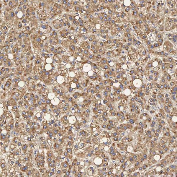
Real IHC data IHC analysis of SEC31A using anti-SEC31A antibody (A04582). SEC31A was detected in a paraffin-embedded section of human liver cancer tissue. Heat mediated antigen retrieval was performed in EDTA buffer (pH 8.0, epitope retrieval solution). The tissue section was blocked with 10% goat serum. The tissue section was then incubated with 2 μg/ml rabbit anti-SEC31A Antibody (A04582) overnight at 4°C. Peroxidase Conjugated Goat Anti-rabbit IgG was used as secondary antibody and incubated for 30 minutes at 37°C. The tissue section was developed using HRP Conjugated Rabbit IgG Super Vision Assay Kit (Catalog # SV0002) with DAB as the chromogen.
Anti-SEC31A Antibody ®
Cat # A04582

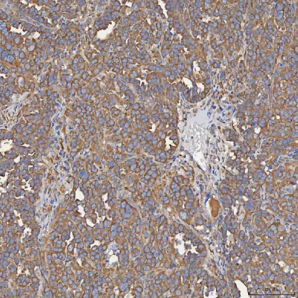
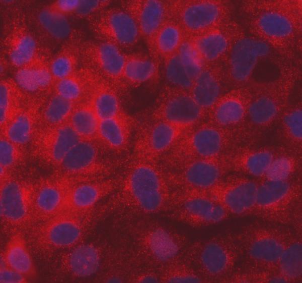
A04582 has IHC images from paraffin sections of human liver cancer, ovarian serous cancer, prostate adenocarcinoma and renal cancer (A04582 IHC captions). A04582 also has IF images from A549 cells and a paraffin section of human ovarian cancer (A04582 IF captions).

Which to pick: Choose A04582 for chromogenic IHC on paraffin sections; its human liver cancer image documents EDTA retrieval at pH 8.0, 2 μg/ml primary antibody and DAB detection (A04582 IHC caption). For IF/ICC, A04582 has images from A549 cells and a human ovarian cancer paraffin section (A04582 IF captions). The catalog lists human, mouse and rat reactivity, but the supplied images show human samples; the IHC captions do not report the fixative (catalog reactivity; A04582 IHC captions).

Each figure is that product's own IHC / IF validation image from its datasheet.

References

  1. UniProt Consortium. UniProt entry O94979 (SC31A_HUMAN, Protein transport protein Sec31A).
  2. Human Protein Atlas. SEC31A tissue IHC expression (reliability: Approved).
  3. Human Protein Atlas. SEC31A subcellular location (ICC-IF): Localized to vesicles and cytosol..
  4. Human Protein Atlas. SEC31A antibody validation summary (1 antibodies).
  5. SEC31a-ATG9a Interaction Mediates the Recruitment of COPII Vesicles for Autophagosome Formation. Advanced science (Weinheim, Baden-Wurttemberg, Germany) 2024 — PMC11600210.
  6. C9orf72-Associated Dipeptide Repeat Expansions Perturb ER-Golgi Vesicular Trafficking, Inducing Golgi Fragmentation and ER Stress, in ALS/FTD. Molecular neurobiology 2024 — PMC11584443.
  7. Cancer-associated fibroblast-derived circKLHL24 drives perineural invasion in pancreatic cancer via dual regulation of the sec31a-CXCL12 axis. Journal of experimental & clinical cancer research : CR 2025 — PMC12502155.
  8. PubMed PMID:10788476 — UniProt-cited evidence.
  9. PubMed PMID:10048485 — UniProt-cited evidence.
  10. PubMed PMID:14702039 — UniProt-cited evidence.