SEC62 / Translocation protein SEC62 · IHC design guide

Design Immunohistochemistry for SEC62

Plan SEC62 chromogenic IHC around the general cytoplasmic tissue staining reported by HPA (HPA tissue IHC) and its ER membrane location (UniProt). This guide covers fixation consistency, antibody dilution and interpretation using the available tissue evidence.

Evidence assembled Oct 2026 · For research use; verify linked source records and product datasheet before use
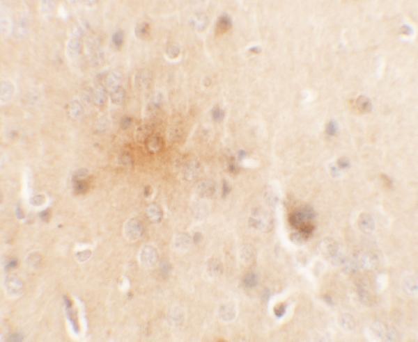
Immunohistochemistry protocol sheet for SEC62 (IHC for SEC62): expected localisation General cytoplasmic tissue staining (HPA tissue IHC), antibody A05906, validated IHC image, and IHC protocol steps
Printable SEC62 IHC protocol sheet — expected localisation General cytoplasmic tissue staining (HPA tissue IHC), antibody A05906, controls and protocol steps. Open the full SEC62 IHC guide →

SEC62 Immunohistochemistry Experimental Design Guide

Expected localisation, validated protocols, controls and antibodies — the at-a-glance facts below, then the full design guide.

Must know before staining
Expected localisation General cytoplasmic tissue staining (HPA tissue IHC)
Staining pattern Cytoplasmic; high in marrow hematopoietic cells (HPA tissue IHC)
Antigen retrieval Citrate pH 6.0 HIER, 95–98 °C, 20 min (rule: cytoplasmic / membrane antigen)
Positive control ⓘ Bone marrow+4 more · see all
Negative control ⓘ Adipose tissue+2 more · see all
Important caveats
Reasons your staining may differ from the expected pattern.
Fixation Keep fixation consistent across sections (standard IHC practice; not target-specific); Matched tissue-IHC evidence does not establish the fixation claim. Validate the specimen-specific method before use. (selected-SKU IHC image A05906)
Caveat Broad cytoplasmic staining limits cell-type specificity (HPA tissue IHC)
Regulation No specific expression regulator annotated (UniProt)
Isoform / epitope 0 annotated isoforms; cytoplasmic vs lumenal epitope matters (UniProt)
Section 1

Recommended SEC62 IHC & IF Protocols

Compare the catalog antibody’s IHC-P protocol with four published SEC62 IHC protocols (PMC9845558; PMC10253301; PMC8036965; PMC10778380).

Recommended immunohistochemistry (IHC-P) protocol parameters
SampleTissue sections; selected-image fixative not specified (standard IHC workflow)
FixationImage fixative and duration unreported (datasheet A05906); verify before use.
Sectioning4–5 µm sections on charged slides (standard)
DeparaffinisationXylene, graded ethanol series to water (standard)
Antigen retrievalHeat-induced epitope retrieval in citrate buffer, pH 6.0, 20 min at 95–98 °C (standard rule: cytoplasmic / membrane antigen)
Peroxidase block3% H2O2, 10 min, room temperature (standard)
Blocking10% normal serum of the secondary host, 30 min, room temperature (standard)
Primary antibodyRabbit anti-SEC62, 5 μg/mL (datasheet A05906)
Primary incubationOvernight at 4 °C (standard)
DetectionHRP-polymer secondary, DAB chromogen 5–10 min (standard)
CounterstainHematoxylin, blue, dehydrate and mount (standard)
Expected resultSEC62-positive staining in hematopoietic cells of bone marrow (HPA tissue IHC: High). HPA tissue profile: General cytoplasmic expression. No signal in the no-primary control.
💡Decision noteStart with citrate pH 6.0 HIER at 95–98 °C for 20 min (page retrieval rule); the published methods also include citrate and Tris-EDTA retrieval (PMC8036965; PMC10778380).
Section 2

What Is the Expected SEC62 Staining Pattern?

SEC62 is an endoplasmic reticulum (ER) membrane protein with two transmembrane segments and cytoplasmic regions on either side of them (UniProt Q99442 topology). In paraffin-section IHC, expect mainly cytoplasmic staining in cells reported as positive by HPA, including glandular cells and cerebellar Purkinje cells (HPA: tissue IHC). HPA rates its tissue IHC profile Approved, with medium consistency between staining and RNA data (HPA: tissue IHC reliability).

What am I looking at on my slide?
Cytoplasmic signal in salivary gland or cervix glandular cells, or cerebellar Purkinje cells (HPA: High in each).This fits HPA's general cytoplasmic IHC profile and UniProt's ER membrane location (HPA: tissue IHC; UniProt Q99442). Compare stained cells and their neighbours; a tissue name alone does not define which cells should be positive.
Predominantly nuclear staining, with little cytoplasmic signal in an expected positive cell population.A nuclear-only pattern does not fit the reported ER location or general cytoplasmic IHC profile (UniProt Q99442; HPA: tissue IHC). Treat it as suspect and review the controls, detection chemistry and antibody conditions before assigning it to SEC62.
Strong staining of adipocytes, skeletal myocytes or urothelial cells (HPA: Not detected in those cells).That conflicts with the specified HPA tissue observations and raises possible cross-reactivity or endogenous detection activity (HPA: tissue IHC). “Not detected” describes HPA's observations; it does not prove every specimen or assay must be negative.
Diffuse colour across cells and surrounding tissue, without a clear cytoplasmic pattern.This is difficult to score against HPA's general cytoplasmic profile (HPA: tissue IHC). As general IHC practice, check background in a primary-antibody omission control and review blocking, washes and detection conditions.
No signal in a sampled HPA High population, such as bone marrow hematopoietic cells (HPA: High).A failed expected-positive control makes a negative study section hard to interpret. As general IHC practice, check section quality, retrieval, antibody dilution and detection; HPA does not establish SEC62-specific fixation sensitivity.
💡Expected SEC62 appearanceCall a result positive when identifiable HPA High cells show clear cytoplasmic staining compatible with ER localization (HPA: tissue IHC; UniProt Q99442); dominant nuclear colour or uniform background is suspect, and HPA intensity ratings are observations rather than a required intensity threshold for every assay.
How each factor affects the staining
Which cells provide comparison?HPA reports High staining in bone marrow hematopoietic cells, Purkinje cells and several glandular populations; adipocytes, skeletal myocytes and urothelial cells were Not detected (HPA: tissue IHC). Compare the named cells, not entire tissues.
Does topology identify the stained epitope?SEC62 has two transmembrane segments, with amino and carboxyl terminal regions facing cytoplasm (UniProt Q99442 topology). The supplied record does not map either antibody's epitope, so topology cannot predict its retrieval needs.
How strong is IHC validation?HPA lists HPA014059 and HPA061450 as IHC Approved, while its tissue profile has medium staining–RNA consistency (HPA: antibodies; HPA: tissue IHC reliability). Use the observed compartment and cell type alongside that qualification.
IF/ICC: what should I see?Mainly ER localization, with additional intermediate-filament localization, is approved in HPA ICC-IF images; HPA014059 is ICC Approved (HPA: subcellular; HPA: antibodies). This answers the localization question only; IF/ICC conditions belong in its separate guide.
Can retrieval or processing explain a discrepancy?As general paraffin IHC practice, retrieval conditions can affect staining and should be checked against the antibody's own instructions. The supplied UniProt and HPA records provide no SEC62-specific fixation or retrieval effect, and no antibody epitope location.
Why is my staining missing, weak or wrong?
SituationLikely causeNext action
Expected positive cells stain weakly or remain blank.The result conflicts with HPA High observations in the selected cell population (HPA: tissue IHC); the assay may also have failed.As general IHC practice, confirm the sampled cell type, then check the antibody instructions, retrieval, dilution and detection using a positive control.
Staining appears mainly nuclear.This disagrees with SEC62's ER membrane location and HPA's general cytoplasmic profile (UniProt Q99442; HPA: tissue IHC).Review counterstain and detection background, then compare with a primary-antibody omission control; do not score nuclear-only colour as the expected SEC62 pattern.
HPA Not detected cells show strong colour.The cell-specific result conflicts with HPA observations; cross-reactivity or endogenous chromogenic activity is possible (HPA: tissue IHC).As general IHC practice, inspect omission and detection-only controls and check whether the signal follows cell boundaries or general background.
Colour is widespread and obscures cell boundaries.The section cannot be read reliably against HPA's cytoplasmic profile (HPA: tissue IHC); nonspecific background is possible.As general IHC practice, review blocking, washing, detection exposure and antibody dilution, then reassess identifiable cells in a repeat section.
Different positive tissues show different staining strengths.HPA reports High and Medium cell populations, and its tissue profile has medium staining–RNA consistency (HPA: tissue IHC).Compare like cell types and controls across sections; record compartment and cell identity before interpreting intensity differences as biological.
An IHC pattern appears inconsistent with the IF/ICC image.HPA reports cytoplasmic tissue IHC and mainly ER localization with additional intermediate-filament localization in ICC-IF (HPA: tissue IHC; HPA: subcellular).Compare the reported compartments while keeping the assays distinct; use the separate IF/ICC guide for its assay conditions and controls.

Sample controls for SEC62 IHC & IF

🧪Run cerebellum first and confirm staining in Purkinje cells (HPA: High in Purkinje cells); use skeletal muscle myocytes as the negative tissue (HPA: Not detected in myocytes). Treat nearby non-Purkinje cells as within-slide background comparators, not confirmed SEC62-negative cells, because their status is not specified in the supplied HPA rows.
Positive control tissue: Bone marrow (Hematopoietic cells, HPA High)
Negative control tissue: Adipose tissue (HPA Not detected)
ICC-IF cell lines (HPA subcellular resource): HPA ICC-IF images show SEC62 in A-431, U-251MG, U2OS, with annotated localisation: Endoplasmic reticulum (approved) (HPA subcellular).
Technical controls: Include no-primary/secondary-only and isotype controls matched to the primary antibody’s host species, immunoglobulin class and clonality, plus SEC62 knockout tissue where available or a validated peptide-block control (standard IHC practice). For cerebellar IF, assess intrinsic autofluorescence with a no-primary section before assigning signal (standard brain IF practice).
⚠️Feasibility: No supplied source reports a SEC62-specific fixation window or establishes whether antigen retrieval is required; the A05906 mouse-brain IHC caption reports 5 μg/mL but leaves the fixative unreported (caption: A05906). The supplied evidence does not establish that frozen sections or IF are easier than paraffin IHC; ICC-IF images document ER localization in A-431, U-251MG and U2OS cells (HPA: subcellular). Cerebellar autofluorescence can complicate IF interpretation (standard brain IF practice).

HPA tissue IHC evidence for SEC62

Comprehensive Human Protein Atlas IHC scoring per tissue (reliability: Approved — Medium consistency between antibody staining and RNA expression data.). Rows are taken directly from the HPA tissue chart — click any row's HPA link to view the source.

Positive expression · recommended positive controls

TissueCell typeLevelEvidenceSource
Bone marrow Hematopoietic cells High Protein (IHC) HPA →
Cerebellum Purkinje cells High Protein (IHC) HPA →
Cervix Glandular cells High Protein (IHC) HPA →
Epididymis Glandular cells High Protein (IHC) HPA →
Salivary gland Glandular cells High Protein (IHC) HPA →

Undetected expression · recommended negative controls

TissueCell typeLevelEvidenceSource
Adipose tissue Adipocytes Not detected Protein (IHC) HPA →
Skeletal muscle Myocytes Not detected Protein (IHC) HPA →
Urinary bladder Urothelial cells Not detected Protein (IHC) HPA →
Section 3

Advanced SEC62 IHC Tips

Troubleshoot SEC62 staining in chromogenic IHC by checking retrieval, compartment, controls and scoring before interpreting differences between samples.

What retrieval should I use when SEC62 staining is weak in paraffin sections?
Start with citrate buffer at pH 6.0 for heat induced epitope retrieval at 95–98 °C for 20 minutes (page retrieval protocol). After cooling, compare a dilution series with a known positive section rather than increasing retrieval time and antibody concentration together (standard IHC practice; HPA: high in cerebellar Purkinje cells). If signal remains weak, test an alternative retrieval buffer on adjacent sections as a fallback, keeping detection and exposure identical (standard IHC practice). Judge improvement by cytoplasmic staining in expected cells and preservation of section structure, since SEC62 is an endoplasmic reticulum membrane protein (UniProt Q99442; HPA: general cytoplasmic expression).
Could fixation explain patchy or absent SEC62 staining?
Target specific sensitivity of SEC62 to fixation is unknown from the supplied evidence; the A05906 mouse brain caption reports antibody use at 5 µg/mL but does not state a fixative (A05906 tissue IHC caption). For paraffin IHC, document fixative, fixation duration, processing schedule and section age for each specimen before comparing staining (standard IHC practice). Test adjacent sections through the same pH 6.0 retrieval and staining run, changing only one processing variable at a time (page retrieval protocol; standard IHC practice). Include a well preserved positive control, because patchiness alone cannot establish whether fixation, tissue damage or staining conditions caused the loss (standard IHC practice).
Where should SEC62 appear, and how should unexpected nuclear staining be assessed?
Expect predominantly cytoplasmic staining compatible with the endoplasmic reticulum membrane, rather than assigning isolated nuclear staining to SEC62 without corroboration (UniProt Q99442: endoplasmic reticulum membrane; HPA: general cytoplasmic expression). SEC62 spans the membrane at residues 197–217 and 235–255, with cytoplasmic regions on either side (UniProt Q99442 topology). Compare the questionable area with neighboring well preserved cells and a positive control stained in the same run (standard IHC practice). If nuclear signal persists while expected cytoplasmic signal disappears, check antibody concentration, secondary detection and omission controls before interpreting that pattern biologically (standard IHC practice).
How can epitope position affect SEC62 staining after retrieval?
Check the antibody's documented immunogen or mapped epitope before interpreting retrieval differences; the supplied record specifies 0 isoforms but provides no epitope location for A05906 (UniProt Q99442; A05906 tissue IHC caption). SEC62 has cytoplasmic regions at residues 1–196 and 256–399, separated by two membrane spans and a short lumenal segment at 218–234 (UniProt Q99442 topology). Several phosphorylation sites are annotated, including residues 158 and 335, but their effects on this antibody's IHC staining are unknown (UniProt Q99442 modified residues). Compare retrieval conditions on adjacent sections and confirm any changed pattern with an independently characterized antibody where available (standard IHC practice).
How should I investigate SEC62 localisation by multiplex IF?
Use a validated marker for the expected cell population, such as Purkinje cells in cerebellum, alongside SEC62 and assess cell identity in separate channels (HPA: high SEC62 staining in cerebellar Purkinje cells; standard IF practice). Choose a fluorophore channel with low background in the specimen, checking unstained tissue for autofluorescence before assigning a weak SEC62 signal (standard IF practice). Permeabilise fixed cells sufficiently to reach the antibody's mapped epitope; SEC62 has substantial cytoplasmic regions and a short lumenal region, so access depends on epitope position (UniProt Q99442 topology; standard IF practice). Compare the resulting pattern with the approved endoplasmic reticulum localisation and assess any filamentous signal separately (HPA subcellular: endoplasmic reticulum and additional intermediate filaments).
What should I check if SEC62 DAB staining is diffuse or widespread?
First inspect a no primary control and verify peroxidase blocking, since endogenous enzyme activity can produce chromogenic signal independent of the primary antibody (standard IHC practice). Reduce primary concentration or incubation strength systematically while retaining a positive section; the selected A05906 mouse brain image reports 5 µg/mL but supplies no general dilution series (A05906 tissue IHC caption; standard IHC practice). Review blocking, washes and DAB development time on paired sections processed together (standard IHC practice). Interpret residual widespread cytoplasmic staining cautiously, because HPA describes general cytoplasmic expression and low tissue RNA specificity, while its IHC reliability is Approved with medium staining to RNA consistency (HPA tissue IHC).
How can I score SEC62 IHC across specimens with different cellularity? ⚠ ANSWER MARKED FOR VERIFICATION
Define the cell population and compartment before scoring, then use a blinded H-score combining staining intensity and percentage of positive cells within that population (standard IHC practice; UniProt Q99442: endoplasmic reticulum membrane). Report 0–300 for an H-score based on intensity categories 0–3, and state the threshold used to call a cell positive (standard IHC practice). Normalise positive cell counts or stained area to the number of eligible cells or analyzed tissue area in mm², respectively, excluding folds and necrosis (standard IHC practice). Keep retrieval, DAB development and image settings consistent, and stratify by cell type because HPA reports different staining levels across tissues and cells (page retrieval protocol; HPA tissue IHC; standard IHC practice).
How do I distinguish convincing SEC62 positivity from staining artefacts?
Favor reproducible cytoplasmic staining in intact expected cells that agrees with SEC62's endoplasmic reticulum membrane localisation (UniProt Q99442; HPA: general cytoplasmic expression). A Purkinje cell rich cerebellar area can serve as a high staining comparator, whereas HPA reports no detected signal in adipocytes or skeletal muscle myocytes (HPA tissue IHC). Treat signal confined to section edges, folds or necrotic areas as suspect and examine a no primary control for endogenous enzyme or detection background (standard IHC practice). Avoid declaring a specimen negative from one weak field: HPA rates its tissue IHC profile Approved with only medium consistency against RNA data, so repeat assessment across well preserved regions and controls (HPA tissue IHC; standard IHC practice).
Boster reagents

Best SEC62 / Translocation protein SEC62 IHC Antibodies

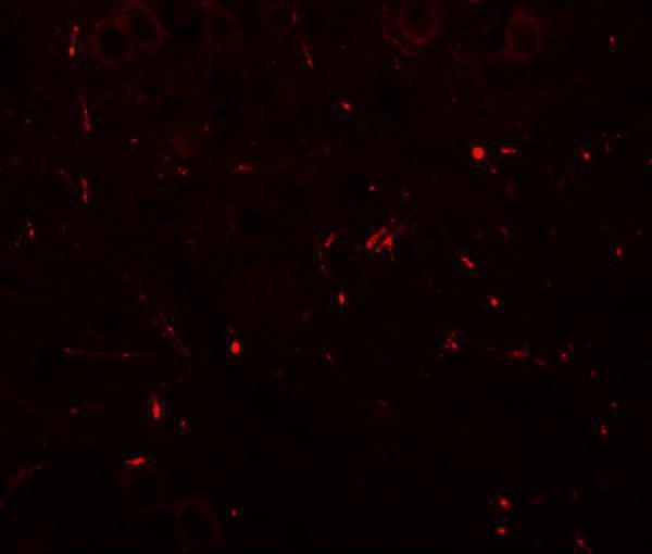
A05906 has mouse brain IHC and IF images (catalog image captions); both antibodies list Human, Mouse and Rat reactivity (catalog: A05906, M05906).

Real IHC data Immunohistochemistry of SEC62 in mouse brain tissue with SEC62 antibody at 5 μg/mL.
Anti-Translocation protein SEC62 SEC62 Antibody
Cat # A05906

A05906 lists IHC-P and IF, with mouse brain images at 5 μg/mL for IHC and 20 μg/mL for IF (catalog applications and image captions). M05906 lists IHC and ICC/IF for Human, Mouse and Rat, but has no sample-specific IHC or IF image in the payload (catalog applications, reactivity and image fields).

Which to pick: For tissue IHC, choose A05906 when a mouse brain example and an IHC-P listing are useful; its caption does not report the fixative (catalog: A05906 IHC image caption and applications). For IF/ICC, M05906 is the rabbit monoclonal with both applications listed, while A05906 has a mouse brain IF image (catalog: M05906 clone and applications; A05906 IF image caption). Both list Human, Mouse and Rat reactivity, but the supplied images document mouse brain only (catalog reactivity and image captions).

Each figure is that product's own IHC / IF validation image from its datasheet.

References

  1. UniProt Consortium. UniProt entry Q99442 (SEC62_HUMAN, Translocation protein SEC62).
  2. Human Protein Atlas. SEC62 tissue IHC expression (reliability: Approved).
  3. Human Protein Atlas. SEC62 subcellular location (ICC-IF): Mainly localized to the endoplasmic reticulum. In addition localized to the intermediate filaments..
  4. Human Protein Atlas. SEC62 antibody validation summary (2 antibodies).
  5. Effect of the 3q26-coding oncogene SEC62 as a potential prognostic marker in patients with ovarian neoplasia. Frontiers in physiology 2022 — PMC9845558.
  6. The 3q Oncogene SEC62 Predicts Response to Neoadjuvant Chemotherapy and Regulates Tumor Cell Migration in Triple Negative Breast Cancer. International journal of molecular sciences 2023 — PMC10253301.
  7. Expression of SEC62 Oncogene in Benign, Malignant and Borderline Melanocytic Tumors-Unmasking the Wolf in Sheep's Clothing?. Cancers 2021 — PMC8036965.
  8. Expression of 3q Oncogene SEC62 Predicts Survival in Head and Neck Squamous Cell Carcinoma Patients Treated with Primary Chemoradiation. Cancers 2023 — PMC10778380.
  9. PubMed PMID:9020021 — UniProt-cited evidence.
  10. PubMed PMID:10683767 — UniProt-cited evidence.
  11. PubMed PMID:10799540 — UniProt-cited evidence.