This website uses cookies to ensure you get the best experience on our website.


- Table of Contents
SEMA4F staining is reported in the retinal nerve fiber layer (HPA tissue IHC), while membrane localization is annotated at the protein level (UniProt). Use this guide to plan paraffin-section IHC with antibody A11873-2 and interpret staining in light of reported off-target binding (datasheet A11873-2; HPA tissue IHC).

Expected localisation, validated protocols, controls and antibodies — the at-a-glance facts below, then the full design guide.
| Expected localisation | Retinal nerve fibers (HPA tissue IHC); membrane expected (UniProt) | |
| Staining pattern | Medium staining in retinal nerve fiber layer; finer pattern unreported (HPA tissue IHC) | |
| Antigen retrieval | EDTA pH 8.0 HIER, heat-mediated (datasheet A11873-2) | |
| Positive control | Retina | |
| Negative control | Caudate+4 more · see all |
| Fixation | Matched tissue-IHC evidence does not establish the fixation claim. Validate the specimen-specific method before use. (selected-SKU IHC image A11873-2) | |
| Caveat | Presumed off-target staining was disregarded (HPA tissue IHC) | |
| Regulation | Regulation not annotated (UniProt) | |
| Isoform / epitope | 2 isoforms; check whether the epitope is extracellular or cytoplasmic (UniProt) |
The catalog antibody’s IHC-P protocol is accompanied by one published SEMA4F IHC protocol using ovarian tumour and adjacent non-tumour tissues (PMC10525231).
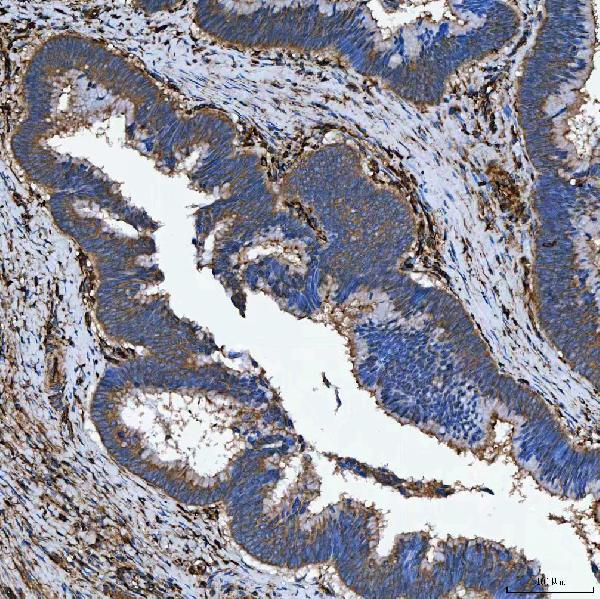
| Sample | Paraffin-embedded human colon adenocarcinoma tissue; fixative not specified (datasheet A11873-2) |
| Fixation | Image fixative and duration unreported (datasheet A11873-2); verify before use. |
| Sectioning | 4–5 µm sections on charged slides (standard) |
| Deparaffinisation | Xylene, graded ethanol series to water (standard) |
| Antigen retrieval | Heat retrieval: EDTA pH 8.0 (datasheet A11873-2); 20 min, 95–100 °C (standard) |
| Peroxidase block | 3% H2O2, 10 min, room temperature (standard) |
| Blocking | 10% goat serum (datasheet A11873-2) |
| Primary antibody | Rabbit anti-SEMA4F, 2-5 μg/ml (datasheet A11873-2) |
| Primary incubation | Overnight at 4 °C (datasheet A11873-2) |
| Detection | HRP-conjugated secondary, DAB chromogen (datasheet A11873-2) |
| Counterstain | Hematoxylin, blue, dehydrate and mount (standard) |
| Expected result | SEMA4F-positive staining in nerve fiber layer of retina (HPA tissue IHC: Medium). HPA tissue profile: Expression in nerve fibers in retina. No signal in the no-primary control. |
SEMA4F is a membrane protein with an extracellular region and a cytoplasmic tail; UniProt also places it at the postsynaptic density, perikaryon and dendrites (UniProt O95754 localization and topology). In tissue IHC, the clearest reported signal is medium staining of retinal nerve fibers (HPA: retina, nerve fiber layer). Treat this as a pattern to compare with controls, since HPA rates its IHC evidence Approved and notes presumed off target binding (HPA: IHC reliability).
| Medium staining follows nerve fibers in the retinal nerve fiber layer, with little nuclear signal (HPA: retina, nerve fiber layer; UniProt O95754 topology). | This matches the reported retinal IHC pattern and is compatible with a membrane-associated protein (HPA: retina, nerve fiber layer; UniProt O95754 localization). Assess staining alongside section morphology and controls; an Approved rating supports use of the observation but does not make every stained structure target-specific (HPA: IHC reliability). |
| Predominantly nuclear staining appears, or a broad intracellular signal obscures cell borders and fibers. | That pattern does not match SEMA4F's annotated membrane, dendritic and synaptic locations (UniProt O95754 localization). Check a control section and the detection background before calling it positive; the location mismatch alone cannot identify the precise artefact (general IHC practice). |
| Strong staining appears in cells HPA reports as undetected, such as caudate glia or colonic glandular cells (HPA: caudate and colon). | Interpret cautiously: those cell-specific HPA observations do not predict a strong signal (HPA: caudate and colon). Consider cross-reactivity or endogenous detection activity, and compare no-primary and tissue controls before assigning SEMA4F positivity (general IHC practice). |
| A uniform haze or widespread chromogen deposit covers tissue structures and makes nerve fibers hard to distinguish. | This is background rather than the localized retinal pattern reported by HPA (HPA: retina, nerve fiber layer). Review blocking, washes, detection reagents and counterstain with a no-primary control; these are general IHC checks, not documented SEMA4F-specific effects (general IHC practice). |
| No discernible nerve fiber signal appears in a retinal section expected to show medium staining (HPA: retina, nerve fiber layer). | The result is inconclusive until tissue quality, antibody application and chromogenic detection have been checked (general IHC practice). HPA's observation provides a comparator, not a guarantee that every retinal specimen or run will be positive (HPA: retina, nerve fiber layer). |
| Tissue and cell context | Retinal nerve fibers provide the clearest supplied IHC reference at medium intensity; HPA reports low staining in hippocampal glia and several other cell groups, and no detected staining in its listed negative groups (HPA: tissue IHC). A low or negative HPA entry is a comparator for that sampled cell group, not proof that an entire organ cannot express SEMA4F. |
| Topology and epitope location | SEMA4F spans residues 660–680, with residues 35–659 extracellular and 681–770 cytoplasmic (UniProt O95754 topology). The supplied record does not locate the IHC antibody's epitope, so topology alone cannot predict which preparation or retrieval condition will reveal it. |
| Protein forms | UniProt lists 2 isoforms, Long and Short, plus a signal peptide at residues 1–34 (UniProt O95754 isoforms and processing). Without the antibody epitope and isoform coverage, the staining record cannot distinguish isoforms or attribute differences between sections to processing. |
| Strength of IHC validation | HPA marks antibody HPA064095 as IHC Approved, while reporting presumed off target binding that was disregarded (HPA: antibody validation and IHC reliability). Use the reported retinal pattern as a qualified reference; this rating does not establish specificity for every tissue or unexpected compartment. |
| IF/ICC evidence boundary | HPA summarizes the subcellular location as membrane but supplies no ICC-IF image-bearing cell lines and no ICC validation entry for the listed antibody (HPA: subcellular summary and antibody validation). This supports a location comparison, not a demonstrated IF/ICC staining pattern or protocol. |
| Situation | Likely cause | Next action |
|---|---|---|
| Retinal nerve fibers show no chromogenic signal (HPA: retina, nerve fiber layer). | A failed run or an unsuitable section is possible; the supplied sources do not identify a SEMA4F-specific fixation or retrieval failure (general IHC practice; HPA: tissue IHC). | Check section integrity and the run's positive and detection controls. Review the catalog antibody's stated IHC-P conditions before changing retrieval or dilution (general IHC practice). |
| Staining is mainly nuclear. | Nuclear predominance conflicts with annotated membrane and neuronal process locations, but does not by itself identify the cause (UniProt O95754 localization). | Compare the same tissue with a no-primary control, inspect morphology and review detection background before scoring nuclei as positive (general IHC practice). |
| Strong signal appears in an HPA-undetected cell group (HPA: listed negative tissues). | Cross-reactivity or endogenous detection activity could account for unexpected staining; HPA also notes presumed off target binding in its IHC assessment (general IHC practice; HPA: IHC reliability). | Check a no-primary control and compare the signal with the retinal reference pattern. Report the unexpected cell type separately rather than treating intensity alone as confirmation (general IHC practice; HPA: retina). |
| Diffuse brown deposit masks tissue architecture. | Background from blocking, washes or detection can obscure localized staining; no target-specific mechanism is established here (general IHC practice). | Review reagent-only or no-primary controls, blocking and wash steps, then reassess whether the retinal nerve fiber layer remains distinguishable (general IHC practice; HPA: retina). |
| A faint signal appears in a tissue HPA labels low (HPA: low-staining cell groups). | Low HPA staining is compatible with a weak observed signal, but a faint deposit can also reflect background (HPA: tissue IHC; general IHC practice). | Score the specified cell group and compartment against matched background controls; avoid converting a weak, unlocalized deposit into a positive call (general IHC practice). |
| Can IF/ICC confirm the IHC pattern? | HPA's membrane summary has no accompanying ICC-IF image-bearing cell lines, and its listed antibody has no ICC validation entry (HPA: subcellular summary and antibody validation). | Use the separate IF/ICC guide for assay design. Here, describe membrane localization as a UniProt-supported expectation and keep IF/ICC claims distinct from the observed retinal IHC result (UniProt O95754 localization; HPA: retina). |


Comprehensive Human Protein Atlas IHC scoring per tissue (reliability: Approved — Presumed off target binding observed and disregarded. External characterization data supports antibody staining.). Rows are taken directly from the HPA tissue chart — click any row's HPA link to view the source.
| Tissue | Cell type | Level | Evidence | Source |
|---|---|---|---|---|
| Retina | Nerve fiber layer | Medium | Protein (IHC) | HPA → |
| Tissue | Cell type | Level | Evidence | Source |
|---|---|---|---|---|
| Caudate | Glial cells | Not detected | Protein (IHC) | HPA → |
| Cerebellum | Cells in granular layer | Not detected | Protein (IHC) | HPA → |
| Cervix | Glandular cells | Not detected | Protein (IHC) | HPA → |
| Colon | Glandular cells | Not detected | Protein (IHC) | HPA → |
| Duodenum | Glandular cells | Not detected | Protein (IHC) | HPA → |
Troubleshoot SEMA4F staining in paraffin sections with chromogenic IHC, using the catalog antibody’s tissue image and independent localisation evidence.
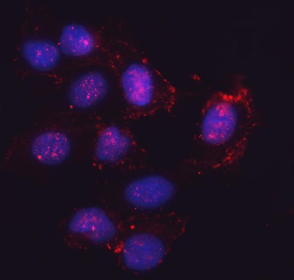
The IHC-validated antibody has images from human paraffin sections and IF images from U2OS cells (catalog image captions); listed reactivity includes human, mouse and rat (catalog reactivity).
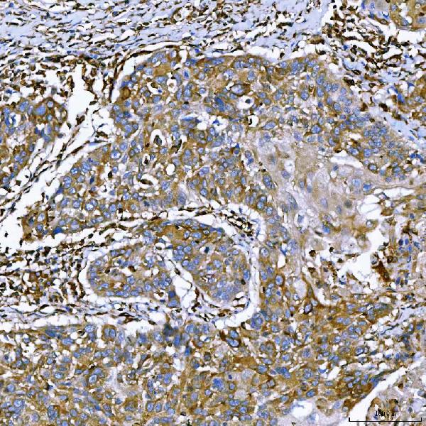
A11873-2 has IHC images from human paraffin sections of colon adenocarcinoma, larynx squamous cell carcinoma, liver cancer and lung adenocarcinoma (catalog IHC image captions). A11873-2 also has an IF image from U2OS cells (catalog IF image caption); human, mouse and rat are listed as reactive species (catalog reactivity).
Which to pick: Choose A11873-2 for chromogenic IHC on paraffin sections: its IHC captions document EDTA retrieval at pH 8.0, 2 μg/ml primary antibody and DAB detection; the fixative is unreported (catalog IHC image captions). For IF/ICC, A11873-2 has an image from U2OS cells at 5 μg/ml (catalog IF image caption). It is also the listed choice for mouse or rat samples based on reactivity, although the supplied images show human tissue and U2OS cells; clonality is unreported (catalog reactivity; catalog image captions; catalog clone field).