This website uses cookies to ensure you get the best experience on our website.


- Table of Contents
Plan SEMA6A paraffin-section IHC around the observed granular cytoplasmic pattern (HPA tissue IHC) and the annotated membrane topology (UniProt). This guide covers staining controls, interpretation, and the catalog antibody’s IHC conditions (datasheet A06967-1).

Expected localisation, validated protocols, controls and antibodies — the at-a-glance facts below, then the full design guide.
| Expected localisation | Granular cytoplasm observed (HPA tissue IHC); cell membrane annotated (UniProt) | |
| Staining pattern | Granular cytoplasmic staining across multiple cell types (HPA tissue IHC) | |
| Antigen retrieval | EDTA pH 8.0 HIER, heat-mediated (datasheet A06967-1) | |
| Positive control | Adrenal gland+4 more · see all | |
| Negative control | None in HPA (detected in all 45 tissues); use no-primary + isotype controls |
| Fixation | Keep fixation consistent across sections (standard IHC practice; not target-specific). Selected-image fixative and duration unreported (datasheet A06967-1); verify before use. | |
| Caveat | Granular tissue staining may differ from membrane expectation (HPA tissue IHC) (UniProt) | |
| Regulation | Regulation not annotated (UniProt) | |
| Isoform / epitope | 2 isoforms; check extracellular versus cytoplasmic epitope (UniProt) |
The catalog antibody’s paraffin IHC protocol (datasheet: A06967-1) is paired with published SEMA6A staining methods from liver, mouse developmental sections, and melanoma (PMC12001175; PMC2647909; PMC9016967).
| Sample | Paraffin-embedded human rectal cancer tissue; fixative not specified (datasheet A06967-1) |
| Fixation | Image fixative and duration unreported (datasheet A06967-1); verify before use. |
| Sectioning | 4–5 µm sections on charged slides (standard) |
| Deparaffinisation | Xylene, graded ethanol series to water (standard) |
| Antigen retrieval | Heat retrieval: EDTA pH 8.0 (datasheet A06967-1); 20 min, 95–100 °C (standard) |
| Peroxidase block | 3% H2O2, 10 min, room temperature (standard) |
| Blocking | 10% goat serum (datasheet A06967-1) |
| Primary antibody | Rabbit anti-SEMA6A, 0.5-1μg/ml (datasheet A06967-1) |
| Primary incubation | Overnight at 4 °C (datasheet A06967-1) |
| Detection | Streptavidin-biotin complex (SABC), DAB chromogen (datasheet A06967-1) |
| Counterstain | Hematoxylin, blue, dehydrate and mount (standard) |
| Expected result | SEMA6A-positive staining in glandular cells of adrenal gland (HPA tissue IHC: High). HPA tissue profile: General cytoplasmic expression with a granular pattern. No signal in the no-primary control. |
SEMA6A is a cell-membrane protein with an extracellular region and cytoplasmic tail (UniProt Q9H2E6 topology). In tissue IHC, HPA reports predominantly granular cytoplasmic staining, including high staining in adrenal glandular cells, cerebellar Purkinje cells and cerebral-cortex neuropil (HPA tissue IHC). Treat this observed pattern as a guide: HPA rates the tissue staining Approved, with medium consistency between antibody staining and RNA expression (HPA tissue IHC reliability).
| Granular cytoplasmic staining in adrenal glandular cells or cerebellar Purkinje cells (HPA tissue IHC). | This matches the reported tissue pattern and high staining level (HPA tissue IHC). Score the named cell population and compare its staining with nearby tissue on the same section (general IHC practice). |
| A membrane-edge signal accompanies granular cytoplasmic staining in the expected cells. | A membrane-associated component is biologically plausible because SEMA6A has one transmembrane segment (UniProt Q9H2E6 topology). The granular cytoplasmic pattern remains the tissue-IHC reference; membrane-only staining needs further validation (HPA tissue IHC). |
| Strong nuclear-only staining replaces the expected granular cytoplasmic pattern. | For paraffin IHC, investigate an off-pattern signal or artefact against the HPA tissue-IHC profile. Do not use nuclear ICC-IF localisation to validate this IHC result: HPA reports nucleoplasm in ICC-IF, while its listed antibodies have Uncertain ICC status (HPA subcellular; HPA antibodies). |
| Intense staining appears chiefly in an unexpected cell population. | Check cell identity against the HPA tissue record; cross-reactivity or endogenous detection activity are possible IHC explanations (HPA tissue IHC; general IHC practice). HPA supplies no formally negative tissue here, so unexpected staining alone cannot prove non-specificity (HPA tissue IHC). |
| Broad, diffuse colour obscures cell boundaries and the granular pattern. | Interpretability is low when background prevents recognition of the reported pattern (HPA tissue IHC). Background from detection reagents, incomplete blocking or excess primary antibody is a general IHC possibility, not a demonstrated SEMA6A-specific effect (general IHC practice). |
| Compartment and epitope location | SEMA6A spans the membrane at residues 650–670, with extracellular residues 19–649 and cytoplasmic residues 671–1030 (UniProt Q9H2E6 topology). The supplied record does not locate the antibody epitope, so it cannot predict whether a given stain will emphasize the cell edge or intracellular material. |
| Tissue and cell selection | HPA reports high staining in adrenal glandular cells, bone-marrow hematopoietic cells, cerebellar Purkinje cells and cerebral-cortex neuropil; skeletal-muscle myocytes, chondrocytes and adipocytes are listed as low (HPA tissue IHC). Low staining is not a validated negative control. |
| Antibody validation strength | HPA031265 is IHC Approved; the other listed antibodies have no IHC status in this payload (HPA antibodies). The tissue profile is Approved with medium RNA–staining consistency, so compare morphology and controls before assigning weak or unusual signal to SEMA6A (HPA tissue IHC reliability; general IHC practice). |
| Molecular features | UniProt lists 2 isoforms, a signal peptide at residues 1–18 and 6 glycosylation sites (UniProt Q9H2E6). These features alone do not establish epitope recognition, shedding, retrieval requirements or a fixation effect for the catalog antibody. |
| Antigen retrieval | Retrieval conditions are an assay variable in paraffin IHC (general IHC practice). Neither the HPA tissue pattern nor UniProt topology establishes SEMA6A-specific retrieval needs or fixation sensitivity; judge retrieval changes using staining pattern and controls. |
| Situation | Likely cause | Next action |
|---|---|---|
| No staining in adrenal glandular cells or cerebellar Purkinje cells (HPA tissue IHC). | The tissue may be unsuitable, or an IHC workflow step may have failed; absence of signal alone does not identify the cause (general IHC practice). | Verify the named cells are present, then check the primary antibody, retrieval, detection reagents and a working positive-control section (general IHC practice). |
| Only weak staining is visible in skeletal-muscle myocytes, chondrocytes or adipocytes (HPA tissue IHC). | HPA lists these cell populations as low, so they provide limited sensitivity checks and are not formal negatives (HPA tissue IHC). | Assess performance in an HPA high-staining population on a suitable section before changing the assay (HPA tissue IHC; general IHC practice). |
| Colour covers most cells, including populations outside the expected pattern. | Primary-antibody background, non-specific secondary binding or endogenous detection activity are possible general IHC causes (general IHC practice). | Compare a primary-omission control, review blocking and detection controls, and adjust antibody concentration only against an interpretable positive control (general IHC practice). |
| Nuclear-only IHC signal dominates the section. | This conflicts with the HPA granular tissue-IHC profile, although HPA separately reports approved nucleoplasmic localisation in ICC-IF (HPA tissue IHC; HPA subcellular). | Review morphology and controls; require independent IHC support before treating nuclear colour as the expected paraffin-section pattern (general IHC practice). |
| Signal is present, but no granular cytoplasmic detail can be resolved. | Excess colour development or diffuse background can conceal the pattern used for interpretation (HPA tissue IHC; general IHC practice). | Review development and counterstain, then compare the same expected cell population under conditions that preserve cellular detail (general IHC practice). |
| Does the reported nucleoplasmic ICC-IF signal define the expected IHC result? | No: ICC-IF and paraffin IHC are separate assays; HPA reports nucleoplasm in ICC-IF but marks the listed antibodies Uncertain for ICC (HPA subcellular; HPA antibodies). | Interpret paraffin IHC against the granular tissue profile. Assess any IF/ICC result with assay-specific controls in its own guide (HPA tissue IHC; general IF/ICC practice). |
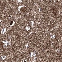


Comprehensive Human Protein Atlas IHC scoring per tissue (reliability: Approved — Medium consistency between antibody staining and RNA expression data.). Rows are taken directly from the HPA tissue chart — click any row's HPA link to view the source.
| Tissue | Cell type | Level | Evidence | Source |
|---|---|---|---|---|
| Adrenal gland | Glandular cells | High | Protein (IHC) | HPA → |
| Appendix | Glandular cells | High | Protein (IHC) | HPA → |
| Bone marrow | Hematopoietic cells | High | Protein (IHC) | HPA → |
| Breast | Glandular cells | High | Protein (IHC) | HPA → |
| Bronchus | Respiratory epithelial cells | High | Protein (IHC) | HPA → |
| Tissue | Cell type | Level | Evidence | Source |
|---|---|---|---|---|
| None in HPA: SEMA6A is detected in all 45 scored tissues. Use a no-primary (secondary-only) and an isotype control instead. | ||||
Troubleshoot SEMA6A staining in paraffin sections by checking retrieval, compartment, cell type and assay controls before assigning biological meaning.
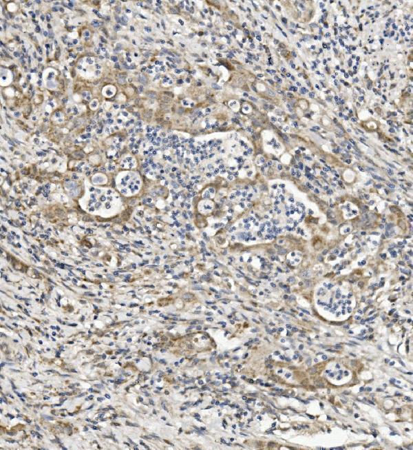
A06967-1 has real IHC data from a paraffin-embedded human rectal cancer section (catalog figure). Its listed reactivity includes human, mouse, and rat (catalog reactivity).
A06967-1 is listed for IHC and for human, mouse, and rat reactivity (catalog applications/reactivity). Its IHC figure shows chromogenic detection in a paraffin-embedded human rectal cancer section after EDTA pH 8 retrieval (A06967-1 figure caption).
Which to pick: Choose A06967-1 for paraffin-section tissue IHC at the listed 0.5–1 μg/ml; its figure used 1 μg/ml, and the fixative is unreported (catalog IHC dilution; A06967-1 figure caption). No IF/ICC application or IF figure is supplied for this SKU, so there is no supported IF/ICC pick here (catalog applications; catalog IF images). For mouse or rat samples, A06967-1 lists reactivity, but its supplied IHC figure shows human tissue only (catalog reactivity; A06967-1 figure caption).