This website uses cookies to ensure you get the best experience on our website.


- Table of Contents
Plan chromogenic SENP3 IHC in paraffin sections and score nuclear and cytoplasmic staining (HPA tissue IHC). Adrenal glandular cells show high staining, while SENP3 was not detected in adipocytes (HPA tissue IHC).

Expected localisation, validated protocols, controls and antibodies — the at-a-glance facts below, then the full design guide.
| Expected localisation | Nuclear and cytoplasmic tissue staining (HPA tissue IHC) | |
| Staining pattern | Nuclear and cytoplasmic staining in most tissues (HPA tissue IHC) | |
| Antigen retrieval | Tris-EDTA pH 9.0 HIER, 95–98 °C, 20 min (rule: nuclear antigen) | |
| Positive control | Adrenal gland+4 more · see all | |
| Negative control | Adipose tissue+4 more · see all |
| Fixation | Keep paraffin-section fixation consistent between samples (standard IHC practice; not target-specific) | |
| Caveat | Staining shows low consistency with RNA levels (HPA tissue IHC) | |
| Regulation | Oxidative stress shifts nucleolar signal (UniProt) | |
| Isoform / epitope | No annotated isoforms or processing variants (UniProt) |
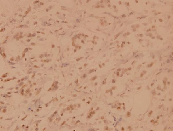
Compare the catalog antibody’s IHC-P protocol with the published SENP3 staining conditions below (PMC10710392).
| Sample | Paraffin-embedded human breast carcinoma tissue; fixative not specified (datasheet A04474Y24) |
| Fixation | Image fixative and duration unreported (datasheet A04474Y24); verify before use. |
| Sectioning | 4–5 µm sections on charged slides (standard) |
| Deparaffinisation | Xylene, graded ethanol series to water (standard) |
| Antigen retrieval | Heat-induced epitope retrieval in Tris-EDTA buffer, pH 9.0, 20 min at 95–98 °C (standard rule: nuclear antigen) |
| Peroxidase block | 3% H2O2, 10 min, room temperature (standard) |
| Blocking | 10% normal serum of the secondary host, 30 min, room temperature (standard) |
| Primary antibody | Rabbit anti-SENP3, 1:50-1:200 (datasheet A04474Y24) |
| Primary incubation | Overnight at 4 °C (standard) |
| Detection | HRP-polymer secondary, DAB chromogen 5–10 min (standard) |
| Counterstain | Hematoxylin, blue, dehydrate and mount (standard) |
| Expected result | SENP3-positive staining in glandular cells of adrenal gland (HPA tissue IHC: High). HPA tissue profile: Nuclear and cytoplasmic expression in most tissues. No signal in the no-primary control. |
SENP3 is mainly nucleolar, with nucleoplasmic localisation and lower cytoplasmic detection described by UniProt; HPA tissue IHC reports nuclear and cytoplasmic staining in most tissues (HPA subcellular; UniProt Q9H4L4; HPA tissue IHC). Adrenal glandular cells stain strongly, while several epithelial, neuronal and tubular cell groups stain at medium levels (HPA tissue IHC). HPA rates the tissue pattern Supported but reports low agreement with RNA expression (HPA tissue IHC).
| Distinct nuclear staining in adrenal glandular cells, sometimes with cytoplasmic colour. | This fits the High adrenal glandular cell signal and nuclear and cytoplasmic tissue pattern (HPA tissue IHC). Score the glandular cells and their compartments separately; HPA's reliability is Supported despite low agreement with RNA expression (HPA tissue IHC). |
| Nuclear staining in bronchial respiratory epithelium or kidney tubule cells at moderate intensity. | Both cell groups are reported at Medium intensity (HPA tissue IHC). A weaker result than adrenal glandular cells can therefore be plausible; compare the named cells rather than assigning one intensity to every cell in the section (HPA tissue IHC). |
| A sharp membrane rim or predominantly extracellular deposit with little nuclear staining. | Treat this as a suspect pattern: SENP3 has no transmembrane segment and is assigned to nuclear, nucleolar, nucleoplasmic and cytoplasmic locations (UniProt Q9H4L4; HPA subcellular). Check tissue morphology and staining controls before calling it SENP3 (general IHC practice). |
| Strong colour in adipocytes or alveolar cells while the expected positive cells are faint. | HPA reports SENP3 as Not detected in those cell groups (HPA tissue IHC). Consider antibody cross-reactivity or endogenous chromogenic detection activity, then compare negative controls and a known-positive cell group; the pattern alone does not identify the cause (general IHC practice). |
| Diffuse colour across nuclei, cytoplasm and tissue spaces, or no signal in adrenal glandular cells. | Diffuse colour obscures the reported cellular pattern; inspect controls for nonspecific staining or detection background (HPA tissue IHC; general IHC practice). If adrenal glandular cells are blank, first check section quality, antibody application and detection controls before interpreting a biological absence (HPA tissue IHC; general IHC practice). |
| Cell group and tissue | Use the named cell group as the comparison unit: adrenal glandular cells are High, while hepatocytes, bronchial respiratory epithelial cells and kidney tubule cells are Medium (HPA tissue IHC). A whole-section average can hide these differences (general IHC practice). |
| Compartment and redox state | SENP3 is reported in nucleoli and nucleoplasm and can redistribute between them during mild oxidative stress (UniProt Q9H4L4; PubMed:19680224). This supports assessing nuclear distribution, but a paraffin IHC pattern alone does not establish the tissue's oxidative state (general IHC interpretation). |
| Evidence strength | HPA labels tissue IHC Supported but notes low consistency between antibody staining and RNA expression (HPA tissue IHC). Treat its listed positive and undetected cell groups as observed staining references, not guarantees for every specimen (HPA tissue IHC; general IHC interpretation). |
| Antibody validation | HPA060290 has Supported IHC and ICC status in the supplied antibody record (HPA antibodies). This supports using its observed pattern as a reference; it does not establish a dilution, retrieval condition or target-specific fixation response (HPA antibodies). |
| Topology and processing | UniProt annotates no transmembrane segment, signal peptide or propeptide, and a chain spanning residues 1–574 (UniProt Q9H4L4). These annotations support skepticism about isolated surface or extracellular staining but do not predict an antigen retrieval requirement (UniProt Q9H4L4). |
| IF/ICC Q: What distribution should be expected? | A: Mainly nucleoli, with additional nucleoplasmic signal in the HPA ICC-IF images (HPA subcellular). That finer localisation can inform interpretation; tissue IHC is described more broadly as nuclear and cytoplasmic (HPA tissue IHC). |
| Situation | Likely cause | Next action |
|---|---|---|
| No colour in adrenal glandular cells. | The expected High signal is absent; the staining run or specimen may have failed, though the image alone cannot assign a cause (HPA tissue IHC; general IHC practice). | Review section integrity, antibody application, detection reagents and a run-level positive control before scoring the cells negative (general IHC practice). |
| Weak signal in bronchial epithelium or kidney tubules. | These groups are reported at Medium, so a weaker signal than adrenal glandular cells can fit the reference pattern (HPA tissue IHC). | Assess the named cells against a positive control and retain the observed intensity in the score; investigate only if the run controls also underperform (HPA tissue IHC; general IHC practice). |
| Colour is widespread outside cell boundaries. | A diffuse deposit does not match HPA's cell-associated nuclear and cytoplasmic description; nonspecific or chromogenic detection background is possible (HPA tissue IHC; general IHC practice). | Compare a reagent-matched negative control and inspect the distribution under higher magnification before changing general blocking, washing or detection settings (general IHC practice). |
| Adipocytes or alveolar cells stain strongly. | HPA lists these cell groups as Not detected; cross-reactivity or endogenous detection activity is possible, but neither is proven by the slide (HPA tissue IHC; general IHC practice). | Check the same run's negative controls and known-positive cells, then review antibody specificity and chromogen background if the discordance persists (general IHC practice). |
| Only membrane-like staining is evident. | This conflicts with the reported intracellular distribution and absence of a transmembrane segment (HPA subcellular; HPA tissue IHC; UniProt Q9H4L4). | Verify that the colour follows cells rather than section edges or deposits; compare controls before treating it as target-specific (general IHC practice). |
| Nuclear pattern differs from an ICC-IF image. | HPA ICC-IF resolves nucleoli and nucleoplasm, whereas its tissue IHC summary reports nuclear and cytoplasmic expression (HPA subcellular; HPA tissue IHC). | Score the compartment visible in the paraffin section and note any resolvable nucleolar detail; avoid inferring oxidative stress from this difference alone (UniProt Q9H4L4; general IHC interpretation). |


Comprehensive Human Protein Atlas IHC scoring per tissue (reliability: Supported — Low consistency between antibody staining and RNA expression data.). Rows are taken directly from the HPA tissue chart — click any row's HPA link to view the source.
| Tissue | Cell type | Level | Evidence | Source |
|---|---|---|---|---|
| Adrenal gland | Glandular cells | High | Protein (IHC) | HPA → |
| Bronchus | Respiratory epithelial cells | Medium | Protein (IHC) | HPA → |
| Caudate | Neuronal cells | Medium | Protein (IHC) | HPA → |
| Cervix | Glandular cells | Medium | Protein (IHC) | HPA → |
| Esophagus | Squamous epithelial cells | Medium | Protein (IHC) | HPA → |
| Tissue | Cell type | Level | Evidence | Source |
|---|---|---|---|---|
| Adipose tissue | Adipocytes | Not detected | Protein (IHC) | HPA → |
| Bone marrow | Hematopoietic cells | Not detected | Protein (IHC) | HPA → |
| Lung | Alveolar cells | Not detected | Protein (IHC) | HPA → |
| Lymph node | Germinal center cells | Not detected | Protein (IHC) | HPA → |
| Ovary | Follicle cells | Not detected | Protein (IHC) | HPA → |
Troubleshoot SENP3 staining in paraffin sections by checking retrieval, compartment-specific signal, controls and scoring before interpreting chromogenic IHC.
The catalog provides SENP3 IHC data from paraffin-embedded human breast carcinoma (A04474Y24 image caption). Both antibodies list human, mouse, and rat reactivity; neither lists IF/ICC (catalog applications/reactivity).
A04474Y24 is the only rendered card; its IHC image shows paraffin-embedded human breast carcinoma at 1:100 (A04474Y24 image caption). Its listed species are human, mouse, and rat, but the supplied image demonstrates human tissue only (A04474Y24 reactivity and image caption).
Which to pick: For paraffin-section IHC, choose A04474Y24: its own caption documents staining in human breast carcinoma, and the fixative is unreported (A04474Y24 image caption). Neither A04474Y24 nor A04474-1 lists IF/ICC, so neither has catalog support for that application (catalog applications). For mouse or rat IHC, either SKU lists reactivity, but only A04474Y24 has a supplied tissue image, and that image is human (catalog reactivity and image alts).