This website uses cookies to ensure you get the best experience on our website.


- Table of Contents
Plan chromogenic IHC for SEPHS1 in paraffin sections using general nuclear staining as the tissue benchmark (HPA tissue IHC). The guide highlights high staining in adipocytes from adipose tissue (HPA tissue IHC) and a catalog antibody with human paraffin section examples (datasheet A10102-1).

Expected localisation, validated protocols, controls and antibodies — the at-a-glance facts below, then the full design guide.
| Expected localisation | Nuclear staining in tissue (HPA tissue IHC) | |
| Staining pattern | Nuclei across diverse tissue cell types (HPA tissue IHC) | |
| Antigen retrieval | EDTA pH 8.0 HIER, heat-mediated (datasheet A10102-1) | |
| Positive control | Adipose tissue+4 more · see all | |
| Negative control | Heart muscle |
| Fixation | Matched tissue-IHC evidence does not establish the fixation claim. Validate the specimen-specific method before use. (selected-SKU IHC image A10102-1) | |
| Caveat | Staining and RNA show medium consistency (HPA tissue IHC) | |
| Regulation | Rises until G2/M, then declines (UniProt) | |
| Isoform / epitope | Four isoforms; check antibody epitope coverage (UniProt) |
The catalog antibody protocol uses EDTA pH 8.0 retrieval (datasheet A10102-1). The published IHC protocols below cover liver tissue, cartilage, and mouse paraffin sections (PMC7903722; PMC8828855; PMC8583877).
| Sample | Paraffin-embedded rat brain tissue; fixative not specified (datasheet A10102-1) |
| Fixation | Image fixative and duration unreported (datasheet A10102-1); verify before use. |
| Sectioning | 4–5 µm sections on charged slides (standard) |
| Deparaffinisation | Xylene, graded ethanol series to water (standard) |
| Antigen retrieval | Heat retrieval: EDTA pH 8.0 (datasheet A10102-1); 20 min, 95–100 °C (standard) |
| Peroxidase block | 3% H2O2, 10 min, room temperature (standard) |
| Blocking | 10% goat serum (datasheet A10102-1) |
| Primary antibody | Rabbit anti-SEPHS1, 2-5 µg/ml (datasheet A10102-1) |
| Primary incubation | Overnight at 4 °C (datasheet A10102-1) |
| Detection | HRP-conjugated secondary, DAB chromogen (datasheet A10102-1) |
| Counterstain | Hematoxylin, blue, dehydrate and mount (standard) |
| Expected result | SEPHS1-positive staining in adipocytes of adipose tissue (HPA tissue IHC: High). HPA tissue profile: General nuclear expression. No signal in the no-primary control. |
SEPHS1 should show predominantly nuclear staining across many cell types in paraffin-section IHC (HPA: general nuclear expression; low tissue specificity). UniProt also lists cytoplasm, cell membrane, nuclear membrane and chromosome locations, so a minor signal outside the nucleus is possible (UniProt P49903: subcellular locations). SEPHS1 has no transmembrane segment (UniProt P49903: topology). HPA rates its tissue IHC evidence Enhanced, with medium agreement between staining and RNA data pending external verification (HPA: reliability).
| Distinct nuclear chromogen in adrenal glandular cells or cerebral cortex neurons (HPA: High in both cell types). | This fits the expected compartment and observed positive cells (HPA: general nuclear expression; High in both). Judge intensity against controls on the same run; High describes HPA's observed staining, not a guaranteed intensity in every specimen. |
| Strong staining confined to cell borders, with little nuclear staining (HPA: general nuclear expression). | The distribution conflicts with the predominant tissue IHC pattern (HPA: general nuclear expression). UniProt lists cell membrane localisation, so do not reject a minor border signal solely on location; assess the nuclear pattern and controls together (UniProt P49903: subcellular locations). |
| Prominent chromogen in cardiomyocytes but absent from adjacent expected positive cells (HPA: cardiomyocytes Not detected). | Check cross-reactivity and endogenous detection activity before calling this SEPHS1. HPA did not detect cardiomyocyte staining; that observation is a comparator, not proof that every heart specimen must be negative (HPA: heart muscle IHC). |
| Diffuse colour across nuclei, cytoplasm and empty tissue spaces, with poor cell boundaries. | Treat the field as background until a control run shows specific localisation. Review blocking, wash stringency and detection controls as general IHC practice; diffuse colour alone does not establish SEPHS1 expression (HPA: general nuclear expression). |
| No nuclear signal in a well-preserved adrenal gland section (HPA: glandular cells High). | First check whether the positive control and detection reagents worked. HPA's High result makes adrenal glandular cells a useful comparator, but its Enhanced rating still has medium RNA agreement pending external verification (HPA: tissue IHC reliability). |
| Cell and tissue context (HPA: tissue IHC). | HPA reports High staining in adipocytes, respiratory epithelial cells, adrenal and duodenal glandular cells, cortical neurons and colonic endothelial cells. It reports Low staining in hippocampal and caudate glia (HPA: tissue IHC); choose a comparator with the relevant cell type. |
| IHC evidence strength (HPA: antibody validation). | HPA037645 and HPA062118 are both rated Enhanced for IHC (HPA: antibody records). HPA describes tissue staining versus RNA agreement as medium and pending external verification (HPA: reliability); interpret an isolated unexpected pattern cautiously. |
| Compartment and topology (UniProt P49903; HPA: tissue IHC). | Nuclear staining has the strongest tissue-pattern support (HPA: general nuclear expression). UniProt also lists cytoplasm and membranes, but reports no transmembrane segment (UniProt P49903: subcellular locations; topology); location alone cannot validate a membrane-dominant stain. |
| Isoforms and cell cycle (UniProt P49903). | Four isoforms are listed, and expression rises through the cell cycle until G2/M before decreasing (UniProt P49903: isoforms; tissue specificity note). Without antibody epitope information, neither an isoform-specific staining pattern nor a fixed intensity threshold can be assigned. |
| IF/ICC Q: Should the IHC pattern transfer directly? | A: Use nucleoplasmic signal as a qualified comparison: HPA calls its ICC-IF location nucleoplasm, uncertain (HPA: subcellular summary). HPA037645 is Uncertain for ICC, and HPA062118 has no ICC rating (HPA: antibody records); this IHC section provides no IF protocol. |
| Situation | Likely cause | Next action |
|---|---|---|
| Positive control lacks nuclear staining (HPA: general nuclear expression). | The IHC run may have failed; absence alone cannot identify a SEPHS1-specific cause. | Check section integrity, detection reagents and the assay's antigen-retrieval settings as general IHC practice. Repeat with a known positive cell population, such as adrenal glandular cells (HPA: High), before scoring study tissue. |
| Nuclear stain is present but weak in a purported positive tissue. | The sampled cell type may have lower observed staining: hippocampal and caudate glia are Low (HPA: tissue IHC). | Identify the actual cell population, then compare it with a matched positive control. Do not apply a High-tissue intensity expectation to glia reported Low by HPA (HPA: tissue IHC). |
| Signal is strongest at cell borders with sparse nuclear stain. | The pattern differs from HPA's general nuclear tissue profile, although UniProt lists cell membrane localisation (HPA: tissue IHC; UniProt P49903: subcellular locations). | Recheck morphology and detection controls; assess whether a clear nuclear component exists. Avoid assigning the border-only pattern to SEPHS1 from compartment alone. |
| Chromogen appears in cardiomyocytes (HPA: Not detected). | Cross-reactivity or endogenous detection activity is possible; HPA reports no cardiomyocyte staining (HPA: heart muscle IHC). | Use appropriate reagent-omission and detection controls as general IHC practice. Compare with a known positive cell type before treating the cardiomyocyte signal as target-specific. |
| Diffuse background obscures nuclei and cell boundaries. | Insufficient blocking, washing or detection control can obscure localisation in chromogenic IHC (general IHC practice). | Review those steps and compare control sections processed together. Score SEPHS1 only where cell-level nuclear staining is distinguishable from background (HPA: general nuclear expression). |
| IF/ICC shows no clear nucleoplasmic enrichment. | HPA's nucleoplasm assignment is uncertain, and its antibody records give ICC as Uncertain or unrated (HPA: subcellular summary; antibody records). | Interpret the IF result within its separate guide and controls. Do not use the IHC Enhanced rating as proof of ICC performance (HPA: antibody validation). |



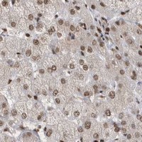


Comprehensive Human Protein Atlas IHC scoring per tissue (reliability: Enhanced — Medium consistency between antibody staining and RNA expression data. Pending external verification.). Rows are taken directly from the HPA tissue chart — click any row's HPA link to view the source.
| Tissue | Cell type | Level | Evidence | Source |
|---|---|---|---|---|
| Adipose tissue | Adipocytes | High | Protein (IHC) | HPA → |
| Adrenal gland | Glandular cells | High | Protein (IHC) | HPA → |
| Breast | Adipocytes | High | Protein (IHC) | HPA → |
| Bronchus | Respiratory epithelial cells | High | Protein (IHC) | HPA → |
| Cerebral cortex | Neuronal cells | High | Protein (IHC) | HPA → |
| Tissue | Cell type | Level | Evidence | Source |
|---|---|---|---|---|
| Heart muscle | Cardiomyocytes | Not detected | Protein (IHC) | HPA → |
Troubleshoot SEPHS1 staining in paraffin sections by checking retrieval, compartment, cell type, controls, and scoring before interpreting chromogenic signal.
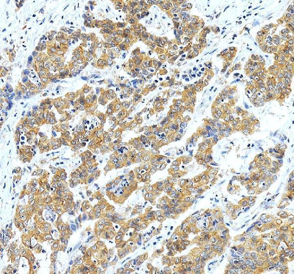
The IHC-validated anti-SEPHS1 antibody has paraffin-section images from rat brain, human liver, and human liver cancer; no IF/ICC image is supplied (catalog: A10102-1 image captions).
A10102-1 will render with an IHC image from a paraffin-embedded rat brain section (catalog: A10102-1 figure caption). Its additional IHC captions show paraffin-embedded human liver and liver cancer sections; listed reactivity is human, mouse, and rat (catalog: A10102-1 image captions and reactivity).
Which to pick: Choose A10102-1 for paraffin-section IHC: its captions document EDTA retrieval at pH 8.0 and 2 µg/ml primary antibody in the pictured rat and human sections; the fixative is unreported (catalog: A10102-1 IHC captions). For cross-species work, it is listed as a rabbit polyclonal reactive with human, mouse, and rat, though the supplied IHC captions show only human and rat tissue (catalog: A10102-1 host, dilution_raw, reactivity, and IHC captions). No IF/ICC option is supported by the supplied application list or images (catalog: A10102-1 applications and if_image_alts).