This website uses cookies to ensure you get the best experience on our website.
- Table of Contents
Facts about Septin-5.
May play a role in platelet secretion (By similarity). .
Human | |
---|---|
Gene Name: | SEPTIN5 |
Uniprot: | Q99719 |
Entrez: | 5413 |
Belongs to: |
---|
TRAFAC class TrmE-Era-EngA-EngB-Septin-like GTPase superfamily |
CDCREL; CDCREL1; CDCREL-1; cell division control related protein 1; Cell division control-related protein 1; HCDCREL-1; peanut-like 1 (Drosophila); peanut-like 1; Peanut-like protein 1; platelet glycoprotein Ib beta chain; PNUTL1H5; septin 5; septin-5
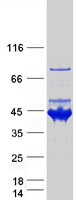
Mass (kDA):
42.777 kDA
Human | |
---|---|
Location: | 22q11.21 |
Sequence: | 22; NC_000022.11 (19714503..19723319) |
Expressed at high levels in the CNS, as well as in heart and platelets (at protein level).
Cytoplasm. Cytoplasm, cytoskeleton. In platelets, found in areas surrounding alpha-granules.
This article will cover the Autoradiography Film, Detection techniques, Application to Western Blots, and Protein Transfer Efficiency by Membrane Staining. It will also give you some tips for using this marker in your own experiments. Boster Bio stocks the SEPTIN5 anti-bodiment. The company offers several different products for this protein. Continue reading to learn more about each of these products!
Boster Bio offers primary antibodies that have high affinity for the SEPTIN5 marker. These antibodies have been validated on immunohistochemistry, Western Blotting, and ELISA techniques. They are recommended to be used in the recombinant SEPTIN5 expression assay. SEPTIN5 is a marker that can be used to diagnose and treat cancer.
This gene can be found in the skin of many mammal species. Sebum production is controlled by the SEPTIN5 genetic. It is found in the skins and hairs of mice, insects, and humans. Its presence in these animals helps researchers better understand their relationship. SEPTIN5 is also expressed on the skin of mammals, even humans. The gene encoding SEPTIN5 is present in several species of mammals, including humans, birds, and reptiles.
Two orthologs for the SEPTIN5 marker were identified in mammals. Septin3b (and septin5b) are expressed at different times in development and the adult brain. These genes are essential for nervous system analysis. There are several detection methods available for the SEPTIN5 mark. Here's a quick description of these markers. The septins are characterized by their coiled-coil domain, phosphoinositide-binding polybasic domain, and a GTP-binding site.
These markers are essential for neuronal growth. The brain of mice was found to contain the soluble SEPTIN5 gene. It is found in the brain where it plays a crucial role in neuronal trafficking and neuronal Polarity. Septin 5 interacts directly with Sec6/Sec8 and syntaxins 1,4 and 5.
The transient transfection effectiveness of the SepTIN5 marker was approximately thirty five percent. A significant amount of Septin 5 was accumulated in the SN4741 cells, resulting in DAergic cell death within 48-72 h. In addition, HA-tagged Septin 5 cells undergo necrotic cell death and nuclear fragmentation. V5-tagged synphilin-1 did not cause cytotoxic cell death in the SN4741 cells.
Increased levels SEPTIN5 may alter synaptic and autocrine neurotransmitter release, which are crucial for neuronal communication. As mentioned, abundant Septin 5 is associated with vesicle structure and is localized in presynaptic terminals. Parkin, which is also a co-receptor protein for Septin 5, is abundantly present in DAergic neuron. For proper synaptic function, strict regulation of SEPTIN5 levels in SN-DAergic neuron is necessary.
Many applications of Western blots for medical research include the diagnosis and treatment or pain management. Western blots can be used to detect the presence of a protein interest in cells or tissues. These studies reveal whether a protein is present in the sample, as indicated by its size or by the presence of antibodies to that protein. This technique is also useful when researching cancer.
Two types of antibodies must be used for western blots. The primary antibody binds directly to the target protein. A secondary antibody is used to detect specific epitopes. There are many antibodies that can detect the target protein. The primary antibody is usually monoclonal. It is important that you choose the right antibody for your application. Not all antibodies are suitable to be used in western blots. Before purchasing a new antibody, you should verify the type of primary antibody that was used.
Another method for detecting proteins in biological samples, protein electrophoresis, is another. This method separates proteins by their charge and molecular weight. The specific application depends on the target protein. A centrifuged sample will remove solids and load only the soluble fraction. If the sample contains the protein of interest, it is possible that the protein is in the insoluble fraction. It is possible to perform a pretreatment step in order to liberate or solubilize proteins before loading them into gels.
With the latest improvements to their Western Blotting system, Boster Bio has significantly improved the efficiency of protein transfer by membrane staining. This system can transfer 10-300kDa protein from polyacrylamide gels onto membranes in 5-10 minutes. The Power Blotter works with commonly used gels, including nitrocellulose and PVDF membranes. It is important to keep in mind that the transfer effectiveness of the Powerblotter can vary depending on the type of membrane used, gel thickness and molecularweight of the target protein.
The method involved applying a DB-71 solution to the membranes and gently shaking them for 10 min. The membranes then had to be rinsed with 50% methanol before being placed in a scanner. The optical density of stained proteins was measured to analyze the images. The results were compared between two experiments. The results were consistent. The new membrane staining system can be used with any type of sample and allows for precise and efficient analysis.
Boster Bio has not only improved the efficiency of protein transfers, but also increased the sensitivity for the detection of transferred proteins. It has been shown that NC membranes increase the ability of antibodies recognize proteins in different samples. Its binding power varies depending on molecular weight. PVDF membranes are more efficient than NC membranes in protein transfer, up to four times.
ECL chemiluminescent detection has many applications. These systems combine high sensitivities with a wide dynamic spectrum and are the preferred choice of many scientists. Electrochemical methods using ECL can be used to quantify a variety of biological materials. These include:
An electrochemical reaction is the basic chemiluminescent process that produces light. The enzymes create a chemical reaction that generates excited intermediates. These excited intermediates then release powerful blue light at 450 nm wavelength. This light is generated only during enzyme reaction with substrate. Substrate exhaustion causes the signal output to cease.
Western blots are widely utilized for the detection of protein samples. It is easy and sensitive to use a chemiluminescent detector system. There are many reagents that you can choose from, but they all have the same basic characteristics. A chemiluminescent reagent is a two component system. It includes a stable peroxide mixture and a secondary protein-specific antibody. Both components are mixed together to create a workable solution. Once the membranes have been incubated with the sample, they are exposed to the antibodies and the chemical reaction that results in light will occur. A sensitive camera will detect the light.
The ECL chemiluminescent detection technique provides highly sensitive and accurate detection of protein from Western blots. Many luminescent assays have low light or short duration. A chemiluminescent reaction uses a luminol in the presence of modified phenols to greatly enhance light emission and kinetics of the reaction. It is one of the most sensitive detection methods and is now the standard in protein labs.
Immunohistochemistry is a common detection method in clinical pathology. This technology has revolutionized cancer diagnosis, classification and prognosis since the 1970s. The field of pathology has seen immunohistochemistry greatly improve since it was originally developed to detect specific molecular markers. This article will focus on the clinical importance of the most common immunohistochemical markers. Listed below are some of the benefits and limitations of immunohistochemistry.
The principle behind immunohistochemistry is that antibodies recognize specific antigens or haptens present in biological tissues. These interactions can then be visualized using fluorescent colors, enzyme-substrate coloring reactions, radioactive elements, and colloidal golden. Biotechnology researchers are making significant progress in tissue fixation and protein conjugation. However, IHC tests have some limitations, such as cross-reactivity and sensitivity of antibodies.
As more people become aware of immunohistochemistry, the field continues to grow. There are now more applications for the technique than ever before. Cancer immunotherapy is a very important area. Many treatments for cancer target specific molecular pathways. To evaluate the response to Erbitux, immunohistochemistry can be used for the protein EGFR. Many commercial systems are available, including Dako pharmDx. These systems allow immunohistochemistry the ability to identify specific tumor targets.
PMID: 9022087 by Zieger B., et al. Alternative expression of platelet glycoprotein Ib(beta) mRNA from an adjacent 5' gene with an imperfect polyadenylation signal sequence.
PMID: 9385360 by McKie J., et al. A human gene similar to Drosophila melanogaster peanut maps to the DiGeorge syndrome region of 22q11.