This website uses cookies to ensure you get the best experience on our website.


- Table of Contents
Plan chromogenic SEPTIN9 IHC in paraffin sections with the catalog antibody at 2–5 μg/ml (datasheet A05279-3). Use the cytoplasmic, occasionally membranous tissue pattern to guide scoring (HPA tissue IHC).

Expected localisation, validated protocols, controls and antibodies — the at-a-glance facts below, then the full design guide.
| Expected localisation | Cytoplasmic, occasionally membranous (HPA tissue IHC) | |
| Staining pattern | Colon glandular cells: cytoplasmic, occasionally membranous (HPA tissue IHC) | |
| Antigen retrieval | EDTA pH 8.0 HIER, heat-mediated (datasheet A05279-3) | |
| Positive control | Bronchus+4 more · see all | |
| Negative control | Adipose tissue+3 more · see all |
| Fixation | Keep fixation consistent across sections. (standard IHC practice; not target-specific) | |
| Caveat | Tissue-specific isoforms may affect epitope detection (UniProt) | |
| Regulation | TGF-β1/EMT changes actin colocalisation (UniProt) | |
| Isoform / epitope | 8 isoforms; epitope coverage is unspecified (UniProt) |
Compare the catalog antibody’s IHC-P protocol (datasheet A05279-3) with the published human brain tissue IHC protocol (PMC5938713).
| Sample | Paraffin-embedded human breast cancer tissue; fixative not specified (datasheet A05279-3) |
| Fixation | Image fixative and duration unreported (datasheet A05279-3); verify before use. |
| Sectioning | 4–5 µm sections on charged slides (standard) |
| Deparaffinisation | Xylene, graded ethanol series to water (standard) |
| Antigen retrieval | Heat retrieval: EDTA pH 8.0 (datasheet A05279-3); 20 min, 95–100 °C (standard) |
| Peroxidase block | 3% H2O2, 10 min, room temperature (standard) |
| Blocking | 10% goat serum (datasheet A05279-3) |
| Primary antibody | Rabbit anti-SEPTIN9, 2-5 μg/ml (datasheet A05279-3) |
| Primary incubation | Overnight at 4 °C (datasheet A05279-3) |
| Detection | HRP-conjugated secondary, DAB chromogen (datasheet A05279-3) |
| Counterstain | Hematoxylin, blue, dehydrate and mount (standard) |
| Expected result | SEPTIN9-positive staining in basal cells of bronchus (HPA tissue IHC: High). HPA tissue profile: General cytoplasmic and occasional membranous expression. No signal in the no-primary control. |
SEPTIN9 is a cytoplasmic cytoskeletal protein with no transmembrane segment (UniProt Q9UHD8 topology and subcellular location). In paraffin sections, expect mainly cytoplasmic staining, sometimes with a membranous appearance (HPA tissue IHC). HPA reports High staining in several epithelial populations, including colon glandular cells and bronchial basal cells (HPA tissue IHC). The tissue IHC profile is Approved, with medium consistency between staining and RNA data (HPA tissue IHC).
| Cytoplasmic signal in colon glandular cells or bronchial basal cells, with occasional membrane-adjacent accentuation. | This matches HPA's High staining in those cells and its general cytoplasmic, occasional membranous profile (HPA tissue IHC). Membrane-adjacent signal is plausible because SEPTIN9 can concentrate at epithelial cell-cell contacts (UniProt Q9UHD8 subcellular location). Judge the stained cells and compartment together, rather than treating every stained area of the section as equivalent. |
| Strong, predominantly nuclear signal with little cytoplasmic staining. | A nuclear-dominant pattern conflicts with the reported cytoplasmic and cytoskeletal localization (UniProt Q9UHD8 subcellular location; HPA tissue IHC). Treat it as suspect until antibody specificity and detection controls are checked. The supplied sources do not establish a nuclear IHC pattern for SEPTIN9. |
| Prominent signal in adipocytes or cardiomyocytes, while expected epithelial cells stain weakly. | HPA reports SEPTIN9 as Not detected in adipocytes and cardiomyocytes, but High in several specified epithelial populations (HPA tissue IHC). Recheck cell identification and controls; unexpected color may reflect cross-reactivity or endogenous detection activity (general chromogenic IHC practice). These HPA entries describe particular cell types, not whole-tissue negatives. |
| Diffuse color covers stroma, empty spaces, or many cell types without a discernible cytoplasmic pattern. | That distribution does not resemble HPA's cell-associated, generally cytoplasmic profile (HPA tissue IHC). Consider excess primary or detection reagent, incomplete blocking, or inadequate washing (general chromogenic IHC practice). Compare with a matched control section before assigning the diffuse color to SEPTIN9. |
| No signal in the glandular cells of a colon control section. | Colon glandular cells are reported as High by HPA, so their absence of staining makes the run difficult to interpret (HPA tissue IHC). Check section quality, the catalog antibody's IHC-P instructions, retrieval and detection setup, and whether another expected structure stained (general IHC practice). A failed positive control cannot establish that an experimental specimen lacks SEPTIN9. |
| Cell type and tissue context | HPA reports High staining in colon, cervix, endometrium, and gallbladder glandular cells, among other listed populations; adipocytes and cardiomyocytes are Not detected (HPA tissue IHC). Select controls by the named cell population within the section. HPA also reports low tissue RNA specificity, which does not make every cell type an equally useful protein-staining control (HPA tissue IHC). |
| Subcellular context | SEPTIN9 is cytoplasmic and cytoskeletal, and lacks a transmembrane segment (UniProt Q9UHD8 topology and subcellular location). HPA describes general cytoplasmic and occasional membranous tissue staining (HPA tissue IHC). Interpret edge accentuation in that context; the supplied evidence does not support treating membrane-only staining as the expected IHC result. |
| Isoforms and antibody epitope | UniProt lists 8 SEPTIN9 isoforms with differential tissue expression (UniProt Q9UHD8 isoforms and tissue specificity). An antibody's epitope determines which isoforms it can detect (general antibody practice). The supplied record gives no epitope or isoform coverage for the catalog antibody, so do not infer isoform identity from staining intensity or distribution. |
| Antibody evidence | HPA042564 is Approved for IHC, whereas no IHC status is supplied for HPA050627; both have Enhanced ICC validation (HPA antibodies). HPA's tissue profile has medium staining-to-RNA consistency (HPA tissue IHC). Use these ratings as context for interpreting a pattern, not as proof that every stained cell is specific. |
| IF/ICC Q&A: Where should SEPTIN9 fluorescence appear? | Mainly along actin filaments, with additional localization to microtubules, the cytokinetic bridge, and primary cilium (HPA subcellular ICC-IF). This is an ICC-IF observation; it does not require resolving those structures in chromogenic paraffin sections. UniProt also places SEPTIN9 in the cytoplasmic cytoskeleton (UniProt Q9UHD8 subcellular location). |
| Retrieval and fixation evidence | No target-specific fixation sensitivity or retrieval condition is established by the supplied UniProt and HPA records. Follow the catalog antibody's IHC-P instructions and evaluate the positive control when setting up a run (general IHC practice). Do not attribute a positive or negative tissue result to a SEPTIN9-specific fixation effect on this evidence. |
| Situation | Likely cause | Next action |
|---|---|---|
| Colon glandular positive control is blank. | Possible run-level retrieval, primary-antibody, or detection failure (general IHC practice); colon glandular cells are High in HPA tissue IHC. | Verify the catalog antibody's IHC-P conditions, reagent sequence, and detection activity; repeat with a control section containing identifiable colon glands (general IHC practice). Interpret experimental negatives only after the positive control works. |
| Signal is mainly nuclear. | The compartment disagrees with cytoplasmic and cytoskeletal localization (UniProt Q9UHD8 subcellular location; HPA tissue IHC). Nonspecific staining or detection artifact is possible (general IHC practice). | Check a matched negative control and a known-positive cell population, then review antibody concentration and detection steps (general IHC practice). Require a reproducible cytoplasmic pattern before calling the section positive. |
| Adipocytes or cardiomyocytes stain strongly. | Those named cell types are Not detected in HPA tissue IHC. Cross-reactivity or endogenous detection activity may contribute to unexpected chromogen (general IHC practice). | Confirm cell identity, compare a primary-omission control, and check the detection blocking steps (general chromogenic IHC practice). Do not label an entire adipose or heart section negative solely from HPA's cell-type entries. |
| A broad brown haze obscures cell boundaries. | Diffuse background can arise from excess reagent, incomplete blocking, or insufficient washing (general chromogenic IHC practice); it obscures HPA's mainly cytoplasmic pattern (HPA tissue IHC). | Inspect a primary-omission control, review blocking and wash steps, and optimize antibody concentration under the catalog IHC-P workflow (general IHC practice). Score only localized cellular signal that remains distinguishable from background. |
| One epithelial population stains while another is faint. | HPA assigns levels to specific cells and reports medium staining-to-RNA consistency (HPA tissue IHC); UniProt reports different tissue expression among SEPTIN9 isoforms (UniProt Q9UHD8 tissue specificity). | Record tissue, cell type, compartment, and intensity separately; compare each with its own HPA entry (HPA tissue IHC). Avoid inferring a particular isoform because antibody epitope coverage is not supplied. |
| Only a thin cell-edge line is visible. | Occasional membranous staining is reported by HPA, and SEPTIN9 can concentrate at epithelial contacts (HPA tissue IHC; UniProt Q9UHD8 subcellular location). An edge-only result is less complete than HPA's general cytoplasmic profile. | Inspect the same cells for cytoplasmic signal and compare with a positive control and negative control (general IHC practice). Record the edge pattern separately if cytoplasmic staining cannot be confirmed. |
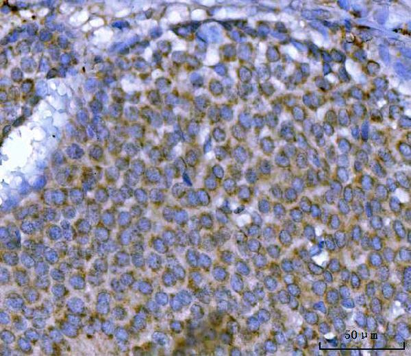
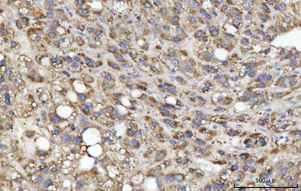
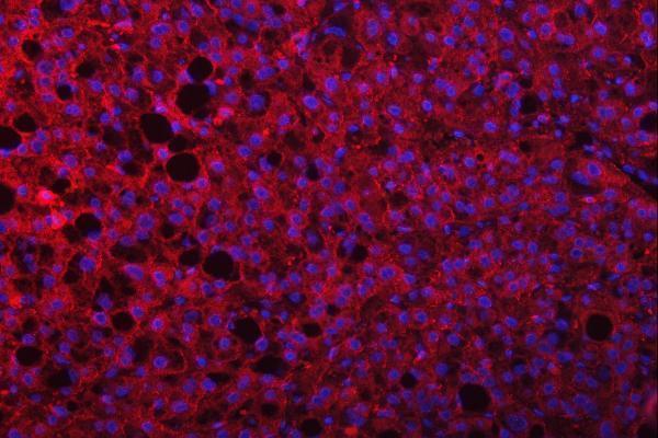
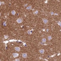


Comprehensive Human Protein Atlas IHC scoring per tissue (reliability: Approved — Medium consistency between antibody staining and RNA expression data.). Rows are taken directly from the HPA tissue chart — click any row's HPA link to view the source.
| Tissue | Cell type | Level | Evidence | Source |
|---|---|---|---|---|
| Bronchus | Basal cells | High | Protein (IHC) | HPA → |
| Cervix | Glandular cells | High | Protein (IHC) | HPA → |
| Colon | Glandular cells | High | Protein (IHC) | HPA → |
| Endometrium | Glandular cells | High | Protein (IHC) | HPA → |
| Esophagus | Squamous epithelial cells | High | Protein (IHC) | HPA → |
| Tissue | Cell type | Level | Evidence | Source |
|---|---|---|---|---|
| Adipose tissue | Adipocytes | Not detected | Protein (IHC) | HPA → |
| Caudate | Glial cells | Not detected | Protein (IHC) | HPA → |
| Heart muscle | Cardiomyocytes | Not detected | Protein (IHC) | HPA → |
| Skeletal muscle | Myocytes | Not detected | Protein (IHC) | HPA → |
Troubleshoot SEPTIN9 chromogenic IHC using the documented paraffin-section workflow, tissue staining patterns, and intracellular localisation (datasheet A05279-3; HPA; UniProt Q9UHD8).
A05279-3 has IHC images from paraffin-embedded human breast, liver, and lung cancers and mouse lymphaden tissue; IF images show paraffin-embedded human breast and liver cancers (catalog image captions).
A05279-3 is listed for IHC and IF in human and mouse samples (catalog applications and reactivity). Its IHC captions document paraffin-embedded human breast, liver, and lung cancers and mouse lymphaden tissue; its IF captions document paraffin-embedded human breast and liver cancers (catalog image captions).
Which to pick: Choose A05279-3 for tissue IHC on paraffin sections, supported by its own IHC captions; the fixative is unreported (catalog IHC image captions). Choose A05279-3 for tissue IF based on its IF captions; ICC validation is unreported (catalog applications and IF image captions). For human and mouse work, A05279-3 lists both species as reactive and has IHC images for each; it is a rabbit antibody, with clonality unreported (catalog reactivity, host, and IHC image captions).