This website uses cookies to ensure you get the best experience on our website.


- Table of Contents
Real validated SERPINA7 Western blot protocols, expected-band and isoform facts, troubleshooting for weak or shifted signal, and recommended anti-SERPINA7 WB antibodies. Everything you need to plan the experiment before you commit precious samples.
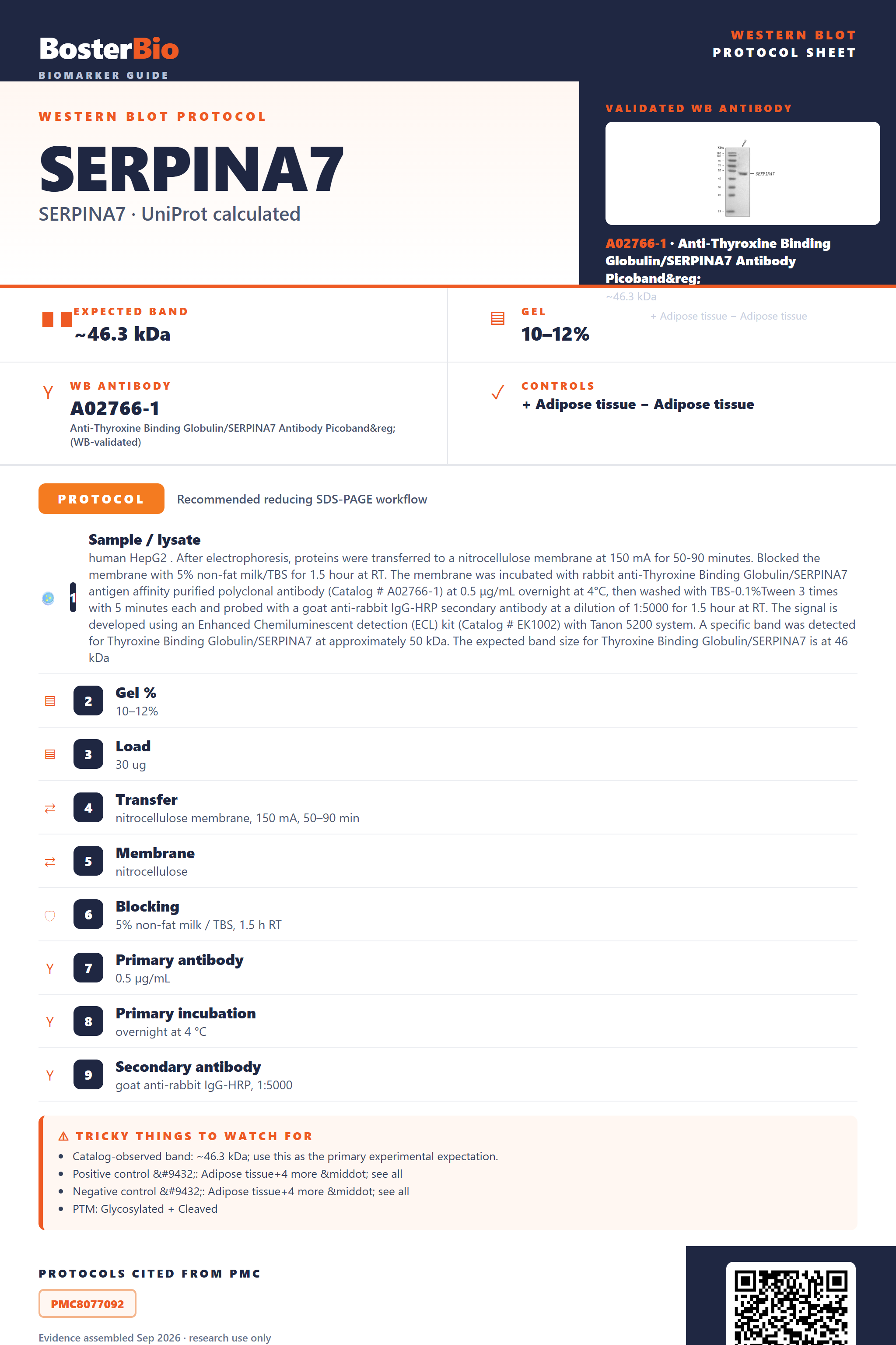
Expected bands, validated protocols, controls and antibodies — the at-a-glance facts below, then the full design guide.
| Expected band | ~46.3 kDa | |
| Observed band | ~50 kDa | |
| Gel | 10–12% | |
| Positive control | Adipose tissue+4 more · see all | |
| Negative control | Adipose tissue+4 more · see all |
| PTM | Glycosylated + Cleaved | |
| Caveat | N-glycosylation increases mass | |
| Regulation | IFN-γ-induced | |
| Isoform | 1 isoform(s) |
Literature-validated Western blot parameters for SERPINA7 — gel percentage, transfer, blocking, antibody incubation and detection, extracted from published methods.
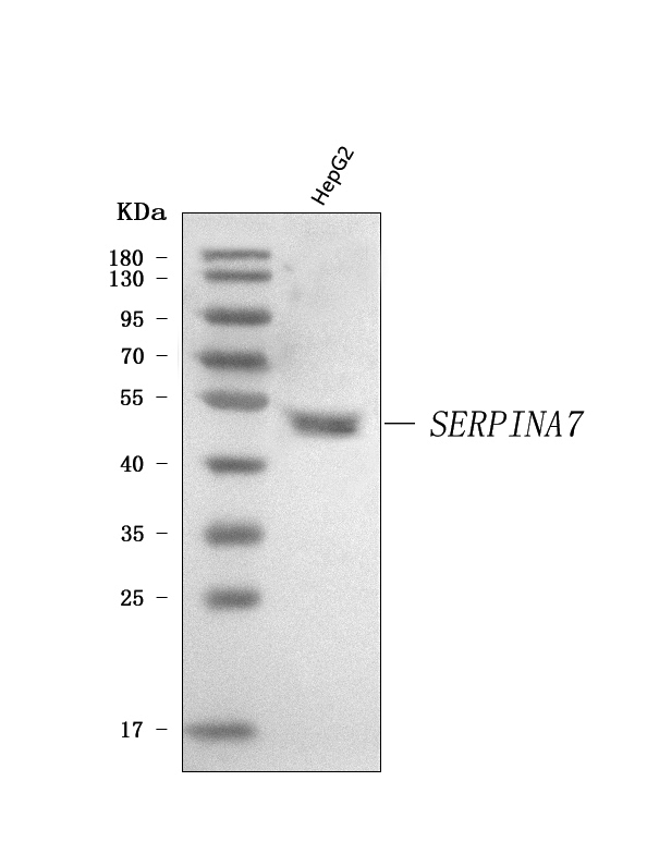
| Sample / lysate | human HepG2 . After electrophoresis, proteins were transferred to a nitrocellulose membrane at 150 mA for 50-90 minutes. Blocked the membrane with 5% non-fat milk/TBS for 1.5 hour at RT. The membrane was incubated with rabbit anti-Thyroxine Binding Globulin/SERPINA7 antigen affinity purified polyclonal antibody (Catalog # A02766-1) at 0.5 μg/mL overnight at 4°C, then washed with TBS-0.1%Tween 3 times with 5 minutes each and probed with a goat anti-rabbit IgG-HRP secondary antibody at a dilution of 1:5000 for 1.5 hour at RT. The signal is developed using an Enhanced Chemiluminescent detection (ECL) kit (Catalog # EK1002) with Tanon 5200 system. A specific band was detected for Thyroxine Binding Globulin/SERPINA7 at approximately 50 kDa. The expected band size for Thyroxine Binding Globulin/SERPINA7 is at 46 kDa |
| Gel % | 10–12% |
| Load | 30 ug |
| Transfer | nitrocellulose membrane, 150 mA, 50–90 min |
| Membrane | nitrocellulose |
| Blocking | 5% non-fat milk / TBS, 1.5 h RT |
| Primary antibody | 0.5 µg/mL |
| Primary incubation | overnight at 4 °C |
| Secondary antibody | goat anti-rabbit IgG-HRP, 1:5000 |
| Wash | TBS-0.1% Tween, 3 × 5 min |
| Detection | ECL |
| Exposure / imaging | Tanon 5200 |
| Observed band | 50 kDa |
SERPINA7/TBG has a 46.3 kDa protein backbone but runs at ~50 kDa on blots because signal-peptide cleavage is outweighed by glycosylation at 5 N-linked sites.
| predominant band at ~50 kDa | the mature, signal-peptide-cleaved, fully N-glycosylated TBG monomer — the expected native band |
| diffuse or smeared band around 48-52 kDa rather than one sharp line | heterogeneous glycan occupancy/branching across the 5 N-linked sites (Asn36, Asn99, Asn116, Asn165, Asn253) broadens the apparent mass |
| sharper band nearer 44-46 kDa | reduced glycan mass, e.g. after deglycosylation treatment or in a non-glycosylated recombinant standard, closer to the unmodified backbone |
| little to no band in whole-cell or tissue lysate | TBG is a secreted plasma protein, so intracellular lysates capture only the pre-secretion pool; serum/plasma is the better sample |
| single clean band without a doublet or extra bands | only one isoform is annotated for SERPINA7, so multiple closely spaced bands are not expected from isoform variation |
| Predicted mass (UniProt) | gives the unmodified 46.3 kDa polypeptide backbone for the 415-aa precursor as the starting reference point |
| Signal peptide (residues 1-20) | cleaved during secretion, so the mature circulating protein starts from a slightly smaller backbone than the full-length 46.3 kDa precursor before glycosylation is added |
| N-glycosylation at Asn36, Asn99, Asn116, Asn165 and Asn253 | five complex N-glycans add substantial mass, shifting the mature band up to the ~50 kDa observed on blots and broadening it into a smear when glycoform occupancy varies |
| Secreted subcellular localization | protein is exported from the cell, so whole-cell lysates give weak signal compared with serum or plasma |
| Non-glycosylated recombinant or bacterial standard | lacks the native glycan load and runs closer to the unmodified ~44-46 kDa backbone mass, lower than native plasma TBG |
| Situation | Likely cause | Next action |
|---|---|---|
| No band in lysate | TBG is secreted into plasma rather than retained intracellularly | use serum or plasma instead of whole-cell lysate, or confirm only the small pre-secretion pool is being probed |
| Band higher than expected | full or partial occupancy across the 5 N-linked glycosylation sites adds more mass than the 46.3 kDa backbone alone | treat a duplicate sample with PNGase F and check for a shift down toward ~44-46 kDa to confirm glycosylation |
| Band lower than expected | sample is non-glycosylated recombinant protein or has been enzymatically deglycosylated | run a native plasma or serum sample in parallel as a fully glycosylated comparator |
| Broad smear instead of sharp band | heterogeneous glycan branching and occupancy across the 5 N-glycosylation sites | resolve on a lower-percentage or gradient gel and allow a longer run/transfer time |
| Fragments below expected size | loss of the 20-residue signal peptide during normal maturation, or proteolytic degradation of the sample | add protease inhibitors during sample preparation and compare against a fresh, properly stored plasma sample |
Comprehensive Human Protein Atlas IHC scoring per tissue. Rows are taken directly from the HPA tissue chart — click any row's HPA link to view the source.
| Tissue | Cell type | Level | Evidence | Source |
|---|---|---|---|---|
| Adipose tissue | adipocytes | Not detected | Protein (IHC) | HPA → |
| Adrenal gland | glandular cells | Not detected | Protein (IHC) | HPA → |
| Appendix | glandular cells | Not detected | Protein (IHC) | HPA → |
| Bone marrow | hematopoietic cells | Not detected | Protein (IHC) | HPA → |
| Breast | adipocytes | Not detected | Protein (IHC) | HPA → |
| Tissue | Cell type | Level | Evidence | Source |
|---|---|---|---|---|
| Adipose tissue | adipocytes | Not detected | Protein (IHC) | HPA → |
| Adrenal gland | glandular cells | Not detected | Protein (IHC) | HPA → |
| Appendix | glandular cells | Not detected | Protein (IHC) | HPA → |
| Bone marrow | hematopoietic cells | Not detected | Protein (IHC) | HPA → |
| Breast | adipocytes | Not detected | Protein (IHC) | HPA → |
Deeper troubleshooting and optimisation questions for SERPINA7, answered from its protein features.
BosterBio's SERPINA7 antibodies are among the best-performing WB antibodies on the market — well cited, thoroughly validated, and orthogonally cross-validated against negative tissues and complementary methods.
The recommended anti-SERPINA7 (Thyroxine Binding Globulin) antibody is a top-performing, widely cited reagent with thorough Western blot validation, including orthogonal confirmation against negative tissue and complementary detection approaches, giving confidence in its specificity and reproducibility for reliable SERPINA7 detection.
Which to pick: Only one Boster SERPINA7 antibody is catalogued, A02766-1, and it includes an actual Western blot validation image (5-20% SDS-PAGE, 70V), so it is both the recommended and the only available choice for WB work on this target.