This website uses cookies to ensure you get the best experience on our website.
- Table of Contents
3 Citations 6 Q&As
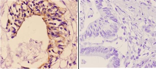
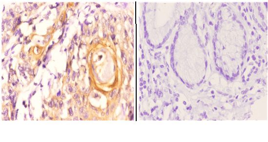
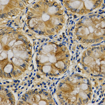
Facts about Serpin B5.
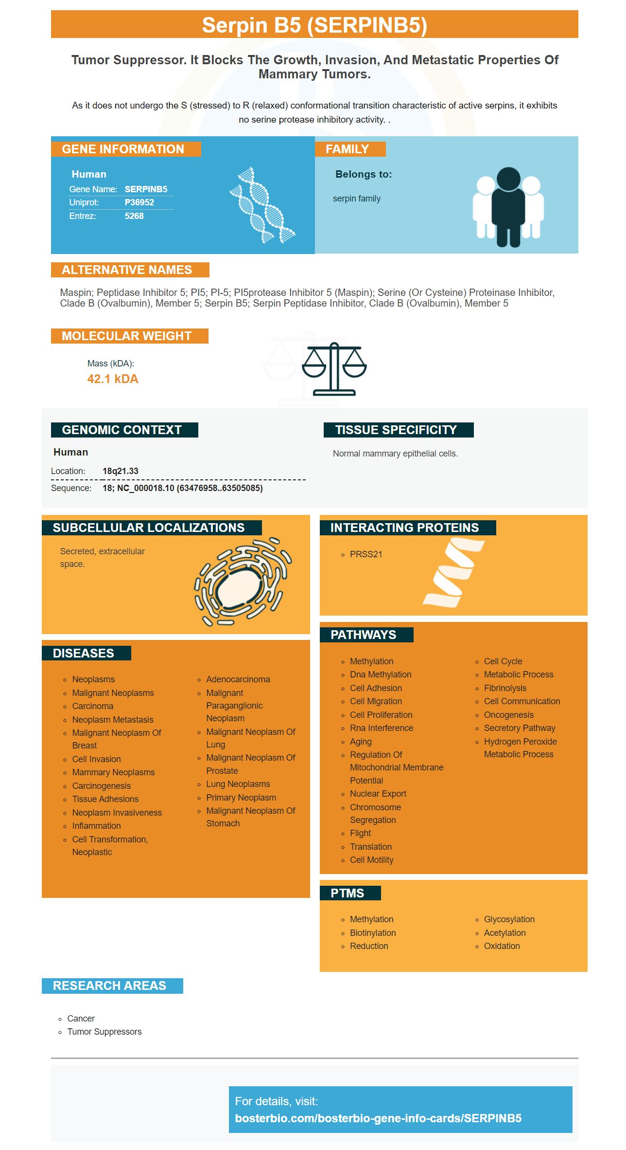
As it does not undergo the S (stressed) to R (relaxed) conformational transition characteristic of active serpins, it exhibits no serine protease inhibitory activity. .
Human | |
---|---|
Gene Name: | SERPINB5 |
Uniprot: | P36952 |
Entrez: | 5268 |
Belongs to: |
---|
serpin family |
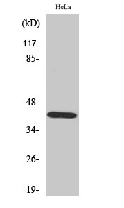
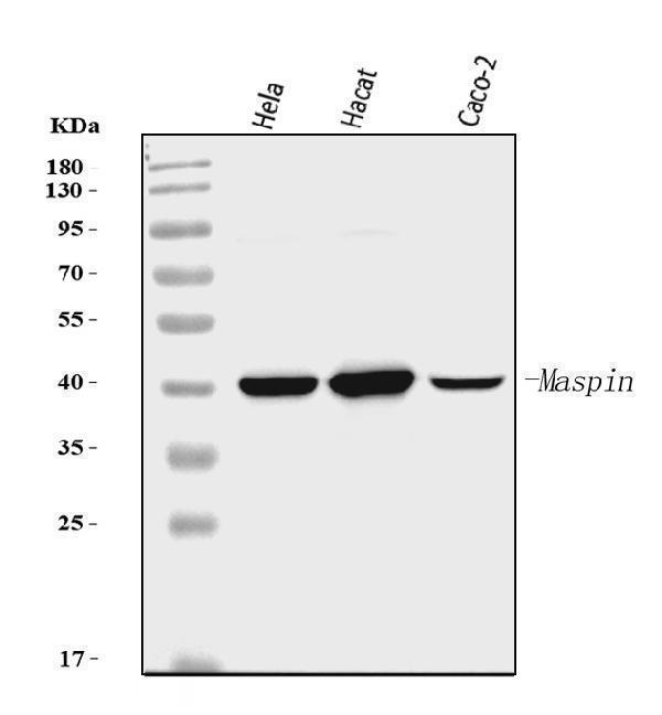
Maspin; Peptidase inhibitor 5; PI5; PI-5; PI5protease inhibitor 5 (maspin); serine (or cysteine) proteinase inhibitor, clade B (ovalbumin), member 5; Serpin B5; serpin peptidase inhibitor, clade B (ovalbumin), member 5
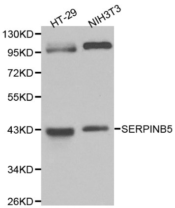
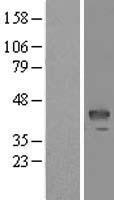
Mass (kDA):
42.1 kDA
Human | |
---|---|
Location: | 18q21.33 |
Sequence: | 18; NC_000018.10 (63476958..63505085) |
Normal mammary epithelial cells.
Secreted, extracellular space.
This article has information about Streptavidin–biotin Complex, SERPINB5Marker, and colorimetric determination. These methods will help you detect SERPINB5 in your experiments. Boster's antibodies have been used in the research community for over 25 years and have been validated for use in Western Blotting, Immunohistochemistry, and ELISA.
SERPINB5 a soluble molecule that can bind cofactors. Serine proteases alter the conformation of serpins. These changes result in a nonstandard hinge zone at the central region. This area traps and inhibits the serine protases. After being released from serine proteases, serpins adopt the native serpin fold. The RCL is close to the strands 4,A and 4,B. They could play a role in the covalent interaction between the target protease.
Six SNPs make up the SERPINB5 genome. They are all located in two blocks of linkage disequilibrium. These SERPINB5 SNPs were genotyped for association analysis in this study. Furthermore, a case-control study was conducted to determine if the SERPINB5 gene is associated with HCC risk. Researchers discovered two haplotypes linked to higher HCC risk in this study.
The SERPINB5 polymorphism was evaluated using a real time PCR system and SDS v3.0. This genotyping fluorescence TaqMan SNP Assay is suitable to analyze SERPINB5 genome polymorphisms. Each reaction contained 10 ng genomic DNA, 0.25 mL TaqMan probe mix, 5 mL TaqMan universalPCR Mastermix, and 5 mL TaqMan universalPCRMaster Mix.
The SERPINB5 genome promoter includes a possible BASH2 binding domain and a putative RCL domain. The RCLdomain influences protein stability and alternative expression efficiency. HCC susceptibility is increased by inhibiting SERPINB5. A loss of SERPINB5 gene expression has been linked to increased malignancy in different cancers.
Streptavidin, a high affinity binding protein, has a unique ligand binding specificity. Biotin bonds to streptavidin using two hydrogen bonds, at N23 & S27. These two positions are responsible for reducing the protein's affinity for biotin by at most eight orders of magnitude. The protein is more apt to accept biotin analogues than its native form. This higher specificity is due to a biotin binding site that exploits the difference between local electrostatic charges. The introduction of a negative charge at S27 breaks two hydrogen bonds, making streptavidin less biotin binding specific.
The Streptavidin-biotin complex is a high affinity DNA-binding protein. To purify proteins it is necessary to include biotin in the DNA. After the DNA molecule has been biotinylated the protein of interest binds with the biotin moiety in streptavidin. The complex can then be isolated by adsorption onto biotin-containing resin.
The Streptavidin/biotin complex can label biological targets, including enzymes and antibodies. The biotin trimer has a high affinity towards streptavidin. It is able to derivatize different reactive elements with a low concentration constant (less then 10 mol/L), and can tag multiple molecules to a single proteins.
The Streptavidin/biotin complex can be made using bacteria expressing the expression vector, pLysE. The cell extract was diluted using 7 M guanidineHCl, pH 1.5. Next, the agarose insoluble fraction of the cell lysate were dialyzed against an acid pH 6.0 solution with Tween-20. To confirm that the mutants bind to the same conditions, diaminobiotin and streptavidin were mixed in a renaturation solution.
ECL is an ECL technique that detects interaction between protein and protein. It uses antibodies conjugated HP to detect the target proteins. The enzyme then reacts with the chemiluminescent substrate to produce excited intermediates, which release strong blue light at 450 nm. The enzyme-substrate reaction is the only time light emits and it ceases when the substrate leaves the enzyme's vicinity.
ECL is less sensitive that traditional chemiluminescent detection. Luminol's half-life is very short and the detection limit is low. Amersham ECL Select chemiluminescent reagent is the most sensitive. It is recommended to be used for protein detection at low and medium abundance levels. It is part in the ECL product range.
ECL provides qualitative data and confirms the presence of target proteins in tissue or cells. Unlike X-ray film, charge-coupled-device (CCD) camera-based digital imaging instruments provide instant image capture and higher resolution. The instruments do not require a darkroom and can be placed alongside other lab equipment. Self-prepared chemiluminescence chemicals are more sensitive, have shorter signal durations and a longer shelf-life.
Several enhanced chemiluminescent substances have been developed to provide high intensity and sensitivity. This is ideal for western blot applications containing high levels of proteins. The Pierce ECL substrate is a high-sensitivity ECL substrate that allows protein detection in picograms and femtograms. This method can detect SERPINB5 with high consistency and reproducibility if properly diluted.
SERPINB5, also referred to as MASPIN in the Serpin gene family, was first identified by breast cancer researchers as a tumor suppressor. Immunohistochemistry staining SERPINB5 demonstrated an increase in expression among patients with worse prognoses. SERPINB5's function varies depending on where it is located in the cell. It is found in breast cancer mainly in the nucleus where it plays a tumour suppressor role. The cellular mechanism SERPINB5 expression occurs in cells that are still not fully understood.
Boster Bio: SERPINB5 monoclonal antibody detects this protein. Boster Bio's antibodies are high-affinity and have been validated on Western Blotting, Immunohistochemistry, and ELISA. The protein can be expressed by many cell types. It is sensitive and specific, which allows researchers to identify proteins in samples containing high levels of protein expression.
PMID: 8290962 by Zou Z., et al. Maspin, a serpin with tumor-suppressing activity in human mammary epithelial cells.
PMID: 7797587 by Pemberton P.A., et al. The tumor suppressor maspin does not undergo the stressed to relaxed transition or inhibit trypsin-like serine proteases. Evidence that maspin is not a protease inhibitory serpin.
*More publications can be found for each product on its corresponding product page