This website uses cookies to ensure you get the best experience on our website.


- Table of Contents
Plan SETD3 staining in paraffin sections around cytoplasmic expression in most tissues and high staining in several glandular cell populations (HPA tissue IHC). The catalog antibody has an IHC dilution of 2–5 μg/ml (datasheet A10965-1); use consistent fixation and appropriate controls (standard IHC practice).

Expected localisation, validated protocols, controls and antibodies — the at-a-glance facts below, then the full design guide.
| Expected localisation | Cytoplasmic tissue staining (HPA tissue IHC) | |
| Staining pattern | Cytoplasmic staining in most tissues; high in several glandular cell populations (HPA tissue IHC) | |
| Antigen retrieval | EDTA pH 8.0 HIER, heat-mediated (datasheet A10965-1) | |
| Positive control | Appendix+4 more · see all | |
| Negative control | Adipose tissue+4 more · see all |
| Fixation | Keep fixation consistent across sections (standard IHC practice; not target-specific) | |
| Caveat | Staining and RNA show medium consistency (HPA tissue IHC) | |
| Regulation | Expression regulation not annotated (UniProt) | |
| Isoform / epitope | 3 isoforms; epitope effects undetermined (UniProt) |
Compare the catalog antibody’s IHC-P protocol (datasheet A10965-1) with the published human tissue microarray and mouse liver protocol (PMC9329778).
| Sample | Paraffin-embedded human breast cancer tissue; fixative not specified (datasheet A10965-1) |
| Fixation | Image fixative and duration unreported (datasheet A10965-1); verify before use. |
| Sectioning | 4–5 µm sections on charged slides (standard) |
| Deparaffinisation | Xylene, graded ethanol series to water (standard) |
| Antigen retrieval | Heat retrieval: EDTA pH 8.0 (datasheet A10965-1); 20 min, 95–100 °C (standard) |
| Peroxidase block | 3% H2O2, 10 min, room temperature (standard) |
| Blocking | 10% goat serum (datasheet A10965-1) |
| Primary antibody | Rabbit anti-SETD3, 2-5 μg/ml (datasheet A10965-1) |
| Primary incubation | Overnight at 4 °C (datasheet A10965-1) |
| Detection | HRP-conjugated secondary, DAB chromogen (datasheet A10965-1) |
| Counterstain | Hematoxylin, blue, dehydrate and mount (standard) |
| Expected result | SETD3-positive staining in glandular cells of appendix (HPA tissue IHC: High). HPA tissue profile: Cytoplasmic expression in most tissues. No signal in the no-primary control. |
In paraffin-section IHC, expect predominantly cytoplasmic SETD3 staining in glandular cells of tissues such as colon and duodenum (HPA: cytoplasmic expression in most tissues; High in these glandular cells). UniProt places SETD3 mainly in the cytoplasm, also in the nucleus, and reports no transmembrane segment (UniProt Q86TU7: localization and topology). Treat tissue predictions as guides: HPA rates its tissue IHC profile Approved, with medium RNA–staining consistency and external verification pending.
| Colon or duodenum glandular cells show clear cytoplasmic chromogen, with recognizable cell boundaries and a clean surrounding field. | This fits the expected IHC distribution (HPA: High in colon and duodenum glandular cells; cytoplasmic expression in most tissues). Compare intensity within the same staining run; HPA’s High category does not prescribe a numerical score or a particular chromogen shade. |
| Signal is confined to nuclei, membranes, or another compartment without discernible cytoplasmic staining. | Question the IHC result and check staining controls and morphology (UniProt Q86TU7: mainly cytoplasmic; HPA: cytoplasmic tissue IHC). Some nuclear localization is recorded by UniProt, so a nuclear component alone does not invalidate an otherwise convincing cytoplasmic pattern. |
| Adipocytes or skeletal myocytes stain prominently while expected glandular cells do not. | Consider antibody cross-reactivity or endogenous detection activity, then review controls and cell identification (HPA: Not detected in adipocytes and skeletal myocytes; High in colon glandular cells). These are cell-level reference observations, not a claim that every cell in those tissues must be negative. |
| Brown signal spreads across stroma, empty spaces, or most cells without clear cellular boundaries. | Treat this as diffuse background until controls show otherwise. In chromogenic IHC, incomplete blocking, residual detection activity, or excessive reagent concentration can obscure cell-level interpretation (general IHC practice). HPA’s cytoplasmic tissue profile does not establish that diffuse staining is SETD3. |
| A colon or duodenum section has no glandular-cell signal, yet morphology appears intact. | Call the run inconclusive until a known-positive control and detection controls are reviewed (HPA: High in colon and duodenum glandular cells). Absence of staining can reflect a failed IHC workflow; this evidence does not identify SETD3-specific fixation sensitivity. |
| Reference tissue and cell type | Select a section containing identifiable glandular cells for a positive control (HPA: High in appendix, colon, duodenum, rectum, small intestine, and stomach glandular cells). Score the named cells, since a tissue label alone does not define every cell’s SETD3 status. |
| Compartment and topology | Prioritize cytoplasmic staining in tissue IHC (HPA: cytoplasmic expression in most tissues; UniProt Q86TU7: mainly cytoplasmic). UniProt also lists the nucleus and no transmembrane segment; it does not support a membrane-only IHC pattern. |
| Strength of tissue evidence | Use HPA levels as reference observations, not universal intensity thresholds (HPA: tissue IHC Approved; medium consistency with RNA; external verification pending). The listed Not detected findings apply to specified cells, including adipocytes and smooth muscle cells (HPA: tissue IHC). |
| Antibody validation and molecular forms | Two listed antibodies have Approved IHC status (HPA: HPA003591 and HPA003639). UniProt lists three isoforms and a phosphoserine at residue 513 (UniProt Q86TU7); the supplied evidence does not identify the antibody epitope or show isoform-dependent staining. |
| IF/ICC Q&A: should mitochondrial signal match tissue IHC? | HPA reports mainly mitochondrial localization in ICC-IF and an Approved ICC result for HPA003591 (HPA: subcellular ICC-IF; antibody status). Interpret that assay separately from HPA’s predominantly cytoplasmic tissue IHC profile; neither record establishes an IF protocol here. |
| Situation | Likely cause | Next action |
|---|---|---|
| No signal in glandular cells of a reference section. | The staining or detection run may have failed; absence alone cannot identify the failed step (HPA: High in colon and duodenum glandular cells). | Check a known-positive control, section integrity, reagent preparation, and detection steps; review retrieval against the validated IHC workflow (general IHC practice). Do not infer SETD3-specific fixation sensitivity from HPA staining levels. |
| Signal is faint in a reference tissue. | Low contrast may reflect assay conditions, while HPA’s High category is a cell-level reference rather than a guaranteed intensity in every run (HPA: High in colon glandular cells). | Compare the control section and detection controls from the same run; confirm antibody dilution and chromogen development against the validated IHC workflow (general IHC practice). |
| The entire section has diffuse brown background. | Nonspecific binding or residual endogenous detection activity can produce widespread chromogen (general chromogenic IHC practice). | Review the no-primary control, blocking, washes, and endogenous-activity controls appropriate to the detection chemistry (general IHC practice). Interpret SETD3 only where cellular staining remains distinct. |
| Nuclei or cell borders dominate the signal. | A compartment mismatch raises concern about specificity or interpretation (HPA: cytoplasmic tissue IHC; UniProt Q86TU7: mainly cytoplasmic, also nuclear). | Verify cell boundaries with the counterstain, compare a reference section, and review controls. Do not reject a modest nuclear component solely because it is nuclear (UniProt Q86TU7: nucleus listed). |
| Adipocytes or smooth muscle cells stain more strongly than reference glandular cells. | Possible cross-reactivity or detection background requires checking; HPA reports those specific cell types as Not detected (HPA: adipocytes and smooth muscle cells). | Confirm cell identity and examine the no-primary and positive controls (general IHC practice). Record staining by cell type rather than assigning a whole tissue a positive or negative result. |
| Tissue IHC appears cytoplasmic, but an ICC-IF image appears mitochondrial. | The supplied assay observations differ (HPA: cytoplasmic tissue IHC; mainly mitochondrial ICC-IF); they do not establish which preparation step accounts for the difference. | Report each result with its assay and controls. Evaluate paraffin-section staining against the tissue IHC reference, and use the separate IF/ICC guide for that assay. |


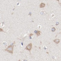

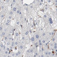
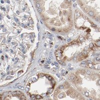


Comprehensive Human Protein Atlas IHC scoring per tissue (reliability: Approved — Medium consistency between antibody staining and RNA expression data. Pending external verification.). Rows are taken directly from the HPA tissue chart — click any row's HPA link to view the source.
| Tissue | Cell type | Level | Evidence | Source |
|---|---|---|---|---|
| Appendix | Glandular cells | High | Protein (IHC) | HPA → |
| Colon | Glandular cells | High | Protein (IHC) | HPA → |
| Duodenum | Glandular cells | High | Protein (IHC) | HPA → |
| Epididymis | Glandular cells | High | Protein (IHC) | HPA → |
| Rectum | Glandular cells | High | Protein (IHC) | HPA → |
| Tissue | Cell type | Level | Evidence | Source |
|---|---|---|---|---|
| Adipose tissue | Adipocytes | Not detected | Protein (IHC) | HPA → |
| Liver | Cholangiocytes | Not detected | Protein (IHC) | HPA → |
| Ovary | Follicle cells | Not detected | Protein (IHC) | HPA → |
| Skeletal muscle | Myocytes | Not detected | Protein (IHC) | HPA → |
| Smooth muscle | Smooth muscle cells | Not detected | Protein (IHC) | HPA → |
Use matched paraffin sections and compartment-aware controls to troubleshoot SETD3 staining; tissue IHC and cell IF report different localisation patterns (HPA tissue IHC; HPA subcellular).
A10965-1 has IHC images from human paraffin-embedded cancer sections and IF images from human breast cancer tissue and U20S cells (catalog image captions); catalog reactivity lists human, mouse and rat (catalog reactivity).
A10965-1 has chromogenic IHC images from paraffin-embedded human breast, liver, ovarian serous and renal cancer sections (IHC image captions). A10965-1 also lists IF and ICC applications, with IF images from U20S cells and paraffin-embedded human breast cancer tissue (catalog applications; IF image captions).
Which to pick: For chromogenic tissue IHC, choose A10965-1: its IHC captions document paraffin sections, EDTA retrieval at pH 8.0, and 2 μg/ml primary antibody with DAB detection; the fixative is unreported (IHC image captions). For IF/ICC, A10965-1 lists both applications and has IF images from U20S cells and human breast cancer tissue at 5 μg/ml (catalog applications; IF image captions). For mouse or rat work, A10965-1 lists species reactivity, but its supplied IHC and IF images show human samples only; it is rabbit hosted, and clonality is unreported (catalog reactivity and host; IHC and IF image captions; catalog clone field).