This website uses cookies to ensure you get the best experience on our website.


- Table of Contents
Plan SEZ6L2 paraffin IHC around cytoplasmic staining in CNS neurons (HPA tissue IHC). This guide covers fixation handling, chromogenic detection, and scoring by cell type and compartment (standard IHC practice).

Expected localisation, validated protocols, controls and antibodies — the at-a-glance facts below, then the full design guide.
| Expected localisation | Predominantly cytoplasmic in tissue (HPA tissue IHC); membrane and ER localisation (UniProt) | |
| Staining pattern | Cytoplasmic staining, mainly in CNS neurons (HPA tissue IHC) | |
| Antigen retrieval | EDTA pH 8.0 HIER, heat-mediated (datasheet A11420-1) | |
| Positive control | Bronchus+4 more · see all | |
| Negative control | Adipose tissue+4 more · see all |
| Fixation | Matched tissue-IHC evidence does not establish the fixation claim. Validate the specimen-specific method before use. (selected-SKU IHC image A11420-1) | |
| Caveat | Antibody staining and RNA show medium consistency (HPA tissue IHC) | |
| Regulation | Expression regulation is unreported (UniProt) | |
| Isoform / epitope | 6 isoforms; extracellular versus cytoplasmic epitope location matters (UniProt) |
The catalog antibody’s IHC-P protocol (datasheet: A11420-1) is paired with a published SEZ6L2 protocol for colorectal cancer tissue and xenograft sections (PMC7171412).
| Sample | Paraffin-embedded mouse brain tissue; fixative not specified (datasheet A11420-1) |
| Fixation | Image fixative and duration unreported (datasheet A11420-1); verify before use. |
| Sectioning | 4–5 µm sections on charged slides (standard) |
| Deparaffinisation | Xylene, graded ethanol series to water (standard) |
| Antigen retrieval | Heat retrieval: EDTA pH 8.0 (datasheet A11420-1); 20 min, 95–100 °C (standard) |
| Peroxidase block | 3% H2O2, 10 min, room temperature (standard) |
| Blocking | 10% goat serum (datasheet A11420-1) |
| Primary antibody | Rabbit anti-SEZ6L2, 2-5 μg/ml (datasheet A11420-1) |
| Primary incubation | Overnight at 4 °C (datasheet A11420-1) |
| Detection | HRP-conjugated secondary, DAB chromogen (datasheet A11420-1) |
| Counterstain | Hematoxylin, blue, dehydrate and mount (standard) |
| Expected result | SEZ6L2-positive staining in ciliated cells (cell body) of bronchus (HPA tissue IHC: High). HPA tissue profile: Cytoplasmic expression in several tissues, mainly in CNS. No signal in the no-primary control. |
SEZ6L2 is annotated at the cell and endoplasmic reticulum membranes, with an extracellular region spanning residues 28–844 and a cytoplasmic tail at 866–910 (UniProt Q6UXD5 topology). In tissue IHC, expect mainly cytoplasmic staining in CNS neurons, with strong staining also reported in selected ciliated epithelia (HPA: tissue IHC profile; High in caudate neurons, bronchial ciliated cells and fallopian tube ciliated cells). HPA rates the tissue pattern Enhanced, with medium consistency against RNA expression (HPA: reliability).
| Strong staining in caudate or cerebral cortex neuronal cells, or in cerebellar molecular layer cells. | This fits the reported CNS pattern; cerebellar molecular layer staining is described as cytoplasmic/membranous (HPA: High in caudate and cerebral cortex neurons; High in cerebellar molecular layer cells). Judge the cell type and distribution together, rather than treating any isolated dark deposit as a positive cell (general IHC practice). |
| Staining concentrates in bronchial ciliated cell bodies or at fallopian tube ciliary tips. | Both are reported strong patterns, but their recorded positions differ (HPA: High in bronchial ciliated cell bodies; High at fallopian tube ciliary tips). A visible signal confined to those positions supports interpretation more strongly than widespread epithelial staining (general IHC practice). |
| A predominantly nuclear pattern appears where a membrane or cytoplasmic pattern is expected. | Nuclear localisation is absent from the supplied localisation records (UniProt Q6UXD5 subcellular location; HPA: cytoplasmic tissue profile). Treat it as discordant; check the counterstain, detection controls and antibody conditions before assigning SEZ6L2 positivity (general IHC practice). |
| Strong signal appears in an HPA-negative cell type, or colour covers tissue without clear cell boundaries. | For example, HPA reports no detection in adipocytes or lung alveolar cells (HPA: Not detected in adipocytes and alveolar cells). Cross-reactivity or endogenous detection activity may explain cell-specific unexpected signal; diffuse colour may reflect background (general IHC practice). Neither appearance alone identifies its cause. |
| A known-positive region has little or no visible stain. | Check that the section contains the specified cells: HPA reports High staining in caudate neurons and bronchial ciliated cells (HPA: tissue IHC). If they are present, review antibody, retrieval and detection controls before interpreting the absence as biology (general IHC practice). HPA's Enhanced rating does not guarantee every run will produce signal (HPA: reliability). |
| Membrane topology and compartment | SEZ6L2 has one transmembrane segment at 845–865 and an extracellular region at 28–844 (UniProt Q6UXD5 topology). Tissue IHC may nevertheless appear mainly cytoplasmic (HPA: tissue IHC profile); do not require a crisp surface outline in every positive cell. |
| Tissue and cell selection | HPA records High staining in CNS neurons and selected ciliated cells, Medium in several glandular populations, and Low in kidney tubule cells (HPA: tissue IHC). Match comparisons to the annotated cell population; a whole-organ label alone can hide the relevant staining pattern. |
| Evidence strength | The tissue pattern is rated Enhanced, with medium consistency between antibody staining and RNA expression (HPA: reliability). HPA lists one rabbit polyclonal antibody, HPA064471, as IHC Enhanced; that rating supports the reported pattern but does not validate every unexpected compartment (HPA: antibodies). |
| Isoforms, processing and epitope | UniProt lists 6 isoforms, a signal peptide at 1–27, and a chain at 28–910 (UniProt Q6UXD5). No antibody epitope is supplied here, so these annotations cannot establish which isoforms the IHC antibody detects or predict its staining after processing. |
| Retrieval conditions | Retrieval is a condition to record and optimise with controls in paraffin-section IHC (general IHC practice). Target-specific fixation sensitivity and an optimal SEZ6L2 retrieval condition are unreported in the supplied UniProt and HPA records. |
| Situation | Likely cause | Next action |
|---|---|---|
| No staining in the expected positive control. | The sampled section may lack the annotated cells, or an IHC step may have failed (HPA: High in caudate neurons and bronchial ciliated cells; general IHC practice). | Confirm the relevant cells on the counterstain, then inspect retrieval, primary antibody and chromogenic detection controls on a known-positive section (general IHC practice). Do not infer loss of SEZ6L2 from a failed control. |
| Strong colour appears in adipocytes or lung alveolar cells. | Those cells are reported as Not detected by HPA; unexpected signal may reflect cross-reactivity or endogenous detection activity (HPA: tissue IHC; general IHC practice). | Compare the expected positive cells, use a primary-omission control and check the detection-system block appropriate to the chromogen (general IHC practice). Investigate persistent cell-specific staining before scoring it as SEZ6L2. |
| Colour is diffuse across tissue or obscures cell boundaries. | Nonspecific background, excessive primary antibody or detection signal are possible technical causes (general IHC practice). The supplied HPA profile describes cellular staining rather than uniform colour (HPA: tissue IHC profile). | Review the primary-omission control, blocking and washes; titrate the antibody and detection time against a known-positive section (general IHC practice). Retain conditions that preserve interpretable cell localisation. |
| The strongest signal looks nuclear. | That compartment conflicts with the supplied membrane and cytoplasmic annotations (UniProt Q6UXD5 subcellular location; HPA: tissue IHC profile). Counterstain overlap or nonspecific signal could mislead interpretation (general IHC practice). | Inspect chromogen and counterstain separately where possible, compare controls, and score cytoplasmic or membrane-associated staining in the expected cells (general IHC practice; HPA: tissue IHC). |
| Only weak signal is seen in a candidate control tissue. | Some listed cells are inherently Low or Medium in the HPA observations; kidney tubule cells are Low, whereas caudate neurons are High (HPA: tissue IHC). | Choose a section containing an HPA High cell population to assess the staining run, and compare the same cell type across sections (HPA: tissue IHC; general IHC practice). Avoid calling a Low population a failed control solely because its signal is faint. |
| Can the tissue IHC pattern define the expected ICC-IF result? | HPA's subcellular summary says Membrane but gives no main location or cell lines with ICC-IF images; HPA064471 has no ICC status in the supplied list (HPA: subcellular record; HPA: antibodies). | Use membrane localisation as a hypothesis and establish ICC-IF localisation with suitable cellular controls on its own guide page (UniProt Q6UXD5 location; general IF practice). Do not treat tissue IHC intensity as an ICC-IF validation result. |
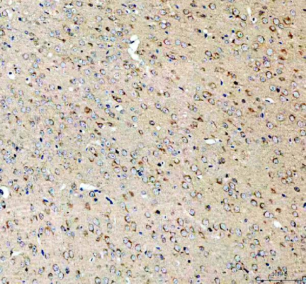
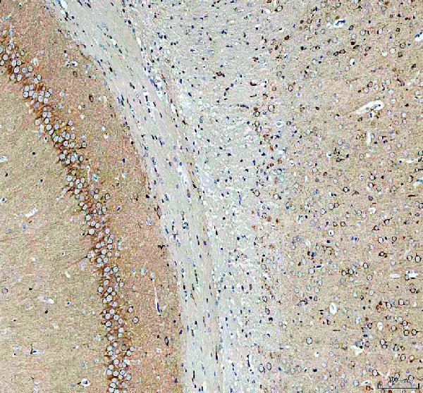


Comprehensive Human Protein Atlas IHC scoring per tissue (reliability: Enhanced — Medium consistency between antibody staining and RNA expression data.). Rows are taken directly from the HPA tissue chart — click any row's HPA link to view the source.
| Tissue | Cell type | Level | Evidence | Source |
|---|---|---|---|---|
| Bronchus | Ciliated cells (cell body) | High | Protein (IHC) | HPA → |
| Caudate | Neuronal cells | High | Protein (IHC) | HPA → |
| Cerebellum | Molecular layer cells - cytoplasm/membrane | High | Protein (IHC) | HPA → |
| Cerebral cortex | Neuronal cells | High | Protein (IHC) | HPA → |
| Fallopian tube | Ciliated cells (tip of cilia) | High | Protein (IHC) | HPA → |
| Tissue | Cell type | Level | Evidence | Source |
|---|---|---|---|---|
| Adipose tissue | Adipocytes | Not detected | Protein (IHC) | HPA → |
| Bone marrow | Hematopoietic cells | Not detected | Protein (IHC) | HPA → |
| Duodenum | Endocrine cells | Not detected | Protein (IHC) | HPA → |
| Esophagus | Squamous epithelial cells | Not detected | Protein (IHC) | HPA → |
| Liver | Cholangiocytes | Not detected | Protein (IHC) | HPA → |
Troubleshoot SEZ6L2 chromogenic IHC by checking retrieval, tissue morphology, cell identity and staining compartment against the available evidence.
The IHC-validated anti-SEZ6L2 antibody has paraffin-section images from mouse and rat brain and human testis (catalog: IHC image captions). No IF/ICC images are supplied (catalog: IF images).
A11420-1 is listed for IHC and reacts with human, mouse and rat samples (catalog: applications and reactivity). Its IHC captions show paraffin sections of mouse and rat brain and human testis using 2 μg/ml primary antibody (catalog: IHC image captions).
Which to pick: Choose A11420-1 for paraffin-section tissue IHC; its mouse brain image used EDTA retrieval at pH 8.0 and 2 μg/ml primary antibody overnight at 4°C (A11420-1 IHC caption). For work across species, it lists human, mouse and rat reactivity, with IHC images from each species; the fixative is unreported (catalog: reactivity; A11420-1 IHC captions). No IF/ICC application or image is supplied for A11420-1, so there is no IF/ICC-validated SKU to recommend from this catalog (catalog: applications and IF images).