SF3A3 / Splicing factor 3A subunit 3 · IHC design guide

Design Immunohistochemistry for SF3A3

Plan SF3A3 staining in paraffin sections using its broadly nuclear tissue profile (HPA tissue IHC) and the catalog antibody’s 2–5 μg/ml IHC range (datasheet A10671-1). Compare nuclear signal with the high staining reported in colon glandular cells and the undetected staining in adipocytes (HPA tissue IHC).

Evidence assembled Oct 2026 · For research use; verify linked source records and product datasheet before use
Immunohistochemistry protocol sheet for SF3A3 (IHC for SF3A3): expected localisation Nuclear staining in tissue (HPA tissue IHC), antibody A10671-1, validated IHC image, and IHC protocol steps
Printable SF3A3 IHC protocol sheet — expected localisation Nuclear staining in tissue (HPA tissue IHC), antibody A10671-1, controls and protocol steps. Open the full SF3A3 IHC guide →

SF3A3 Immunohistochemistry Experimental Design Guide

Expected localisation, validated protocols, controls and antibodies — the at-a-glance facts below, then the full design guide.

Must know before staining
Expected localisation Nuclear staining in tissue (HPA tissue IHC)
Staining pattern Nuclear staining across many cell types (HPA tissue IHC)
Antigen retrieval EDTA pH 8.0 HIER, heat-mediated (datasheet A10671-1)
Positive control ⓘ Adrenal gland+4 more · see all
Negative control ⓘ Adipose tissue
Important caveats
Reasons your staining may differ from the expected pattern.
Fixation Keep section fixation consistent (standard IHC practice; not target-specific)
Caveat Adipocytes are not detected despite the broad profile (HPA tissue IHC)
Regulation Ubiquitous expression (UniProt)
Isoform / epitope No annotated isoforms; one 1–501 chain (UniProt)
Section 1

Recommended SF3A3 IHC & IF Protocols

The catalog antibody protocol is paired with one published SF3A3 IHC protocol for paraffin sections (PMC8973551: Immunohistochemistry methods).

Recommended immunohistochemistry (IHC-P) protocol parameters
SampleParaffin-embedded human larynx squamous cell carcinoma tissue; fixative not specified (datasheet A10671-1)
FixationImage fixative and duration unreported (datasheet A10671-1); verify before use.
Sectioning4–5 µm sections on charged slides (standard)
DeparaffinisationXylene, graded ethanol series to water (standard)
Antigen retrievalHeat retrieval: EDTA pH 8.0 (datasheet A10671-1); 20 min, 95–100 °C (standard)
Peroxidase block3% H2O2, 10 min, room temperature (standard)
Blocking10% goat serum (datasheet A10671-1)
Primary antibodyRabbit anti-SF3A3, 2-5 μg/ml (datasheet A10671-1)
Primary incubationOvernight at 4 °C (datasheet A10671-1)
DetectionHRP-conjugated secondary, DAB chromogen (datasheet A10671-1)
CounterstainHematoxylin, blue, dehydrate and mount (standard)
Expected resultSF3A3-positive staining in glandular cells of adrenal gland (HPA tissue IHC: High). HPA tissue profile: Ubiquitous nuclear expression. No signal in the no-primary control.
💡Decision noteStart with heat-mediated EDTA retrieval at pH 8.0 (datasheet A10671-1); the published protocol does not specify retrieval (PMC8973551: Immunohistochemistry methods).
Section 2

What Is the Expected SF3A3 Staining Pattern?

SF3A3 should stain nuclei across many cell types, consistent with its ubiquitous expression and lack of a transmembrane segment (UniProt Q12874). Nuclear speckles are its principal location (UniProt Q12874; HPA: supported ICC-IF location). Tissue IHC shows ubiquitous nuclear expression, with high staining in several named cell types (HPA: tissue IHC; Supported, pending retesting).

What am I looking at on my slide?
Nuclear staining in colon glandular cells or bronchial respiratory epithelial cells.This fits the reported compartment and cell distribution; both cell types have high HPA tissue staining (UniProt Q12874: nucleus; HPA: High in colon glandular and bronchial respiratory epithelial cells). Judge intensity within the same staining run, since HPA levels do not define an absolute chromogen threshold.
Predominantly cytoplasmic staining with little nuclear signal.Treat this as a localization mismatch, especially if nuclei in a known-positive tissue remain unstained (UniProt Q12874: nucleus and nuclear speckle; HPA: ubiquitous nuclear tissue pattern). Review the negative control, antibody conditions and counterstain before assigning the cytoplasmic signal to SF3A3 (general IHC practice).
Strong adipocyte staining alongside weak or absent staining in expected-positive cells.HPA reports SF3A3 as not detected in adipocytes, while several other cell types stain highly (HPA: adipocytes Not detected; colon glandular cells High). This discordance raises possible cross-reactivity or endogenous detection activity; it does not establish either cause without controls (general IHC interpretation).
A diffuse wash of chromogen covers nuclei and surrounding tissue.The background obscures whether the expected nuclear pattern is present (HPA: ubiquitous nuclear tissue pattern). Compare a negative control and inspect blocking, wash and detection conditions; these are general IHC checks, not documented SF3A3-specific effects.
No nuclear signal in a section containing HPA high-staining cell types.An absent signal in colon glandular cells or bone marrow hematopoietic cells conflicts with their reported high staining (HPA: High in both cell types). First assess tissue preservation and the staining run with appropriate controls; HPA does not establish that a particular retrieval or fixation change will restore SF3A3 signal.
💡Expected SF3A3 appearanceCall a section positive when its expected cell types show predominantly nuclear chromogen, with strong staining plausible in HPA high-staining cells; isolated cytoplasmic signal or strong adipocyte staining is suspect (HPA: tissue IHC; UniProt Q12874: nucleus).
How each factor affects the staining
Cell type and tissue contextSF3A3 is ubiquitous, yet observed intensity varies: HPA lists high staining in colon glandular cells and bone marrow hematopoietic cells, low staining in liver cholangiocytes, and no detection in adipocytes (UniProt Q12874: ubiquitous; HPA: tissue IHC). Interpret a weak field by its constituent cells.
Antibody evidenceTwo listed antibodies have Supported tissue IHC status; only HPA032054 also has Supported ICC status (HPA: antibody validation). The tissue profile is Supported for consistency with RNA expression and is pending retesting (HPA: tissue IHC). These ratings support pattern interpretation without proving that every positive cell is specific.
Molecular location and processingThe annotated chain spans residues 1–501, with no signal peptide, propeptide or transmembrane segment (UniProt Q12874). These annotations support a nuclear expectation and give no basis for predicting secreted or membrane staining. Epitope position and target-specific retrieval sensitivity are not supplied.
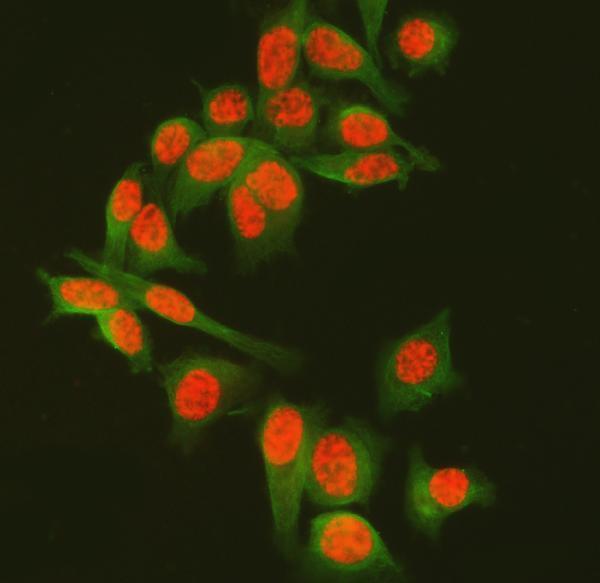
IF/ICC Q: Where should the signal appear?A: Mainly in nuclear speckles, with additional nucleoplasmic signal (HPA: ICC-IF subcellular localization; UniProt Q12874: nucleus speckle). HPA lists ICC-IF images for A-431, U-251MG and U2OS, and Supported ICC status for HPA032054 (HPA: subcellular and antibody records). This localization can guide interpretation; IF/ICC conditions are outside this IHC-P section.
Why is my staining missing, weak or wrong?
SituationLikely causeNext action
Nuclei are blank in a known-positive tissue.The run may have failed, or the sampled cells may differ from the reported high-staining population; HPA records high staining for colon glandular cells (HPA: tissue IHC).Check a known-positive section and run controls, then review the catalog antibody's IHC-P conditions and general retrieval workflow. No SF3A3-specific fixation or retrieval effect is established.
Chromogen is strongest in cytoplasm.This disagrees with the reported nuclear localization and could reflect nonspecific detection (UniProt Q12874: nucleus; HPA: tissue IHC).Compare negative controls and examine whether nuclear staining appears in a high-staining cell type. Adjust blocking, washing or detection only as general IHC troubleshooting.
Adipocytes stain strongly.HPA reports adipocytes as Not detected; cross-reactivity or endogenous detection activity is possible, but unconfirmed (HPA: adipocytes Not detected).Compare a negative control and another validated antibody where available; inspect whether neighboring expected-positive nuclei retain the reported pattern (HPA: antibody validation and tissue IHC).
Background is broad and masks cellular detail.Excess detection background can make nuclear localization unreadable (general IHC practice).Inspect the negative control, blocking, washes and chromogen development; reassess only after nuclei and surrounding tissue can be distinguished (general IHC practice).
Cholangiocytes look weak beside other cell types.Low cholangiocyte staining is reported and need not indicate a failed run (HPA: liver cholangiocytes Low).Score cholangiocytes separately and check a high-staining cell population in a control section before changing conditions (HPA: tissue IHC; general IHC practice).
Two antibody results disagree.Supported status reflects available validation, while the tissue profile remains pending retesting (HPA: antibody validation; tissue IHC reliability).Compare nuclear localization, cell identity and controls for each run. Treat an isolated discordant pattern as unresolved rather than assigning it to SF3A3.

Sample controls for SF3A3 IHC & IF

🧪Run bone marrow first: hematopoietic cells should show nuclear staining (HPA: High in bone marrow hematopoietic cells; UniProt: nucleus and nuclear speckles). Use adipose tissue as the negative tissue, where adipocytes are listed as not detected; within the marrow slide, counterstained cells lacking nuclear signal provide background comparators, though no marrow cell type is established as a biological negative (HPA: Not detected in adipocytes; HPA: High in bone marrow hematopoietic cells; UniProt: ubiquitous).
Positive control tissue: Adrenal gland (Glandular cells, HPA High)
Negative control tissue: Adipose tissue (HPA Not detected)
ICC-IF cell lines (HPA subcellular resource): HPA ICC-IF images show SF3A3 in A-431, U-251MG, U2OS, with annotated localisation: Nuclear speckles (supported) (HPA subcellular).
Technical controls: Include a no-primary, secondary-only slide, matched nonimmune rabbit IgG isotype control, and a validated SF3A3 knockout sample if available (selected IHC caption: rabbit primary antibody; standard IHC control practice). Quench endogenous peroxidase in bone marrow, since myeloid-cell peroxidase can produce DAB background (selected IHC caption: HRP/DAB detection; standard IHC practice).
⚠️Feasibility: A target-specific fixation window or fixation effect is unreported, and the selected A10671-1 paraffin-section caption does not state the fixative (selected IHC caption: fixative not stated). The reported IHC procedure uses heat-mediated EDTA retrieval at pH 8.0; this documents a working retrieval condition, not a comparison establishing retrieval dependence (selected IHC caption: EDTA pH 8.0). Neither frozen sections nor tissue IF are established as easier by the supplied evidence; bone marrow myeloid peroxidase can complicate chromogenic scoring, while HPA ICC-IF images support nuclear-speckle localization in cultured cells (HPA: ICC-IF images in A-431, U-251MG and U2OS; HPA: nuclear speckles supported; standard IHC practice).

HPA tissue IHC evidence for SF3A3

Comprehensive Human Protein Atlas IHC scoring per tissue (reliability: Supported — High consistency between antibody staining and RNA expression data. Pending retesting.). Rows are taken directly from the HPA tissue chart — click any row's HPA link to view the source.

Positive expression · recommended positive controls

TissueCell typeLevelEvidenceSource
Adrenal gland Glandular cells High Protein (IHC) HPA →
Bone marrow Hematopoietic cells High Protein (IHC) HPA →
Bronchus Respiratory epithelial cells High Protein (IHC) HPA →
Caudate Glial cells High Protein (IHC) HPA →
Cerebellum Cells in molecular layer High Protein (IHC) HPA →

Undetected expression · recommended negative controls

TissueCell typeLevelEvidenceSource
Adipose tissue Adipocytes Not detected Protein (IHC) HPA →
Section 3

Advanced SF3A3 IHC Tips

Troubleshoot SF3A3 staining by checking nuclear localisation, section processing, antibody controls and cell-specific scoring (UniProt Q12874; HPA tissue IHC).

How should I adjust retrieval when SF3A3 nuclear staining is weak?
Start with heat-mediated retrieval in EDTA at pH 8.0 (datasheet A10671-1). The demonstrated paraffin-section workflow then used 2 μg/ml primary antibody overnight at 4°C, so match those conditions before attributing a weak signal to retrieval (datasheet A10671-1). Compare a modest change in heating time on adjacent sections while holding antibody concentration, detection and DAB development constant (standard IHC practice). If staining remains weak, test citrate at pH 6.0 as a fallback alongside the EDTA condition, with a no-primary control to reveal added background (standard IHC practice). Score nuclear signal because SF3A3 localises to the nucleus and nuclear speckles (UniProt Q12874 localisation).
Could fixation explain absent SF3A3 staining in paraffin sections?
Target-specific sensitivity of SF3A3 to fixation is unknown from the supplied evidence; the selected paraffin-section caption does not state a fixative (datasheet A10671-1). Record the fixative and processing history for each specimen, then compare sections processed alike before changing retrieval or antibody concentration (standard IHC practice). In a controlled trial, vary one processing step at a time and assess both nuclear staining and preservation of tissue morphology (standard IHC practice; UniProt Q12874 localisation). Include a no-primary control and a consistently stained reference section in each run to distinguish processing variation from detection background (standard IHC practice). Do not infer fixation tolerance from the reported ubiquitous nuclear staining pattern (HPA tissue IHC).
What SF3A3 pattern should I expect across cells and nuclei?
Expect predominantly nuclear staining, with nuclear speckles and additional nucleoplasmic localisation reported for SF3A3 (UniProt Q12874 localisation; HPA subcellular). HPA describes ubiquitous nuclear tissue expression and reports high staining in colon glandular cells and bronchial respiratory epithelial cells (HPA tissue IHC). Compare suspected negative cells against neighbouring nuclei on the same section while checking that the nuclear counterstain and tissue structure remain intact (standard IHC practice). Cytoplasmic-only or diffuse extracellular colour should prompt review of blocking, washing and detection controls before it is scored as SF3A3 (UniProt Q12874 localisation; standard IHC practice). Do not require every nucleus to show resolvable speckles with chromogenic detection (standard IHC microscopy practice).
Could isoforms or epitope changes explain uneven nuclear staining?
The supplied record annotates 0 isoforms and a single chain spanning residues 1–501; it does not map the catalog antibody’s epitope (UniProt Q12874 record). SF3A3 has annotated modified residues, including phosphoserines at 54, 121, 295 and 299, but their effect on this antibody’s staining is unknown (UniProt Q12874 modified residues). Compare adjacent sections with identical retrieval and detection settings before attributing uneven nuclear colour to an epitope change (standard IHC practice). If another independently validated SF3A3 antibody is available, concordant nuclear patterns can help investigate epitope dependence, while no-primary controls assess detection background (standard IHC practice). Record the antibody and retrieval conditions with each comparison (standard IHC practice).
How can I investigate SF3A3 localisation by multiplex IF?
Use IF as a separate validation experiment, since this page’s demonstrated antibody workflow is chromogenic IHC on a paraffin section (datasheet A10671-1). Pair SF3A3 with a marker for the expected cell population, such as an epithelial marker when examining squamous epithelial cells, and include a nuclear counterstain (HPA tissue IHC: high in cervical squamous epithelial cells; standard IF practice). Choose spectrally separated fluorophores and inspect unstained tissue so autofluorescence does not masquerade as nuclear signal (standard IF practice). SF3A3 is nuclear and has no transmembrane segment, so optimise permeabilisation for access to its nuclear epitope without assuming that the unmapped antibody epitope has a known location (UniProt Q12874 localisation and topology; standard IF practice).
How do I reduce widespread DAB signal without losing nuclear SF3A3?
Run a no-primary section to identify colour from the detection system, and inspect the DAB reaction before treating widespread brown staining as SF3A3 (standard IHC practice). The demonstrated workflow used 10% goat serum blocking, 2 μg/ml primary antibody overnight at 4°C, and a peroxidase-conjugated secondary for 30 minutes at 37°C (datasheet A10671-1). Add a peroxidase block and optimise washing and DAB development as general chromogenic IHC steps (standard IHC practice). If background persists, titrate primary antibody on matched sections while preserving the demonstrated EDTA pH 8.0 retrieval as the reference condition (datasheet A10671-1; standard IHC practice). Judge improvement by nuclear signal relative to surrounding background (UniProt Q12874 localisation; standard IHC practice).
How should I quantify SF3A3 across sections with different cell composition? ⚠ ANSWER MARKED FOR VERIFICATION
Define the cell population and section regions before scoring, then measure nuclear staining rather than total brown area because SF3A3 localises to nuclei (UniProt Q12874 localisation; standard IHC practice). Report the percentage of positive nuclei and, when intensity matters, an H-score from 0–300 using the same thresholds across sections (standard IHC quantification practice). Normalise positive counts to all evaluable nuclei in the chosen population, or report positive-cell density per mm² of viable tissue when cell abundance is the outcome (standard IHC quantification practice). Exclude folds and damaged areas, keep exposure and DAB development consistent, and record the analysed area and cell population (standard IHC practice).
When is apparent SF3A3 positivity likely to be artefactual?
Treat staining in nuclei as plausible SF3A3 signal, especially where a cell population is expected to stain; HPA reports ubiquitous nuclear expression but also reports adipocytes as not detected (UniProt Q12874 localisation; HPA tissue IHC). Examine apparent cytoplasmic or extracellular signal against a no-primary control before calling it positive (UniProt Q12874 localisation; standard IHC practice). Strong colour restricted to section edges, folds or necrotic regions should trigger review of section quality and DAB development (standard IHC practice). If the no-primary section also develops colour, investigate endogenous peroxidase activity and the peroxidase block before interpreting cell-specific staining (standard IHC practice). Compare candidate positives with intact neighbouring nuclei and the expected tissue distribution (HPA tissue IHC; standard IHC practice).
Boster reagents

Best SF3A3 / Splicing factor 3A subunit 3 IHC Antibodies

A10671-1 has IHC images from paraffin-embedded human cancer sections and IF images from PC-3 cells and human intestinal cancer tissue (catalog image captions). Human, mouse and rat reactivity is listed (catalog).

Real IHC data IHC analysis of SF3A3 using anti-SF3A3 antibody (A10671-1). SF3A3 was detected in a paraffin-embedded section of human larynx squamous cell carcinoma tissue. Heat mediated antigen retrieval was performed in EDTA buffer (pH 8.0, epitope retrieval solution). The tissue section was blocked with 10% goat serum. The tissue section was then incubated with 2 μg/ml rabbit anti-SF3A3 Antibody (A10671-1) overnight at 4°C. Peroxidase Conjugated Goat Anti-rabbit IgG was used as secondary antibody and incubated for 30 minutes at 37°C. The tissue section was developed using HRP Conjugated Rabbit IgG Super Vision Assay Kit (Catalog # SV0002) with DAB as the chromogen.
Anti-SF3A3 Antibody ®
Cat # A10671-1

A10671-1 will render with IHC images from paraffin-embedded human larynx squamous cell carcinoma, lung adenocarcinoma, ovarian serous cancer and prostate adenocarcinoma sections (catalog IHC image captions). Its IF images show PC-3 cells and a paraffin-embedded human intestinal cancer section (catalog IF image captions).

Which to pick: Choose A10671-1 for chromogenic tissue IHC: its own paraffin-section captions document EDTA retrieval at pH 8.0 and 2 μg/ml primary antibody; the fixative is unreported (A10671-1 IHC image captions). For IF/ICC, A10671-1 has supplied cell and tissue images, while M10671 lists IF/ICC and IHC applications but has no supplied images (catalog IF image captions; catalog applications and image alts). Both SKUs list human, mouse and rat reactivity, but the supplied tissue images document human samples only (catalog reactivity; A10671-1 image captions).

Each figure is that product's own IHC / IF validation image from its datasheet.

References

  1. UniProt Consortium. UniProt entry Q12874 (SF3A3_HUMAN, Splicing factor 3A subunit 3).
  2. Human Protein Atlas. SF3A3 tissue IHC expression (reliability: Supported).
  3. Human Protein Atlas. SF3A3 subcellular location (ICC-IF): Mainly localized to the nuclear speckles. In addition localized to the nucleoplasm..
  4. Human Protein Atlas. SF3A3 antibody validation summary (2 antibodies).
  5. SF3A3 in Liver Hepatocellular Carcinoma: Oncogenic Role and Prognostic Significance. Oncology 2026 — PMC13423322.
  6. Interaction of STIL with FOXM1 regulates SF3A3 transcription in the hepatocellular carcinoma development. Cell division 2025 — PMC11740530.
  7. CircSCAP interacts with SF3A3 to inhibit the malignance of non-small cell lung cancer by activating p53 signaling. Journal of experimental & clinical cancer research : CR 2022 — PMC8973551.
  8. Exploring the level of metabolic reprogramming and the role of prognostic factor SF3A3 in hepatocellular carcinoma through integrated single-cell landscape analysis. PloS one 2025 — PMC12111341.
  9. PubMed PMID:8022796 — UniProt-cited evidence.
  10. PubMed PMID:7816610 — UniProt-cited evidence.
  11. PubMed PMID:16710414 — UniProt-cited evidence.