This website uses cookies to ensure you get the best experience on our website.


- Table of Contents
Plan SF3B1 chromogenic IHC on paraffin sections using the catalog antibody at a starting dilution of 1:50 (datasheet M00507-1). Assess nuclear and cytoplasmic staining in the tissue context, accounting for transient redistribution during mitosis (HPA tissue IHC; UniProt).

Expected localisation, validated protocols, controls and antibodies — the at-a-glance facts below, then the full design guide.
| Expected localisation | Nuclear and cytoplasmic tissue staining (HPA tissue IHC) | |
| Staining pattern | Nuclear and cytoplasmic staining in most tissues (HPA tissue IHC) | |
| Antigen retrieval | EDTA pH 8.0 HIER, heat-mediated (datasheet M00507-1) | |
| Positive control | Bone marrow+4 more · see all | |
| Negative control | Adipose tissue |
| Fixation | Keep paraffin-section fixation consistent across samples. (standard IHC practice; not target-specific) | |
| Caveat | Mitosis can disperse SF3B1 into the cytoplasm (UniProt) | |
| Regulation | No specific expression regulator annotated (UniProt) | |
| Isoform / epitope | 2 isoforms; epitope coverage is unspecified (UniProt) |
The catalog antibody protocol is supplemented by published SF3B1 IHC methods for ovarian cancer and mouse tissue (PMC10682409; PMC6025833).
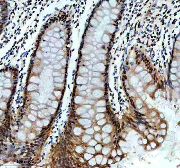
| Sample | Paraffin-embedded human colon tissue; fixative not specified (datasheet M00507-1) |
| Fixation | Image fixative and duration unreported (datasheet M00507-1); verify before use. |
| Sectioning | 4–5 µm sections on charged slides (standard) |
| Deparaffinisation | Xylene, graded ethanol series to water (standard) |
| Antigen retrieval | Heat retrieval: EDTA pH 8.0 (datasheet M00507-1); 20 min, 95–100 °C (standard) |
| Peroxidase block | 3% H2O2, 10 min, room temperature (standard) |
| Blocking | 10% goat serum (datasheet M00507-1) |
| Primary antibody | Rabbit monoclonal (clone 18S60) anti-SF3B1, 1:50 (datasheet M00507-1) |
| Primary incubation | Overnight at 4 °C (datasheet M00507-1) |
| Detection | HRP-conjugated secondary, DAB chromogen (datasheet M00507-1) |
| Counterstain | Hematoxylin, blue, dehydrate and mount (standard) |
| Expected result | SF3B1-positive staining in hematopoietic cells of bone marrow (HPA tissue IHC: High). HPA tissue profile: Cytoplasmic and nuclear expression in most tissues. No signal in the no-primary control. |
SF3B1 is a nuclear spliceosome protein enriched in nuclear speckles and has no transmembrane segment (UniProt O75533). Tissue IHC shows nuclear and cytoplasmic staining in most tissues, including strong staining in selected hematopoietic, neuronal, and glandular cells (HPA: tissue IHC). HPA rates the tissue pattern Approved, with medium consistency between staining and RNA expression (HPA: reliability).
| Clear nuclear staining in hematopoietic cells, neurons, or glandular cells. | This fits high staining reported in bone marrow hematopoietic cells, cerebral cortex and hippocampal neurons, and several glandular tissues (HPA: tissue IHC). Nuclear staining also fits SF3B1 localisation (UniProt O75533). |
| Staining is predominantly membranous, with little nuclear signal. | Treat this as a suspect pattern: SF3B1 has no transmembrane segment and is assigned to the nucleus (UniProt O75533). Review morphology and controls before scoring it as SF3B1. |
| Strong staining appears in adipose tissue adipocytes. | This conflicts with the HPA adipose tissue result, where adipocytes were not detected (HPA: adipose tissue IHC). Investigate cross-reactivity or endogenous detection activity; breast adipocytes are a separate low-staining observation (HPA: breast IHC). |
| Color spreads across tissue, including areas without clear cellular boundaries. | Diffuse background cannot establish cellular localisation. Check the no-primary control, blocking, washing, and detection chemistry before interpreting the slide (general IHC practice). |
| No signal appears in a known high-staining tissue. | First check whether the expected cells are present; HPA reports high staining in bone marrow hematopoietic cells and several neuronal and glandular populations (HPA: tissue IHC). Then assess the assay controls (general IHC practice). |
| Nuclear localisation and topology | SF3B1 localises to the nucleus and nuclear speckles and lacks a transmembrane segment (UniProt O75533). Use cell boundaries and a nuclear counterstain to judge compartment; a membrane-only pattern needs investigation (general IHC practice). |
| Cell cycle | During mitosis, SF3B1 transiently disperses from nuclear speckles into the cytoplasm (UniProt O75533). Interpret cytoplasmic signal in mitotic cells in that context; HPA also reports cytoplasmic and nuclear tissue IHC staining (HPA: tissue IHC). |
| Strength of tissue evidence | HPA rates tissue IHC Approved but describes only medium consistency with RNA data (HPA: reliability). Its antibody HPA050275 is Approved for IHC and Enhanced for ICC (HPA: antibody validation); those ratings do not validate every new assay condition. |
| IF/ICC: what pattern is expected? | Enhanced nuclear speckles are the main location in HPA cell images (HPA: subcellular ICC-IF). UniProt also assigns SF3B1 to nuclear speckles (UniProt O75533). This is an IF/ICC localisation reference, not an IHC protocol. |
| Isoforms and modifications | UniProt lists two isoforms and multiple modified residues (UniProt O75533). Their effects on staining cannot be predicted from these records; do not assign a compartment or tissue pattern to either isoform without further evidence. |
| Situation | Likely cause | Next action |
|---|---|---|
| Positive tissue has no visible nuclear stain. | The expected cell population may be absent, or the assay may have failed; high staining is cell-specific (HPA: tissue IHC). | Confirm the relevant cells on the section, then review positive and no-primary controls, retrieval, antibody dilution, and detection steps (general IHC practice). |
| Only membrane outlines stain. | That pattern conflicts with nuclear localisation and the lack of a transmembrane segment (UniProt O75533). | Check the no-primary control and cell morphology; reassess specificity before reporting SF3B1-positive cells (general IHC practice). |
| Adipose tissue adipocytes stain strongly. | HPA reports no detected staining in adipose tissue adipocytes (HPA: adipose tissue IHC); cross-reactivity or detection background is possible. | Compare with a no-primary control and a known high-staining tissue in the same run (general IHC practice). |
| Diffuse brown signal obscures nuclei. | Excess background or endogenous enzyme activity can obscure compartment calls (general IHC practice). | Review blocking, washing, detection controls, and counterstain intensity before scoring localisation (general IHC practice). |
| Some cells show cytoplasmic signal. | HPA reports cytoplasmic as well as nuclear tissue staining; mitotic dispersion is also described (HPA: tissue IHC; UniProt O75533). | Check cell morphology and the accompanying nuclear pattern. Do not label all cytoplasmic signal an artifact solely because SF3B1 is nuclear (HPA: tissue IHC). |
| Different tissues have unequal staining intensity. | HPA reports high staining in selected cell populations, medium staining in skin keratinocytes, and low staining in thyroid glandular cells (HPA: tissue IHC). | Score the specified cell population and compartment in each tissue; compare with HPA levels as context, not a universal intensity cutoff (HPA: tissue IHC). |


Comprehensive Human Protein Atlas IHC scoring per tissue (reliability: Approved — Medium consistency between antibody staining and RNA expression data.). Rows are taken directly from the HPA tissue chart — click any row's HPA link to view the source.
| Tissue | Cell type | Level | Evidence | Source |
|---|---|---|---|---|
| Bone marrow | Hematopoietic cells | High | Protein (IHC) | HPA → |
| Cerebral cortex | Neuronal cells | High | Protein (IHC) | HPA → |
| Cervix | Glandular cells | High | Protein (IHC) | HPA → |
| Gallbladder | Glandular cells | High | Protein (IHC) | HPA → |
| Hippocampus | Neuronal cells | High | Protein (IHC) | HPA → |
| Tissue | Cell type | Level | Evidence | Source |
|---|---|---|---|---|
| Adipose tissue | Adipocytes | Not detected | Protein (IHC) | HPA → |
Troubleshoot SF3B1 staining by checking retrieval, nuclear localisation, controls and cell level scoring before interpreting chromogenic IHC (UniProt O75533 localisation; standard IHC practice).
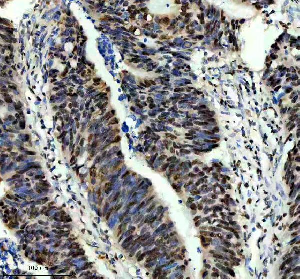
M00507-1 has human paraffin-section IHC images (catalog IHC captions) and lists IF/ICC and human, mouse and rat reactivity (catalog applications/reactivity); no IF image is supplied (catalog IF images).
M00507-1 has IHC images from paraffin-embedded human colon, colon cancer and liver sections (M00507-1 IHC captions). IF/ICC is listed for M00507-1, with human, mouse and rat reactivity, but no IF image is supplied (catalog applications/reactivity and IF images).
Which to pick: For tissue IHC, choose the rabbit monoclonal M00507-1: its own captions show human paraffin sections stained after EDTA pH 8.0 retrieval at 1:50 (catalog host/clone; M00507-1 IHC captions). The captions do not report the fixative (M00507-1 IHC captions). For IF/ICC or mouse and rat samples, M00507-1 lists those applications and species, although the supplied images document human IHC only (catalog applications/reactivity; M00507-1 IHC captions and IF images).