This website uses cookies to ensure you get the best experience on our website.


- Table of Contents
Plan SF3B3 chromogenic IHC around its general nuclear tissue pattern (HPA tissue IHC). The catalog antibody A08240-3 has a reported IHC concentration of 2–5 μg/mL (datasheet).

Expected localisation, validated protocols, controls and antibodies — the at-a-glance facts below, then the full design guide.
| Expected localisation | General nuclear staining in tissue sections (HPA tissue IHC) | |
| Staining pattern | Nuclear staining across many cell types (HPA tissue IHC) | |
| Antigen retrieval | EDTA pH 8.0 HIER, heat-mediated (datasheet A08240-3) | |
| Positive control | Adipose tissue+4 more · see all | |
| Negative control | None in HPA (detected in all 45 tissues); use no-primary + isotype controls |
| Fixation | Keep fixation consistent across sections (standard IHC practice; not target-specific) | |
| Caveat | Soft-tissue fibroblasts show low staining (HPA tissue IHC) | |
| Regulation | No specific regulator reported (UniProt) | |
| Isoform / epitope | 3 isoforms; epitope differences unreported (UniProt) |
The catalog antibody’s IHC-P protocol (datasheet A08240-3) is accompanied by one published paraffin-section protocol (PMC7999425).
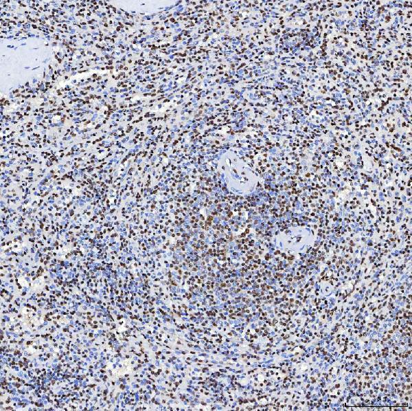
| Sample | Paraffin-embedded human spleen tissue; fixative not specified (datasheet A08240-3) |
| Fixation | Image fixative and duration unreported (datasheet A08240-3); verify before use. |
| Sectioning | 4–5 µm sections on charged slides (standard) |
| Deparaffinisation | Xylene, graded ethanol series to water (standard) |
| Antigen retrieval | Heat retrieval: EDTA pH 8.0 (datasheet A08240-3); 20 min, 95–100 °C (standard) |
| Peroxidase block | 3% H2O2, 10 min, room temperature (standard) |
| Blocking | 10% goat serum (datasheet A08240-3) |
| Primary antibody | Rabbit anti-SF3B3, 2-5 μg/ml (datasheet A08240-3) |
| Primary incubation | Overnight at 4 °C (datasheet A08240-3) |
| Detection | HRP-conjugated secondary, DAB chromogen (datasheet A08240-3) |
| Counterstain | Hematoxylin, blue, dehydrate and mount (standard) |
| Expected result | SF3B3-positive staining in adipocytes of adipose tissue (HPA tissue IHC: High). HPA tissue profile: General nuclear expression. No signal in the no-primary control. |
SF3B3 is a nuclear spliceosome protein with no transmembrane segment (UniProt Q15393: location, function, topology). In paraffin-section IHC, expect a general nuclear pattern across many cell types (HPA tissue IHC: Supported; general nuclear expression). HPA reports high staining in adipocytes, glandular cells, hematopoietic cells, respiratory epithelial cells, glial cells and Purkinje cells, while soft-tissue fibroblasts are low (HPA tissue IHC).
| Nuclear chromogen is clear in adipocytes, glandular cells or hematopoietic cells. | This matches the reported compartment and high-staining cell types (UniProt Q15393: nucleus; HPA tissue IHC: general nuclear expression; High in these cell types). Judge intensity against other cells in the same section; HPA's levels describe observed staining, not a universal cutoff. |
| Strong cytoplasmic staining dominates while nuclei remain pale. | This conflicts with the reported nuclear pattern (UniProt Q15393: nucleus; HPA tissue IHC: general nuclear expression). Consider nonspecific binding or chromogen background before calling it SF3B3. A weak cytoplasmic tint alone does not establish a new localisation. |
| An unexpected cell population stains strongly, with little nuclear contrast. | Check its identity and compartment before interpreting the signal. Cross-reactivity or endogenous detection activity can produce misleading staining (general IHC practice). HPA reports low staining in soft-tissue fibroblasts, but low is not equivalent to absent (HPA tissue IHC: Low in fibroblasts). |
| Chromogen spreads across tissue or blank areas without defined nuclei. | Treat this as background until staining can be assigned to cells. Inadequate blocking, residual detection activity or excess reagent can cause diffuse signal (general IHC practice). The expected result has nuclear contrast (HPA tissue IHC: general nuclear expression). |
| Nuclei are unstained in a section expected to contain high-staining cells. | Adipocytes, bone-marrow hematopoietic cells and bronchial respiratory epithelial cells are reported High (HPA tissue IHC). If those cells are present and identifiable, first check the IHC run and controls (general IHC practice); one negative slide does not overturn the reported pattern. |
| Cell type and tissue | HPA describes low tissue specificity at the RNA level and general nuclear protein staining (HPA tissue IHC). Use its reported High cell types as practical positive references, including adrenal glandular cells and cerebellar Purkinje cells; do not treat the Low fibroblast entry as a negative control (HPA tissue IHC). |
| Compartment and protein topology | SF3B3 belongs to the nuclear U2 snRNP spliceosome and has no transmembrane segment or signal peptide (UniProt Q15393: function, subcellular location, topology, processing). These annotations support nuclear interpretation; they do not establish how fixation or antigen retrieval affects staining. |
| Isoforms and antibody recognition | UniProt lists 3 SF3B3 isoforms (UniProt Q15393: isoforms). The supplied record gives no antibody epitope or isoform-specific IHC result, so apparent differences between tissues cannot be assigned to an isoform from staining alone. |
| Antibody validation | HPA lists IHC as Supported for HPA042986 and ICC as Supported for HPA041134 (HPA antibodies). These are application-specific entries; the ICC rating does not validate an IHC result. The supplied HPA tissue profile rates its IHC evidence Supported for consistency with RNA expression (HPA tissue IHC). |
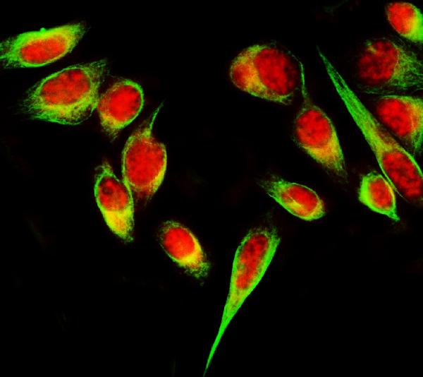
| Q: What should IF/ICC show? | A: HPA supports nucleoplasm, nucleoli and nucleoli-rim localisation, with images listed for MCF-7, SiHa and U2OS (HPA subcellular ICC-IF). These finer IF compartments need not be separately resolved in chromogenic paraffin IHC (general IHC practice). |
| Situation | Likely cause | Next action |
|---|---|---|
| No nuclear signal in a reported High cell population | The run may have failed, or the chosen IHC conditions may be unsuitable (general IHC practice). | Verify cell identity, positive and negative controls, retrieval, antibody dilution and detection steps (general IHC practice). The HPA High designation is an observed reference, not a guaranteed result under every protocol (HPA tissue IHC). |
| Predominantly cytoplasmic staining | Nonspecific binding or background is possible (general IHC practice); the reported location is nuclear (UniProt Q15393; HPA tissue IHC). | Compare with a negative control and inspect nuclear detail; adjust blocking or antibody concentration if background persists (general IHC practice). |
| Strong signal in cells expected to stain weakly | Cell identification, cross-reactivity or endogenous detection activity may explain the discrepancy (general IHC practice). | Confirm the cell type and nuclear compartment; check detection controls before comparing with HPA's Low soft-tissue fibroblast entry (HPA tissue IHC; general IHC practice). |
| Diffuse colour obscures nuclei | Nonspecific reagent binding, inadequate blocking or excess chromogen may reduce contrast (general IHC practice). | Review negative-control staining, blocking, reagent concentration and chromogen development; score only identifiable nuclear signal (general IHC practice; HPA tissue IHC: general nuclear expression). |
| Nuclear signal varies between tissue regions | Different cell populations can have different reported staining levels; a technical gradient is also possible (HPA tissue IHC; general IHC practice). | Compare like cell types across regions and inspect controls and section quality before assigning a biological difference (general IHC practice). |

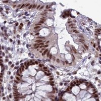


Comprehensive Human Protein Atlas IHC scoring per tissue (reliability: Supported — High consistency between antibody staining and RNA expression data.). Rows are taken directly from the HPA tissue chart — click any row's HPA link to view the source.
| Tissue | Cell type | Level | Evidence | Source |
|---|---|---|---|---|
| Adipose tissue | Adipocytes | High | Protein (IHC) | HPA → |
| Adrenal gland | Glandular cells | High | Protein (IHC) | HPA → |
| Appendix | Glandular cells | High | Protein (IHC) | HPA → |
| Bone marrow | Hematopoietic cells | High | Protein (IHC) | HPA → |
| Breast | Adipocytes | High | Protein (IHC) | HPA → |
| Tissue | Cell type | Level | Evidence | Source |
|---|---|---|---|---|
| None in HPA: SF3B3 is detected in all 45 scored tissues. Use a no-primary (secondary-only) and an isotype control instead. | ||||
Use nuclear staining, the selected antibody’s spleen section, and matched controls to troubleshoot SF3B3 chromogenic IHC (UniProt Q15393; datasheet A08240-3).
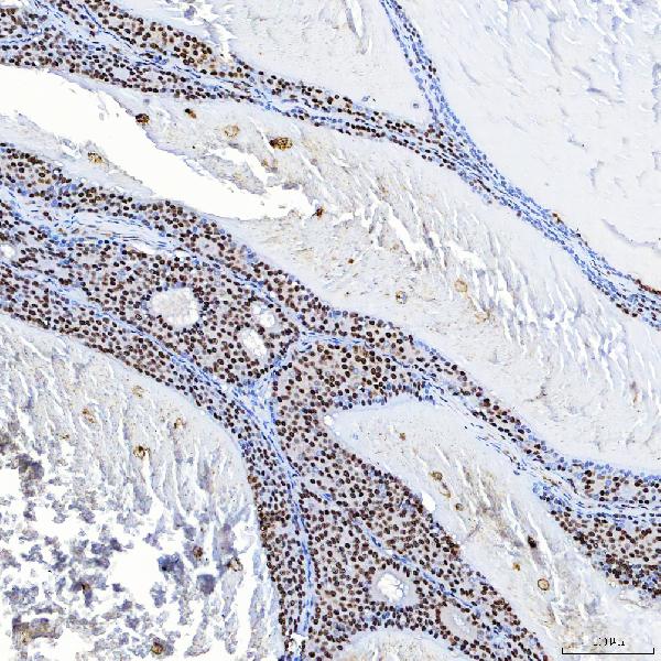
A08240-3 has IHC images from human paraffin sections and IF images from PC-3 cells, human ovarian cancer, and rat brain (catalog image captions). M08240-1 lists IHC and IF/ICC (catalog applications).
A08240-3 shows IHC in paraffin-embedded human spleen, breast cancer, colon adenocarcinoma, and larynx squamous cell carcinoma, plus IF in PC-3 cells, human ovarian cancer, and rat brain (catalog image captions). M08240-1 lists IHC and ICC/IF and human, mouse, and rat reactivity, but has no supplied IHC or IF image captions (catalog applications/reactivity/image alts).
Which to pick: For tissue IHC, choose A08240-3: its own human spleen caption documents a paraffin section, EDTA retrieval at pH 8.0, and 2 μg/ml primary antibody overnight at 4°C; the fixative is unreported (A08240-3 IHC image caption). For IF/ICC, A08240-3 has images from PC-3 cells and human ovarian cancer and rat brain sections (A08240-3 IF image captions); M08240-1 is a rabbit monoclonal, clone 19S30, that lists ICC/IF but has no supplied image captions (catalog clone/applications/image alts). For cross-species work, both list human, mouse, and rat reactivity (catalog reactivity), while A08240-3 also has a rat brain IF image (A08240-3 IF image caption).