This website uses cookies to ensure you get the best experience on our website.
- Table of Contents
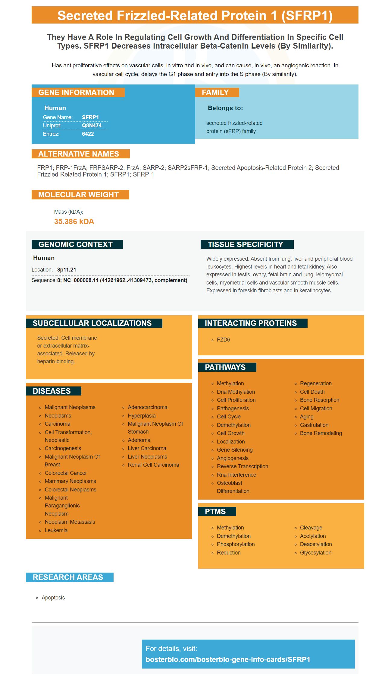
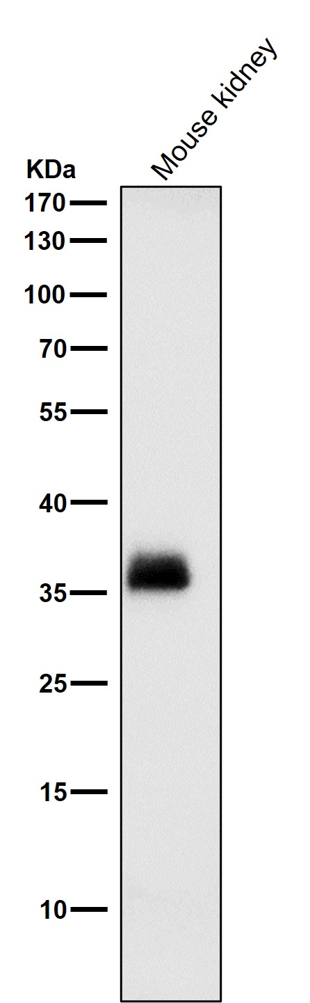
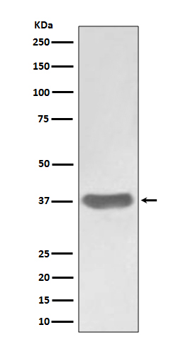
Facts about Secreted frizzled-related protein 1.
Has antiproliferative effects on vascular cells, in vitro and in vivo, and can cause, in vivo, an angiogenic reaction. In vascular cell cycle, delays the G1 phase and entry into the S phase (By similarity).
Human | |
---|---|
Gene Name: | SFRP1 |
Uniprot: | Q8N474 |
Entrez: | 6422 |
Belongs to: |
---|
secreted frizzled-related protein (sFRP) family |
FRP1; FRP-1FrzA; FRPSARP-2; FrzA; SARP-2; SARP2sFRP-1; Secreted apoptosis-related protein 2; secreted frizzled-related protein 1; sFRP1; sFRP-1
Mass (kDA):
35.386 kDA
Human | |
---|---|
Location: | 8p11.21 |
Sequence: | 8; NC_000008.11 (41261962..41309473, complement) |
Widely expressed. Absent from lung, liver and peripheral blood leukocytes. Highest levels in heart and fetal kidney. Also expressed in testis, ovary, fetal brain and lung, leiomyomal cells, myometrial cells and vascular smooth muscle cells. Expressed in foreskin fibroblasts and in keratinocytes.
Secreted. Cell membrane or extracellular matrix-associated. Released by heparin-binding.
If you're interested in knowing more about SFRP1 or Dicer then you're in the right place. This article will discuss the features of this marker and its clinical applications and more. This article is suitable for both academics and researchers. We will discuss its applications in clinical and dicer settings, and aid you in understanding its function in the biology of cancer cells.
This is a reliable marker for a variety of research projects, but what are the most effective uses of the SFRP1 gene in humans? A previous study shows that SFRP1 can be used to target cancer cells. The SFRP1 gene is regulated by Wnt signaling, and a decrease of this gene inhibits the growth of tumors. In this study, researchers discovered that SFRP1 restores expression of b-catenin an important protein in the cell.
This marker is found in about half of PCa tumors. Only a handful of clinical trials have been conducted to confirm the presence of this gene. This fusion could have significant implications for the development of new drugs in the absence of androgens. It is possible that the coexpression of SFRP1 with TMPRSS2-ERG may increase the odds of survival for patients suffering from this disease.
The SFRP1 promoter is a the d2EGFP. This is an unstable variant of wild-type GFP. The SFRP1 promoter was then cloned to a expression vector for the pCAGIG gene. The primer sequences were used to control the positive (H3K9me3, RNA Polymerase II In Actin promoter) and HP1a was employed as a negative control. The SFRP1 promoter was enriched with Pax6+NPs, while the Tbr2+NPs did not show any increase.
SFRP1 is a member of the SFRP protein family. It has a cysteine rich domain and a possible Wnt-binding location. It may regulate Wnt signaling or be involved in polarization of photoreceptor cells in the retina. SFRP1 is expressed in a variety of tissues, with the most abundant expression in the retina and in the heart. This SFRP1 marker is used for research purposes only. It is not for the purpose of diagnostics or for resale.
The expression of Dicer is linked to that of SFRP1 an indicator of tumors. The correlation was found in 40 CCA samples with a Pearson correlation coefficient of -0.47. This correlation, however does not mean that the expression of Dicer is reduced in cancer cells. This also suggests that there is a relationship between the two markers. Further research is needed to determine the connection between these two proteins.
Dicer is a member the RNase III family endoribonucleases. It regulates the methylation of CpG islands in mammalian cell. Its exact mechanism of action is not understood. Dicer is present in cholangiocarcinoma cells and promotes H3K9 trimethylation. It was found to be negatively associated with SFRP1 expression within CCA cells. Furthermore it was found that high Dicer expression was associated with lymph node metastasis.
High Dicer expression was associated with an increased cancer risk. It was also associated with more tumors and lymph node metastasis. However, high Dicer expression did not correlate with other parameters like age or sex, nor with the severity of the pathology. In addition, the four-year overall survival rate was significantly lower than that of those with low Dicer expression. This study, therefore, provides convincing evidence for the value of using the Dicer SFRP1 marker as a powerful instrument in cancer research.
First, we must comprehend the mechanism behind promoter methylation to know more about it. The mechanism for methylation in the biological system is similar to the regulation and suppression of genes. The latter is based on DNA methylation. The DNA methylation signals can be found in the promoter. This information is crucial for gene silencing. It also affects gene activity. It is crucial to know how promoter methylation works in cells.
The process of methylation is crucial to regulate gene expression. DNA methylation is an essential epigenetic mechanism that regulates the expression of certain genes. For instance, 50 percent of the cytosine sequences of plants are methylated. Fungi also show methylation of repetitive DNA sequences. Additionally, small interfering RNA is also known as DNMTs, methylates transposons. In addition to DNA methylation, DNA methylation affects gene expression and is also associated with various diseases.
Previous studies have suggested that DNA methylation is a key process in human development and aging. Researchers Adkins RM and Sanchez FJ have further substantiated this conclusion. These studies have shown that DNA methylation levels correlate with age across human tissues and cell types. Further research is needed to discover the significance of this process. This study is fortunate to have an exciting future ahead of it.
Certain types of cancer can be detected by the SFRP1 gene. Overexpression of this gene is associated with poor outcomes for patients and lymphoma metastasis. In metastatic gastric and metastatic kidney cancer cells, SFRP1 was upregulated, and the knockdown of small interfering RNA of SFRP1 reduces cell invasion. It has also shown potential as a way to determine early detection of ovarian cancer.
A study found that SFRP1-related genes had a distinct gene expression profile. In cells that were cultured in the primary stage, SFRP1 was expressed more than WNT-associated genes. Contrarily, WNT5A and CCND1 were upregulated in adenocarcinoma cells. These results were used in the development of the gene network using CluePedia terms as well as GO terms.
In addition, SFRP1 expression is associated with a low prognosis in several kinds of cancer. SFRP1 expression has been linked to recurrences in renal and colorectal cancer. In addition, tumors expressing low levels of SFRP1 have a lower overall survival rate and higher risk of the invasion of vascular tissue. Clinical applications of the SFRP1 marker can aid in identify targeted therapies for cancers with this gene.
The expression of SFRP1 mRNA was high in a pancancer panel consisting of 21 cancer types. This suggests that SFRP1 expression might be a valuable biomarker for patients with cancers of the upper gastrointestinal tract. These cases require further study to determine the clinical relevance and clinical value of the SFRP1 marker. The research findings suggest that SFRP1 expression could be a factor in improving the outcome of patients.
Western blotting has identified SFRP1 as a novel protein. It is required for a robust neuroinflammatory response and contributes to astrocyte-to-microglia crosstalk. These results suggest a direct role for SFRP1 in AD pathogenesis. This research has provided new methods to detect SFRP1 in the brain. These methods are discussed below.
TERT-siSFRP1 cells show an expression pattern distinguished by the expression of CD44/CD24 cell surface markers. These cells were then placed in agarose/DMEM coated dishes to prevent cell attachment. FACs analysis was carried out after 24 hours to determine if viable cells were positive for propidium Iodide. Two separate experiments were carried out with similar results. Bars represent mean + SEM percent cell death.
The SFRP1 gene is expressed in a broad range of human tissues. The loss of this protein is likely to cause TERT cells of 76 N to enter EMT. Therefore, additional studies are necessary to fully understand how the mechanism behind the loss of SFRP1.
In addition to the studies on functional, SFRP1 has been implicated in cell specification, proliferation and differentiation in the development and is crucial for adult tissues. Additionally, hypermethylation of SFRP1 promoter is associated with poor prognosis in cancer cells, but these mice remain viable and tumor-free. Trevant et al. (2008) also reported that the loss of SFRP1 did not cause tumors.
The SFRP1 gene is among the first genes to be known to regulate cell-growth. To understand the role of SFRP1 within the brain and in the development and progression of various diseases, researchers have utilized epigenetic and drug approaches. In addition, researchers have discovered that SFRP1 may help in preventing cancer by regulating the AR gene. However, more research is needed to establish whether SFRP1 is a tumor suppressor.
The SFRP1 marker was associated with cancer risk factors including CRC. This was confirmed by TCGA data. Recombinant plasmids containing SFRP1 and SFRP2 fragments were found to have an increase in promoter activity. In these tumors, the activity of the promoter of SFRP1 or SFRP2 was inversely linked to the degree of DNA methylation. However, SFRP1 methylation was not significantly associated with mRNA expression.
Proliferation in prostate cancer cells is inhibited by the absence of the sFRP1. Loss of sFRP1 inhibits transcription of AR and androgen receptor kinases, thereby stopping androgen-dependent proliferative growth of prostate cancer cells. In addition, sFRP1 inhibits the colony formation of LNCaP-r cells within media containing G418. These results suggest that SFRP1 has an anti-prostate-cancer effect.
AR is inhibited by the sFRP1 gene product. This mechanism is not dependent on Wnt Signalling, or b-catenin, as is the case with. This mechanism is independent of its ability to bind Wnt ligands that are endogenous. Further studies will need to analyze the role of these molecules in AR suppression. It's a thrilling moment in science. So what is the marker SFRP1?
PMID: 9192640 by Finch P.W., et al. Purification and molecular cloning of a secreted, Frizzled-related antagonist of Wnt action.
PMID: 9391078 by Melkonyan H.S., et al. SARPs: a family of secreted apoptosis-related proteins.