This website uses cookies to ensure you get the best experience on our website.


- Table of Contents
Plan chromogenic paraffin section IHC for SFXN3 using the catalog antibody at 2–5 μg/ml (datasheet A14648-1). Use high staining in hepatocytes as a positive reference (HPA tissue IHC), and interpret cytoplasmic and nuclear tissue staining in light of the mitochondrial membrane annotation (HPA tissue IHC; UniProt).

Expected localisation, validated protocols, controls and antibodies — the at-a-glance facts below, then the full design guide.
| Expected localisation | Tissue IHC shows cytoplasmic and nuclear staining; the protein is annotated at the mitochondrial membrane (HPA tissue IHC; UniProt) | |
| Staining pattern | High staining in hepatocytes; cytoplasmic and nuclear overall (HPA tissue IHC) | |
| Antigen retrieval | EDTA pH 8.0 HIER, heat-mediated (datasheet A14648-1) | |
| Positive control | Hippocampus+4 more · see all | |
| Negative control | Adipose tissue+4 more · see all |
| Fixation | Keep fixation consistent across paraffin sections. (standard IHC practice; not target-specific) | |
| Caveat | Nuclear IHC staining differs from the mitochondrial membrane annotation (HPA tissue IHC; UniProt) | |
| Regulation | Expression regulation is not annotated (UniProt) | |
| Isoform / epitope | No isoforms annotated; epitope orientation is unreported (UniProt) |
The catalog antibody’s IHC-P protocol is accompanied by a published SFXN3 thyroid-section protocol (PMC10290451).
| Sample | Paraffin-embedded human liver cancer tissue; fixative not specified (datasheet A14648-1) |
| Fixation | Image fixative and duration unreported (datasheet A14648-1); verify before use. |
| Sectioning | 4–5 µm sections on charged slides (standard) |
| Deparaffinisation | Xylene, graded ethanol series to water (standard) |
| Antigen retrieval | Heat retrieval: EDTA pH 8.0 (datasheet A14648-1); 20 min, 95–100 °C (standard) |
| Peroxidase block | 3% H2O2, 10 min, room temperature (standard) |
| Blocking | 10% goat serum (datasheet A14648-1) |
| Primary antibody | Rabbit anti-SFXN3, 2-5 μg/ml (datasheet A14648-1) |
| Primary incubation | Overnight at 4 °C (datasheet A14648-1) |
| Detection | HRP-conjugated secondary, DAB chromogen (datasheet A14648-1) |
| Counterstain | Hematoxylin, blue, dehydrate and mount (standard) |
| Expected result | SFXN3-positive staining in neuronal cells of hippocampus (HPA tissue IHC: High). HPA tissue profile: Cytoplasmic and nuclear expression in most tissues. No signal in the no-primary control. |
SFXN3 is a mitochondrial membrane protein with 4 transmembrane segments (UniProt Q9BWM7 topology). In paraffin IHC, assess cytoplasmic staining in cells reported as high, including kidney tubular cells, hepatocytes and lung macrophages (HPA: High). HPA also reports nuclear staining across most tissues; that observation needs cautious interpretation alongside mitochondrial localisation (HPA: tissue profile; HPA: ICC-IF mitochondria). Tissue IHC reliability is Approved, with medium consistency against RNA data (HPA: tissue IHC).
| Cytoplasmic, possibly granular staining in kidney tubular cells, hepatocytes or lung macrophages (HPA: High in each). | This fits the reported high IHC signal and the predicted mitochondrial compartment (HPA: tissue IHC; UniProt Q9BWM7 localisation). Granularity is an expectation from mitochondrial localisation, not a separately established HPA tissue IHC pattern. |
| Predominantly nuclear staining with little cytoplasmic signal (HPA: nuclear expression reported in most tissues). | HPA tissue IHC includes nuclear staining, while UniProt and HPA ICC-IF place SFXN3 in mitochondria (HPA: tissue profile; UniProt Q9BWM7; HPA: ICC-IF). Treat nuclear-only staining as unresolved; compare controls before assigning it to SFXN3. |
| Strong staining in adipocytes or marrow hematopoietic cells (HPA: Not detected in those cells). | This disagrees with the listed tissue IHC references and raises possible cross-reactivity or detection artefact (HPA: tissue IHC; general IHC practice). HPA reports low tissue specificity, so an unexpected cell alone does not establish a false positive (HPA: RNA specificity). |
| Diffuse color across cells and tissue spaces, obscuring cell boundaries (general IHC practice). | This is background rather than an interpretable cell pattern (general IHC practice). Blocking, washing or detection chemistry may contribute (general IHC practice); the supplied HPA and UniProt records do not identify a SFXN3-specific cause. |
| No signal in kidney tubular cells, hepatocytes or lung macrophages (HPA: High in each). | The result conflicts with HPA reference staining but does not prove that the tissue lacks SFXN3 (HPA: tissue IHC). Check the IHC-validated antibody, positive control and detection run before scoring the sample negative (HPA: HPA008028 IHC Approved; general IHC practice). |
| Compartment reference | SFXN3 is assigned to the mitochondrial membrane, and HPA ICC-IF reports enhanced mitochondrial localisation (UniProt Q9BWM7; HPA: ICC-IF). Cytoplasmic IHC signal should be judged in that context; paraffin IHC need not resolve individual mitochondria (general IHC practice). |
| Membrane topology | The 4 annotated transmembrane segments support a membrane-associated target (UniProt Q9BWM7 topology). Their positions do not identify this antibody's epitope or establish a particular antigen retrieval condition (UniProt Q9BWM7 topology; general IHC practice). |
| Tissue and cell reference | HPA reports high staining in several specific cell populations, low staining in others, and no detection in selected cells (HPA: tissue IHC). Compare the same cell types when scoring; the reference levels are not guaranteed intensities for every IHC run (general IHC practice). |
| Evidence strength by application | HPA008028 is IHC Approved; HPA008028 and HPA048105 are ICC Enhanced (HPA: antibody validation). ICC validation supports the mitochondrial localisation assessment but does not upgrade tissue IHC beyond its Approved, medium-consistency status (HPA: antibody validation; HPA: tissue IHC). |
| Situation | Likely cause | Next action |
|---|---|---|
| High-reference cells show no detectable stain (HPA: High in kidney tubules, hepatocytes and lung macrophages). | The run may have a reagent, retrieval or detection problem (general IHC practice); HPA intensity alone does not identify which step failed (HPA: tissue IHC). | Inspect a positive tissue control and reagent controls, then review retrieval and detection against the IHC-validated antibody's instructions (general IHC practice; HPA: HPA008028 IHC Approved). |
| Nuclear signal dominates the slide (HPA: nuclear expression in most tissues). | HPA tissue IHC and mitochondrial localisation evidence differ on this compartment (HPA: tissue profile; HPA: ICC-IF; UniProt Q9BWM7). | Score cytoplasmic and nuclear compartments separately; review matched controls before calling nuclear-only signal specific (general IHC practice). |
| Adipocytes or marrow hematopoietic cells stain strongly (HPA: Not detected in those cells). | Cross-reactivity or detection activity is possible (general IHC practice), though HPA's reference pattern cannot diagnose the cause in a new specimen (HPA: tissue IHC). | Compare a high-reference tissue and a no-primary control; interpret the unexpected cells only after assessing background (HPA: tissue IHC; general IHC practice). |
| Color is diffuse or tissue-wide (general IHC practice). | Background may arise from blocking, washing or the detection system (general IHC practice); no SFXN3-specific background mechanism is supplied. | Review wash and blocking steps and a no-primary control; for enzyme detection, assess endogenous activity according to the detection chemistry (general IHC practice). |
| Only faint staining appears in a high-reference cell population (HPA: High in kidney tubules, hepatocytes and lung macrophages). | A weak run or differences from the HPA reference may explain the result (general IHC practice; HPA: tissue IHC). No target-specific fixation sensitivity is established here. | Check tissue integrity, positive control performance and the antibody's IHC instructions before interpreting faint signal as low expression (general IHC practice). |
| Can IF/ICC clarify a disputed compartment? | HPA ICC-IF reports enhanced mitochondrial localisation, whereas its tissue IHC profile includes cytoplasmic and nuclear staining (HPA: ICC-IF; HPA: tissue profile). | Use the separate IF/ICC guide for that application; compare its localisation evidence with the IHC result without treating it as an IHC protocol (HPA: ICC-IF; general IHC practice). |


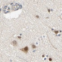

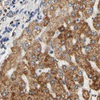


Comprehensive Human Protein Atlas IHC scoring per tissue (reliability: Approved — Medium consistency between antibody staining and RNA expression data.). Rows are taken directly from the HPA tissue chart — click any row's HPA link to view the source.
| Tissue | Cell type | Level | Evidence | Source |
|---|---|---|---|---|
| Hippocampus | Neuronal cells | High | Protein (IHC) | HPA → |
| Kidney | Cells in tubules | High | Protein (IHC) | HPA → |
| Liver | Hepatocytes | High | Protein (IHC) | HPA → |
| Lung | Macrophages | High | Protein (IHC) | HPA → |
| Placenta | Trophoblastic cells | High | Protein (IHC) | HPA → |
| Tissue | Cell type | Level | Evidence | Source |
|---|---|---|---|---|
| Adipose tissue | Adipocytes | Not detected | Protein (IHC) | HPA → |
| Bone marrow | Hematopoietic cells | Not detected | Protein (IHC) | HPA → |
| Cervix | Squamous epithelial cells | Not detected | Protein (IHC) | HPA → |
| Esophagus | Squamous epithelial cells | Not detected | Protein (IHC) | HPA → |
| Oral mucosa | Squamous epithelial cells | Not detected | Protein (IHC) | HPA → |
Troubleshoot SFXN3 staining in paraffin sections by checking retrieval, compartment, cell identity and controls before comparing signal across samples.
A14648-1 is listed for human IHC and IF, with images from human paraffin sections for both applications (catalog applications, reactivity, and image captions).
A14648-1 is listed for human IHC, with images of paraffin sections from human liver cancer, lung adenocarcinoma, spleen, and thyroid cancer tissue (catalog applications, reactivity, and IHC image captions). Its IF image shows a human renal cancer paraffin section (IF image caption).
Which to pick: For tissue IHC, choose A14648-1, a rabbit antibody with human reactivity and documented paraffin-section IHC images (catalog metadata and IHC image captions); the fixative is unreported (IHC image captions). For IF, A14648-1 has a paraffin-section image; ICC validation is unreported (catalog applications and IF image caption). No cross-species option is documented: A14648-1 lists human reactivity only (catalog reactivity).