SFXN4 / Sideroflexin-4 · IHC design guide

Design Immunohistochemistry for SFXN4

Plan SFXN4 chromogenic IHC in paraffin sections using the catalog antibody at 2–5 μg/ml (datasheet A12029-2). Compare cytoplasmic staining in muscle cells with appropriate controls, bearing in mind the medium concordance between staining and RNA expression (HPA tissue IHC).

Evidence assembled Oct 2026 · For research use; verify linked source records and product datasheet before use
Immunohistochemistry protocol sheet for SFXN4 (IHC for SFXN4): expected localisation Cytoplasmic tissue staining (HPA tissue IHC); mitochondrial inner membrane (UniProt), antibody A12029-2, validated IHC image, and IHC protocol steps
Printable SFXN4 IHC protocol sheet — expected localisation Cytoplasmic tissue staining (HPA tissue IHC); mitochondrial inner membrane (UniProt), antibody A12029-2, controls and protocol steps. Open the full SFXN4 IHC guide →

SFXN4 Immunohistochemistry Experimental Design Guide

Expected localisation, validated protocols, controls and antibodies — the at-a-glance facts below, then the full design guide.

Must know before staining
Expected localisation Cytoplasmic tissue staining (HPA tissue IHC); mitochondrial inner membrane (UniProt)
Staining pattern Cytoplasmic staining, high in muscle cells (HPA tissue IHC)
Antigen retrieval EDTA pH 8.0 HIER, heat-mediated (datasheet A12029-2)
Positive control ⓘ Heart muscle+4 more · see all
Negative control ⓘ Cervix+1 more · see all
Important caveats
Reasons your staining may differ from the expected pattern.
Fixation Keep fixation consistent across sections. (standard IHC practice; not target-specific)
Caveat Staining has medium RNA concordance; verify with controls (HPA tissue IHC)
Regulation No expression regulator annotated (UniProt)
Isoform / epitope 3 isoforms; antibody epitope coverage is unknown (UniProt)
Section 1

Recommended SFXN4 IHC & IF Protocols

The catalog antibody protocol and 2 published IHC protocols provide starting points for SFXN4 staining in paraffin sections (datasheet A12029-2; PMC9675821; PMC11576178).

Recommended immunohistochemistry (IHC-P) protocol parameters
SampleParaffin-embedded human colon cancer tissue; fixative not specified (datasheet A12029-2)
FixationImage fixative and duration unreported (datasheet A12029-2); verify before use.
Sectioning4–5 µm sections on charged slides (standard)
DeparaffinisationXylene, graded ethanol series to water (standard)
Antigen retrievalHeat retrieval: EDTA pH 8.0 (datasheet A12029-2); 20 min, 95–100 °C (standard)
Peroxidase block3% H2O2, 10 min, room temperature (standard)
Blocking10% goat serum (datasheet A12029-2)
Primary antibodyRabbit anti-SFXN4, 2-5 μg/ml (datasheet A12029-2)
Primary incubationOvernight at 4 °C (datasheet A12029-2)
DetectionHRP-conjugated secondary, DAB chromogen (datasheet A12029-2)
CounterstainHematoxylin, blue, dehydrate and mount (standard)
Expected resultSFXN4-positive staining in cardiomyocytes of heart muscle (HPA tissue IHC: High). HPA tissue profile: General cytoplasmic expression, high abundance in muscle cells. No signal in the no-primary control.
💡Decision noteStart with heat-mediated EDTA pH 8.0 retrieval for the catalog antibody, as specified by its datasheet; neither published excerpt states a retrieval method (datasheet A12029-2; PMC9675821; PMC11576178).
Section 2

What Is the Expected SFXN4 Staining Pattern?

SFXN4 is an inner mitochondrial membrane protein with five transmembrane segments (UniProt Q6P4A7 topology). In paraffin-section IHC, expect cytoplasmic staining, especially in cardiomyocytes, skeletal myocytes and smooth muscle cells, where HPA reports High staining (HPA tissue IHC). HPA rates its tissue IHC profile Approved, with medium consistency against RNA expression and pending external verification (HPA tissue IHC).

What am I looking at on my slide?
Cytoplasmic stain in cardiomyocytes, skeletal myocytes or smooth muscle cells.This fits the reported High muscle-cell staining (HPA tissue IHC). Its cytoplasmic appearance is compatible with mitochondrial SFXN4, although routine chromogenic IHC cannot resolve the inner membrane (UniProt Q6P4A7; standard IHC practice).
Predominantly nuclear stain, with little cytoplasmic stain, in a muscle-cell positive control.This departs from the reported tissue IHC profile (HPA tissue IHC). HPA also reports nucleoplasm in ICC-IF, but marks that location uncertain; a nuclear IHC result alone should therefore prompt a control and detection check rather than a localization claim (HPA subcellular).
Strong stain in cells outside the expected pattern, especially without muscle-cell staining on the same run.Compare the named cell type with HPA's tissue-level entries before calling it unexpected: several glandular and hematopoietic populations show Medium staining (HPA tissue IHC). If the discrepancy persists, consider cross-reactivity or endogenous detection activity (standard IHC practice).
Diffuse color across tissue and areas without cells, with poor separation between cell populations.A pattern that does not track cells is difficult to score as SFXN4. Uneven detection background, insufficient blocking or washing, and chromogen deposition are general IHC possibilities; assess them with appropriate controls (standard IHC practice).
No stain in cardiomyocytes, skeletal myocytes or smooth muscle cells.These are HPA High populations, so a blank result warrants a run-level check before biological interpretation (HPA tissue IHC). Review tissue preservation, retrieval, antibody and detection steps against the validated IHC-P workflow (standard IHC practice).
💡Expected SFXN4 appearanceA convincing positive is discernible cytoplasmic chromogenic staining in muscle cells, strongest in HPA High cardiomyocytes, skeletal myocytes or smooth muscle cells; dominant isolated nuclear color or diffuse cell-free color is a warning pattern (HPA tissue IHC; HPA subcellular; standard IHC practice).
How each factor affects the staining
Compartment and optical resolutionUniProt places SFXN4 in the mitochondrial inner membrane, while HPA describes general cytoplasmic tissue IHC staining (UniProt Q6P4A7; HPA tissue IHC). Read chromogenic cytoplasmic signal as compatible with that location, not as proof of membrane-level localization (standard IHC practice).
Choice of positive and low-signal tissueHeart, skeletal and smooth muscle cells are High; adrenal and appendix glandular cells, among others, are Medium (HPA tissue IHC). Cervical glandular and vaginal squamous cells are Not detected in the supplied HPA entries; those observations are context-specific, not universal negative controls (HPA tissue IHC).
Antibody evidenceThe HPA antibody HPA020872 has Approved IHC status; HPA018028 has Supported ICC status and no supplied IHC status (HPA antibodies). HPA's tissue profile has medium RNA–stain consistency and awaits external verification, so corroborate a surprising pattern before assigning SFXN4 specificity (HPA tissue IHC).
Antigen retrieval and processingTarget-specific fixation effects are not established by the supplied assay evidence. Verify with a matched IHC source before attributing a result to fixation.
Isoforms and epitope coverageUniProt lists three SFXN4 isoforms and five transmembrane segments (UniProt Q6P4A7). Without a supplied antibody epitope or isoform-reactivity map, these annotations cannot predict which isoforms the IHC antibody detects or explain a missing tissue signal.
IF/ICC Q: What location should fluorescence show?A: Mitochondrial localization is supported; nucleoplasmic and vesicular localizations are marked uncertain (HPA subcellular). HPA reports ICC-IF images in A-431, HEK293 and U-251MG, but this section makes no IF protocol recommendation (HPA subcellular).
Why is my staining missing, weak or wrong?
SituationLikely causeNext action
High muscle-cell positive control is blank.A run-level failure is plausible because those populations are reported High (HPA tissue IHC).Check section integrity, retrieval, primary-antibody use and detection controls against the antibody's IHC-P instructions; do not infer SFXN4 absence from this slide alone (standard IHC practice).
Nuclear color dominates the positive control.Nucleoplasm is an uncertain ICC-IF location, whereas tissue IHC is described as generally cytoplasmic (HPA subcellular; HPA tissue IHC).Review counterstain and chromogen separation, then repeat with detection controls; treat persistent nuclear staining as unresolved until independently corroborated (standard IHC practice).
Many unexpected cell types stain strongly.Some nonmuscle cells have genuine Medium HPA entries; remaining discordance may reflect cross-reactivity or endogenous detection activity (HPA tissue IHC; standard IHC practice).Identify cells before scoring, compare them with HPA's named populations, and inspect a no-primary detection control for endogenous color (standard IHC practice).
Color is diffuse or appears outside cells.Background deposition, weak washing or detection activity can obscure a cellular pattern (standard IHC practice).Compare no-primary and tissue controls, then review blocking, washing and chromogen development within the established IHC workflow (standard IHC practice).
A cervical glandular or vaginal squamous sample stains.HPA lists those specific cell populations as Not detected, but its IHC profile is Approved with pending external verification (HPA tissue IHC).Confirm the stained cell identity and rule out background with controls before treating the result as a biological exception or an antibody-specific signal (standard IHC practice).
Only an ICC-IF result appears mitochondrial, while tissue IHC is broad cytoplasmic.The methods resolve compartments differently; HPA supports mitochondrial ICC-IF localization and describes general cytoplasmic tissue IHC staining (HPA subcellular; HPA tissue IHC).Interpret each image at its own resolution and use the IHC tissue pattern for paraffin-section scoring; do not require visible mitochondrial outlines in chromogenic IHC (standard IHC practice).

Sample controls for SFXN4 IHC & IF

🧪Run heart muscle first: cardiomyocytes should stain (HPA: High in cardiomyocytes). Run cervix as the negative tissue (HPA: Not detected in glandular cells); on the heart slide, compare cardiomyocytes with adjacent noncardiomyocyte areas for background staining without assuming those areas are biologically negative.
Positive control tissue: Heart muscle (Cardiomyocytes, HPA High)
Negative control tissue: Cervix (HPA Not detected)
ICC-IF cell lines (HPA subcellular resource): HPA ICC-IF images show SFXN4 in A-431, HEK293, U-251MG, with annotated localisation: Nucleoplasm (uncertain), Mitochondria (supported) (HPA subcellular).
Technical controls: Include a no-primary, secondary-only control, a host- and clonality-matched rabbit IgG isotype control, and a biological negative such as SFXN4 knockout tissue or peptide-blocked primary antibody if the immunizing peptide is available (standard IHC practice; selected-SKU tissue-IHC caption: rabbit primary). Quench endogenous peroxidase and assess blood-associated background in heart sections before interpreting DAB signal (standard chromogenic IHC practice).
⚠️Feasibility: A target-specific fixation window or fixation effect is unreported in the supplied evidence, and the selected-SKU tissue-IHC caption does not report a fixative. The paraffin-section caption documents heat-mediated EDTA retrieval at pH 8.0 (selected-SKU tissue-IHC caption); it does not establish a required retrieval condition or fixation. Tissue IF or frozen sections cannot be judged easier from the supplied evidence; heart muscle may have autofluorescent lipofuscin that needs checking if IF is attempted (standard IF practice). The selected A12029-2 tissue-IHC caption documents paraffin sections, but does not specify the fixative (selected-SKU IHC image A12029-2).

HPA tissue IHC evidence for SFXN4

Comprehensive Human Protein Atlas IHC scoring per tissue (reliability: Approved — Medium consistency between antibody staining and RNA expression data. Pending external verification.). Rows are taken directly from the HPA tissue chart — click any row's HPA link to view the source.

Positive expression · recommended positive controls

TissueCell typeLevelEvidenceSource
Heart muscle Cardiomyocytes High Protein (IHC) HPA →
Skeletal muscle Myocytes High Protein (IHC) HPA →
Smooth muscle Smooth muscle cells High Protein (IHC) HPA →
Adrenal gland Glandular cells Medium Protein (IHC) HPA →
Appendix Glandular cells Medium Protein (IHC) HPA →

Undetected expression · recommended negative controls

TissueCell typeLevelEvidenceSource
Cervix Glandular cells Not detected Protein (IHC) HPA →
Vagina Squamous epithelial cells Not detected Protein (IHC) HPA →
Section 3

Advanced SFXN4 IHC Tips

Troubleshoot SFXN4 staining by checking retrieval, compartment, cellular controls, and detection artefacts before comparing chromogenic IHC results across sections.

Which antigen retrieval should I start with for paraffin sections stained for SFXN4?
Start with heat-mediated retrieval in EDTA at pH 8.0 for paraffin sections (datasheet A12029-2). The selected tissue image used that retrieval before overnight incubation at 4°C with 2 μg/ml antibody (datasheet A12029-2). If staining is weak, first check retrieval heating, cooling, and section adherence across a control section; these procedural variables can change chromogenic signal (standard IHC practice). Only then compare a different retrieval buffer on adjacent sections, keeping antibody concentration and DAB development constant (standard IHC practice). SFXN4 has 5 annotated transmembrane segments, but topology alone does not establish which retrieval condition exposes this antibody’s epitope (UniProt Q6P4A7 topology).
How should I troubleshoot weak staining when fixation history is uncertain?
Record the fixative, fixation interval, and tissue processing for every section before comparing SFXN4 staining (standard IHC practice). The selected image identifies a paraffin-embedded human colon cancer section but does not state its fixative, so target-specific fixation sensitivity remains unknown (datasheet A12029-2). Compare sections with documented processing under the same EDTA pH 8.0 retrieval and 2 μg/ml primary antibody conditions used in that image (datasheet A12029-2). Include a known staining control and assess tissue preservation alongside signal, because processing differences can affect IHC interpretation (standard IHC practice). Do not infer fixation tolerance from SFXN4 localisation, topology, or tissue staining patterns (UniProt Q6P4A7; HPA tissue IHC).
Where should convincing SFXN4 chromogenic staining appear within positive cells?
Evaluate SFXN4 IHC as cytoplasmic staining with a distribution compatible with mitochondria, while recognising that DAB does not resolve individual inner membranes (HPA tissue IHC; UniProt Q6P4A7; standard IHC practice). UniProt places SFXN4 in the mitochondrial inner membrane and annotates 5 transmembrane segments (UniProt Q6P4A7 topology). HPA tissue IHC reports general cytoplasmic expression and high abundance in muscle cells, including cardiomyocytes and myocytes (HPA tissue IHC). If nuclear staining dominates, review morphology and detection controls before calling it specific: HPA lists nucleoplasm as uncertain, while mitochondrial localisation is supported (HPA subcellular). Compare the same cell compartment across matched sections (standard IHC practice).
Could isoforms or membrane topology explain inconsistent SFXN4 staining?
Check the antibody’s documented immunogen or mapped epitope before attributing discordant IHC staining to splice forms; the supplied caption does not identify that epitope (datasheet A12029-2). SFXN4 has 3 annotated isoforms and 5 transmembrane segments, so epitope position could affect accessibility, although this antibody’s isoform coverage is unknown (UniProt Q6P4A7). The record lists acetylation at residues 2 and 197, but supplies no evidence that either modification changes IHC binding (UniProt Q6P4A7). Compare adjacent sections using the documented EDTA pH 8.0 retrieval while holding detection settings constant (datasheet A12029-2; standard IHC practice). Treat an isoform-specific explanation as provisional until supported by epitope information or an independent assay (standard IHC practice).
How can I use IF to check the cellular source of SFXN4 IHC staining?
Use IF as a complementary localisation check and multiplex SFXN4 with a marker for the expected cell type, such as a muscle-cell marker when examining muscle-rich tissue (HPA tissue IHC; standard IF practice). Include a mitochondrial reference marker to assess the supported mitochondrial localisation, while interpreting apparent nucleoplasmic or vesicular signal cautiously because HPA labels those locations uncertain (HPA subcellular). Choose fluorophores in channels with low measured tissue autofluorescence and include single-label controls for spectral bleed-through (standard IF practice). Match permeabilisation to whether the antibody’s epitope faces an accessible membrane surface or lies behind the mitochondrial membranes; its epitope side is unspecified here (UniProt Q6P4A7 topology; standard IF practice).
What should I check when SFXN4 DAB staining is diffuse or unexpectedly widespread?
Inspect a no-primary control for endogenous peroxidase, nonspecific secondary binding, and pigment that could resemble DAB signal (standard IHC practice). Apply an appropriate peroxidase block and keep DAB development comparable between sections; these are general chromogenic workflow controls, not evidence of SFXN4-specific staining (standard IHC practice). The selected image used 10% goat serum blocking and a peroxidase-conjugated secondary incubated for 30 minutes at 37°C (datasheet A12029-2). Review whether diffuse signal follows damaged areas or section edges, and compare its cellular pattern with the reported general cytoplasmic expression (standard IHC practice; HPA tissue IHC). Optimise background against a matched positive control rather than removing plausible mitochondrial signal (UniProt Q6P4A7; standard IHC practice).
How should I score SFXN4 staining across sections with different cell composition? ⚠ ANSWER MARKED FOR VERIFICATION
Define the cell population and compartment before scoring, then use an H-score combining intensity and percentage of positive cells or report both measures separately (standard IHC practice). Normalise positive-cell counts to the number of evaluable cells of that type, or report stained-cell density per mm² of viable tissue (standard IHC practice). Record staining in muscle cells separately when present, because HPA reports high abundance in cardiomyocytes, skeletal myocytes, and smooth muscle cells (HPA tissue IHC). Keep section thickness, retrieval, DAB development, and image analysis thresholds consistent across the comparison (standard IHC practice). Exclude necrotic and damaged regions by a predefined rule, and report the denominator with every score (standard IHC practice).
When is an apparent SFXN4-positive cell likely to be an artefact?
Call a cell positive only when signal has plausible cytoplasmic distribution, preserved morphology, and intensity above the matched background control (HPA tissue IHC; standard IHC practice). SFXN4 is assigned to the mitochondrial inner membrane, while HPA supports mitochondrial localisation and marks nucleoplasmic localisation uncertain (UniProt Q6P4A7; HPA subcellular). Strong staining confined to nuclei, tissue edges, or necrotic areas therefore warrants review before interpretation (HPA subcellular; standard IHC practice). Check a no-primary section and peroxidase blocking when brown deposits could reflect endogenous enzyme activity rather than antibody binding (standard IHC practice). Compare cellular identity with reported high staining in muscle cells, while recognising that HPA’s tissue IHC reliability is Approved with pending external verification (HPA tissue IHC).
Boster reagents

Best SFXN4 / Sideroflexin-4 IHC Antibodies

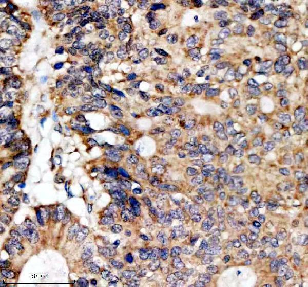
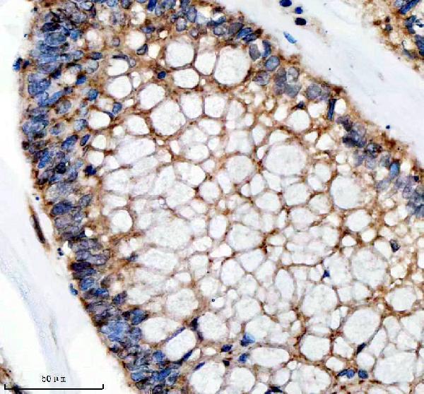
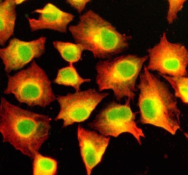
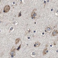
A12029-2 has IHC images from paraffin sections of human colon and ovarian cancers and rat brain, plus IF images from A549 cells and rat brain sections (catalog image captions).

Real IHC data IHC analysis of SFXN4 using anti-SFXN4 antibody (A12029-2). SFXN4 was detected in a paraffin-embedded section of human colon cancer tissue. Heat mediated antigen retrieval was performed in EDTA buffer (pH 8.0, epitope retrieval solution). The tissue section was blocked with 10% goat serum. The tissue section was then incubated with 2 μg/ml rabbit anti-SFXN4 Antibody (A12029-2) overnight at 4°C. Peroxidase Conjugated Goat Anti-rabbit IgG was used as secondary antibody and incubated for 30 minutes at 37°C. The tissue section was developed using HRP Conjugated Rabbit IgG Super Vision Assay Kit (Catalog # SV0002) with DAB as the chromogen.
Anti-SFXN4 Antibody ®
Cat # A12029-2

A12029-2 is the sole SKU; its IHC captions show human colon and ovarian cancer and rat brain paraffin sections stained at 2 μg/ml after EDTA pH 8.0 retrieval (A12029-2 IHC captions). Its IF captions show A549 cells and rat brain sections, and the catalog lists IF and ICC applications (A12029-2 IF captions; catalog applications).

Which to pick: For tissue IHC, choose A12029-2 for paraffin sections using its documented retrieval and staining conditions; the fixative is unreported (A12029-2 IHC captions). For IF/ICC, A12029-2 is listed for both applications at 5 μg/ml, with IF images from A549 cells and rat brain sections (catalog applications/dilutions; A12029-2 IF captions). For cross-species work, the catalog lists Human, Mouse and Rat reactivity, while its IHC images document human and rat samples; verify mouse tissue staining with appropriate controls (catalog reactivity; A12029-2 IHC captions).

Each figure is that product's own IHC / IF validation image from its datasheet.

References

  1. UniProt Consortium. UniProt entry Q6P4A7 (SFXN4_HUMAN, Sideroflexin-4).
  2. Human Protein Atlas. SFXN4 tissue IHC expression (reliability: Approved).
  3. Human Protein Atlas. SFXN4 subcellular location (ICC-IF): Mainly localized to the nucleoplasm and mitochondria. In addition localized to vesicles..
  4. Human Protein Atlas. SFXN4 antibody validation summary (2 antibodies).
  5. Expression and Clinical Significance of Sideroflexin 4 (SFXN4) in Colorectal Cancer. In vivo (Athens, Greece) 2025 — PMC12588242.
  6. Complementary anti-cancer pathways triggered by inhibition of sideroflexin 4 in ovarian cancer. Scientific reports 2022 — PMC9675821.
  7. A novel hypoxia- and lactate metabolism-related prognostic signature to characterize the immune landscape and predict immunotherapy response in osteosarcoma. Frontiers in immunology 2024 — PMC11576178.
  8. PubMed PMID:14756423 — UniProt-cited evidence.
  9. PubMed PMID:15164054 — UniProt-cited evidence.
  10. PubMed PMID:15489334 — UniProt-cited evidence.