This website uses cookies to ensure you get the best experience on our website.


- Table of Contents
Plan chromogenic SGO2 IHC in paraffin sections using nuclear staining in testicular pachytene spermatocytes as a benchmark (HPA tissue IHC). Start with the IHC-validated antibody at 1:100–1:300 (datasheet), then score staining by cell type and nuclear location.

Expected localisation, validated protocols, controls and antibodies — the at-a-glance facts below, then the full design guide.
| Expected localisation | Nuclear staining in seminiferous ducts (HPA tissue IHC) | |
| Staining pattern | Pachytene spermatocytes: nuclear staining (HPA tissue IHC) | |
| Antigen retrieval | Tris-EDTA pH 9.0 HIER, 95–98 °C, 20 min (rule: nuclear antigen) | |
| Positive control | Testis+4 more · see all | |
| Negative control | Adipose tissue+4 more · see all |
| Fixation | Matched tissue-IHC evidence does not establish the fixation claim. Validate the specimen-specific method before use. (selected-SKU IHC image A30763) | |
| Caveat | Centromeric signal can disappear at anaphase (UniProt) | |
| Regulation | Centromeric signal lost at anaphase (UniProt) | |
| Isoform / epitope | 3 isoforms; epitope coverage unspecified (UniProt) |
The catalog antibody’s IHC-P protocol (datasheet) is accompanied by four published SGO2 IHC protocols (PMC10414046; PMC12264263; PMC8448842; PMC6554469).
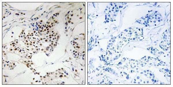
| Sample | Paraffin-embedded human breast carcinoma tissue; fixative not specified (datasheet A30763) |
| Fixation | Image fixative and duration unreported (datasheet A30763); verify before use. |
| Sectioning | 4–5 µm sections on charged slides (standard) |
| Deparaffinisation | Xylene, graded ethanol series to water (standard) |
| Antigen retrieval | Heat-induced epitope retrieval in Tris-EDTA buffer, pH 9.0, 20 min at 95–98 °C (standard rule: nuclear antigen) |
| Peroxidase block | 3% H2O2, 10 min, room temperature (standard) |
| Blocking | 10% normal serum of the secondary host, 30 min, room temperature (standard) |
| Primary antibody | Rabbit anti-SGO2, 1:100 - 1:300 (datasheet A30763) |
| Primary incubation | Overnight at 4 °C (standard) |
| Detection | HRP-polymer secondary, DAB chromogen 5–10 min (standard) |
| Counterstain | Hematoxylin, blue, dehydrate and mount (standard) |
| Expected result | SGO2-positive staining in pachytene spermatocytes of testis (HPA tissue IHC: High). HPA tissue profile: Nuclear expression in seminiferous ducts. No signal in the no-primary control. |
SGO2 is a nuclear, centromere and kinetochore protein with no transmembrane segment (UniProt Q562F6 localization and topology). In paraffin section IHC, expect nuclear staining in seminiferous ducts, strongest in pachytene spermatocytes (HPA: tissue profile; High in pachytene spermatocytes). HPA rates its tissue IHC evidence Enhanced, while noting medium consistency between antibody staining and RNA expression; interpret other positive cells with that qualification (HPA: reliability).
| Distinct nuclear staining in pachytene spermatocytes within seminiferous ducts, with relatively little surrounding haze (HPA: nuclear expression in seminiferous ducts; High in pachytene spermatocytes). | This is the clearest tissue level match for SGO2. Nuclear localization agrees with the protein record; centromere or kinetochore localization is phase dependent, so every positive nucleus need not show discrete dots (UniProt Q562F6 localization; HPA: tissue profile). |
| Predominantly cytoplasmic, membranous or extracellular color where nuclear signal is absent (UniProt Q562F6 localization and topology). | That distribution does not match the annotated nuclear and chromosomal compartments or the HPA tissue profile. Check counterstain alignment, chromogen deposits and the detection controls before scoring it as SGO2 (UniProt Q562F6 localization; HPA: tissue profile; general IHC practice). |
| Strong staining in adipocytes or bone marrow hematopoietic cells, especially alongside a weak testis result (HPA: Not detected in adipose adipocytes and bone marrow hematopoietic cells; High in pachytene spermatocytes). | The tissue pattern conflicts with these HPA observations. Consider antibody cross reactivity or endogenous detection activity, then compare a no primary control and the known positive section under matched conditions (HPA: tissue IHC; general IHC practice). |
| Diffuse brown color across nuclei, cytoplasm and section background, without a recognizable cell population (HPA: nuclear tissue profile). | This lacks the cell and compartment selectivity expected from the HPA profile. Review blocking, washes, reagent concentration and section handling; diffuse color alone does not establish SGO2 expression (HPA: tissue profile; general IHC practice). |
| No nuclear signal in pachytene spermatocytes while morphology and counterstain remain assessable (HPA: High in pachytene spermatocytes). | The expected positive control has failed. Check antibody identity and IHC validation, retrieval conditions, primary and detection steps, and a concurrently processed control before calling the specimen negative (HPA: tissue IHC and antibody validation; general IHC practice). |
| Cell population and cell cycle (HPA: High in pachytene spermatocytes; UniProt Q562F6 localization). | Choose seminiferous ducts containing identifiable pachytene spermatocytes for the positive readout. UniProt describes centromeric localization that changes through division, so absence of obvious puncta in every nucleus is not, by itself, a failed IHC result (HPA: tissue IHC; UniProt Q562F6 localization). |
| Strength of tissue evidence (HPA: Enhanced IHC reliability; medium staining and RNA consistency). | HPA reports Medium staining in appendix and duodenal endocrine cells, fallopian tube non ciliated cells, and rectal enterocytes. These are possible secondary comparisons, but the Enhanced rating still carries the stated RNA consistency limitation (HPA: tissue IHC and reliability). |
| Assay specific localization (HPA: tissue IHC profile and ICC IF subcellular summary). | HPA tissue IHC describes nuclear expression in seminiferous ducts, whereas ICC IF reports mainly nucleoplasm with additional nuclear bodies. Use the latter as context, not as a requirement for resolving nuclear bodies in chromogenic paraffin sections (HPA: tissue IHC; HPA: ICC IF). |
| Antibody validation and isoforms (HPA: antibody validation; UniProt Q562F6 isoforms). | HPA lists Enhanced IHC for HPA035163 and no IHC status for HPA040817; both have Enhanced ICC status. UniProt lists three isoforms, but these records alone do not establish which isoforms an antibody detects in a section (HPA: antibodies; UniProt Q562F6 isoforms). |
| Situation | Likely cause | Next action |
|---|---|---|
| Testis positive control has no nuclear staining (HPA: High in pachytene spermatocytes). | The selected field may lack identifiable pachytene cells, or an IHC reagent step may have failed (HPA: testis cell profile; general IHC practice). | Review morphology in seminiferous ducts, then check the primary antibody, detection reagents and a concurrently processed positive control (HPA: tissue profile; general IHC practice). |
| Signal is weak in a secondary tissue but clear in testis (HPA: High in pachytene spermatocytes; Medium in listed secondary cells). | HPA records lower staining levels in those listed cells; weak staining alone does not imply an assay failure (HPA: tissue IHC). | Score each named cell population separately and retain the testis section as the stronger positive reference (HPA: tissue IHC; general IHC practice). |
| Color is mainly outside nuclei (UniProt Q562F6 localization; HPA: nuclear tissue profile). | Chromogen deposition, background or antibody cross reactivity may mimic signal outside the expected compartment (general IHC practice). | Compare nuclear counterstain and a no primary control; review washes and detection conditions before accepting that pattern (general IHC practice). |
| Negative control cells show strong color (HPA: Not detected in adipose adipocytes and bone marrow hematopoietic cells). | Background binding or endogenous detection activity is possible; HPA's Not detected calls are observations, not an absolute absence of protein (HPA: tissue IHC; general IHC practice). | Check the no primary control and use the detection system's appropriate endogenous activity block; compare matched sections (general IHC practice). |
| All compartments show diffuse staining (HPA: nuclear tissue profile). | Insufficient blocking or washing, or overly concentrated reagents, can reduce IHC contrast (general IHC practice). | Review blocking and washes, and titrate the primary antibody and detection reagents against the positive control (general IHC practice). |
| An ICC IF image shows nuclear bodies, but paraffin IHC shows broader nuclear color (HPA: ICC IF nucleoplasm and nuclear bodies; tissue IHC nuclear profile). | The two HPA assay summaries describe different readouts and do not establish that nuclear bodies must resolve in paraffin IHC (HPA: ICC IF; HPA: tissue IHC). | Judge the paraffin result against the documented nuclear staining and named tissue cell populations; consult the separate IF guide for ICC IF interpretation (HPA: tissue IHC; HPA: ICC IF). |


Comprehensive Human Protein Atlas IHC scoring per tissue (reliability: Enhanced — Medium consistency between antibody staining and RNA expression data.). Rows are taken directly from the HPA tissue chart — click any row's HPA link to view the source.
| Tissue | Cell type | Level | Evidence | Source |
|---|---|---|---|---|
| Testis | Pachytene spermatocytes | High | Protein (IHC) | HPA → |
| Appendix | Endocrine cells | Medium | Protein (IHC) | HPA → |
| Duodenum | Endocrine cells | Medium | Protein (IHC) | HPA → |
| Fallopian tube | Non-ciliated cells | Medium | Protein (IHC) | HPA → |
| Rectum | Enterocytes | Medium | Protein (IHC) | HPA → |
| Tissue | Cell type | Level | Evidence | Source |
|---|---|---|---|---|
| Adipose tissue | Adipocytes | Not detected | Protein (IHC) | HPA → |
| Adrenal gland | Glandular cells | Not detected | Protein (IHC) | HPA → |
| Bone marrow | Hematopoietic cells | Not detected | Protein (IHC) | HPA → |
| Breast | Adipocytes | Not detected | Protein (IHC) | HPA → |
| Caudate | Glial cells | Not detected | Protein (IHC) | HPA → |
Use nuclear staining and cell identity to evaluate SGO2 chromogenic IHC, with the catalog image and tissue atlas as separate evidence sources.
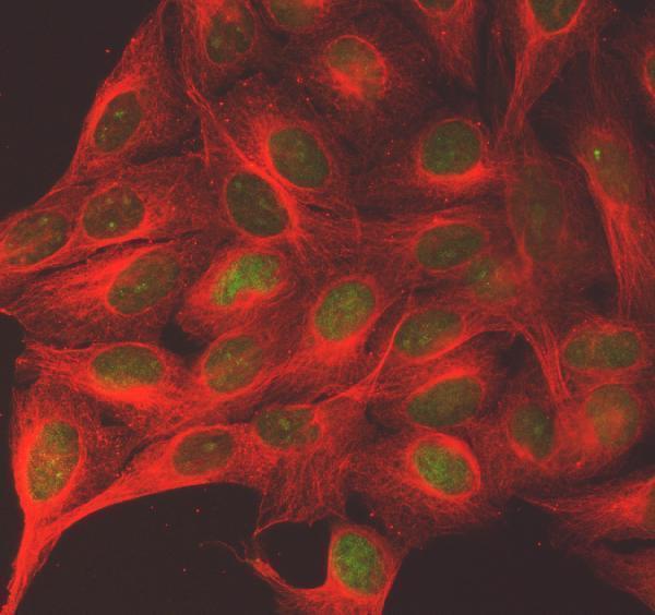
The catalog includes anti-SGO2 antibodies with paraffin-section IHC data in human breast carcinoma (A30763 image caption) and IF data in U20S cells (A08632 image caption); listed reactivity spans human, mouse and rat (catalog).
A30763 lists IHC and IF for human, mouse and rat, with an IHC image of paraffin-embedded human breast carcinoma (catalog applications, reactivity and A30763 image caption). A08632 lists IF/ICC for human samples, with an IF image of U20S cells (catalog applications, reactivity and A08632 image caption).
Which to pick: Choose A30763 for paraffin-section IHC: it lists IHC at 1:100–1:300 and has a paraffin-section IHC image; the caption does not report the fixative (catalog dilution and A30763 image caption). For IF/ICC, A08632 has a U20S cell IF image at 5 μg/ml, while A30763 also lists IF/ICC (A08632 image caption; catalog applications). For mouse or rat samples, A30763 is the listed polyclonal option with those species in its reactivity list; its displayed IHC image is from a human sample (A30763 dilution_raw, catalog reactivity and A30763 image caption).