This website uses cookies to ensure you get the best experience on our website.


- Table of Contents
Plan SGSH staining in paraffin sections using lung macrophages or kidney collecting ducts as high-staining references (HPA tissue IHC). Evaluate granular cytoplasmic staining by cell type, with adipocytes as a documented unstained comparison (HPA tissue IHC).

Expected localisation, validated protocols, controls and antibodies — the at-a-glance facts below, then the full design guide.
| Expected localisation | Granular cytoplasm (HPA tissue IHC); lysosomal protein (UniProt) | |
| Staining pattern | Granular cytoplasmic staining in several cell types (HPA tissue IHC) | |
| Antigen retrieval | EDTA pH 8.0 HIER, heat-mediated (datasheet A04608-1) | |
| Positive control | Adrenal gland+4 more · see all | |
| Negative control | Adipose tissue+4 more · see all |
| Fixation | Keep fixation conditions consistent across sections. (standard IHC practice; not target-specific) | |
| Caveat | Lung macrophages stain strongly; score cell types separately (HPA tissue IHC) | |
| Regulation | No expression regulator documented (UniProt) | |
| Isoform / epitope | No annotated isoforms; mature chain 21–502 (UniProt) |
The catalog antibody’s IHC-P protocol is paired with published SGSH staining methods for mouse paraffin sections and human bronchial biopsies (PMC6881609; PMC6374378).
| Sample | Paraffin-embedded human appendiceal adenocarcinoma tissue; fixative not specified (datasheet A04608-1) |
| Fixation | Image fixative and duration unreported (datasheet A04608-1); verify before use. |
| Sectioning | 4–5 µm sections on charged slides (standard) |
| Deparaffinisation | Xylene, graded ethanol series to water (standard) |
| Antigen retrieval | Heat retrieval: EDTA pH 8.0 (datasheet A04608-1); 20 min, 95–100 °C (standard) |
| Peroxidase block | 3% H2O2, 10 min, room temperature (standard) |
| Blocking | 10% goat serum (datasheet A04608-1) |
| Primary antibody | Rabbit anti-SGSH, 2-5 μg/ml (datasheet A04608-1) |
| Primary incubation | Overnight at 4 °C (datasheet A04608-1) |
| Detection | HRP-conjugated secondary, DAB chromogen (datasheet A04608-1) |
| Counterstain | Hematoxylin, blue, dehydrate and mount (standard) |
| Expected result | SGSH-positive staining in glandular cells of adrenal gland (HPA tissue IHC: High). HPA tissue profile: Granular cytoplasmic expression in several cell types. No signal in the no-primary control. |
SGSH is a lysosomal enzyme with no transmembrane segment (UniProt P51688 topology). In paraffin-section IHC, expect granular cytoplasmic staining in selected cell types, including kidney collecting ducts, lung macrophages and several glandular epithelia (HPA tissue IHC). HPA rates the tissue staining pattern Enhanced because antibody staining is highly consistent with RNA expression data (HPA tissue IHC reliability).
| Granular cytoplasmic staining is prominent in kidney collecting ducts or lung macrophages. | This fits lysosomal SGSH localisation (UniProt P51688) and the High staining reported for those cells (HPA tissue IHC). Assess the named cells rather than treating every cell in the section as an equivalent positive control (HPA tissue IHC). |
| Staining is predominantly nuclear or outlines the plasma membrane. | That compartment conflicts with the annotated lysosomal location and lack of a transmembrane segment (UniProt P51688 topology). Check the counterstain and compare a no-primary control before assigning the signal to SGSH (general IHC practice). |
| Strong staining appears in adipocytes, skeletal-muscle myocytes or salivary-gland cells. | HPA reports SGSH as Not detected in those respective cell populations (HPA tissue IHC). Review morphology and controls for cross-reactivity or endogenous chromogenic activity (general IHC practice); a single unexpected field does not establish a new SGSH expression pattern. |
| A diffuse deposit covers multiple compartments or obscures cell boundaries. | The expected HPA description is granular cytoplasmic expression, so diffuse staining cannot be scored as that pattern (HPA tissue IHC). Inspect the no-primary control and reagent background, then reassess blocking, washes and detection conditions (general IHC practice). |
| No staining is visible in kidney collecting ducts on a run intended to detect SGSH. | Collecting ducts are reported High by HPA, making them a useful tissue control (HPA tissue IHC). Verify that the ducts are present, then review antibody application, detection and run controls (general IHC practice); absence in one run alone cannot establish biological loss. |
| Compartment and molecular form | SGSH is annotated in lysosomes, has a signal peptide at residues 1–20 and lacks a transmembrane segment (UniProt P51688 topology and processing). Interpret punctate or granular cytoplasmic staining in that context; those annotations do not predict a specific chromogenic intensity. |
| Tissue and cell selection | HPA reports High staining in adrenal, epididymal, gallbladder and prostate glandular cells, kidney collecting ducts, lung macrophages and placental syncytiotrophoblast cell bodies; breast glandular cells are Medium (HPA tissue IHC). Match the assessment to the cell population, not just the organ. |
| Low and undetected reference populations | Several populations are Low, including cerebral-cortex neurons and rectal glandular cells, while adipocytes and skeletal-muscle myocytes are Not detected (HPA tissue IHC). Low or undetected populations offer different interpretive expectations and should not be treated as interchangeable positive controls. |
| Strength of the IHC evidence | The tissue profile is rated Enhanced for consistency between antibody staining and RNA expression; two listed rabbit polyclonal antibodies, HPA023436 and HPA023451, each have Enhanced IHC status (HPA tissue IHC; HPA antibody validation). This supports the reported pattern without proving every signal in a new run is specific. |
| IF/ICC: is the same pattern independently shown? | HPA provides no main subcellular ICC-IF location or cell-line images, and lists no ICC validation for either antibody (HPA subcellular; HPA antibody validation). Its HUVEC/TERT2 RNA value is an expression measurement, not an IF localisation result (HPA subcellular). |
| Situation | Likely cause | Next action |
|---|---|---|
| A known-positive kidney section has no signal in collecting ducts. | The expected High collecting-duct signal is missing (HPA tissue IHC); the cause cannot be determined from the negative image alone. | Confirm duct morphology and check a successful run control, antibody application and chromogenic detection steps (general IHC practice). Record the run as inconclusive until the control performs. |
| The whole section shows diffuse brown staining. | A broad deposit does not match the granular cytoplasmic HPA profile (HPA tissue IHC); background from detection reagents or insufficient washing is possible (general IHC practice). | Examine a no-primary control, check endogenous detection activity, and review blocking, washing and chromogen development (general IHC practice). Score only clearly localised cellular signal. |
| Adipocytes or skeletal-muscle myocytes stain strongly. | These populations are reported Not detected (HPA tissue IHC). Cell misidentification or nonspecific signal is possible (general IHC practice). | Verify the stained cell type against morphology and counterstain; compare the no-primary control and a positive cell population on the same run (general IHC practice). |
| Signal is mainly nuclear or traces cell surfaces. | This location conflicts with lysosomal SGSH and its lack of a transmembrane segment (UniProt P51688 topology). | Check focus, counterstain and control staining, then reassess whether granular cytoplasmic signal is present in an HPA-positive population (general IHC practice; HPA tissue IHC). |
| A weak field is being called SGSH-negative. | HPA levels differ by population: breast glandular cells are Medium, while cerebral-cortex neurons are Low (HPA tissue IHC). A weak field alone cannot resolve expression from run performance. | Identify the exact cell population and compare it with a High reference population and run controls before interpreting a negative result (HPA tissue IHC; general IHC practice). |
| An IF/ICC image is being used to validate the IHC call. | The supplied HPA subcellular record has no ICC-IF location or images, and the listed antibodies have no ICC status (HPA subcellular; HPA antibody validation). | Base this paraffin-section call on morphology, IHC controls and the HPA tissue pattern (HPA tissue IHC; general IHC practice). Treat IF/ICC localisation as unverified by the supplied record. |

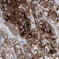
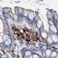

Comprehensive Human Protein Atlas IHC scoring per tissue (reliability: Enhanced — High consistency between antibody staining and RNA expression data.). Rows are taken directly from the HPA tissue chart — click any row's HPA link to view the source.
| Tissue | Cell type | Level | Evidence | Source |
|---|---|---|---|---|
| Adrenal gland | Glandular cells | High | Protein (IHC) | HPA → |
| Epididymis | Glandular cells | High | Protein (IHC) | HPA → |
| Gallbladder | Glandular cells | High | Protein (IHC) | HPA → |
| Kidney | Collecting ducts | High | Protein (IHC) | HPA → |
| Lung | Macrophages | High | Protein (IHC) | HPA → |
| Tissue | Cell type | Level | Evidence | Source |
|---|---|---|---|---|
| Adipose tissue | Adipocytes | Not detected | Protein (IHC) | HPA → |
| Bone marrow | Hematopoietic cells | Not detected | Protein (IHC) | HPA → |
| Bronchus | Basal cells | Not detected | Protein (IHC) | HPA → |
| Caudate | Glial cells | Not detected | Protein (IHC) | HPA → |
| Oral mucosa | Squamous epithelial cells | Not detected | Protein (IHC) | HPA → |
Troubleshoot SGSH staining by checking retrieval, cellular pattern, controls, and cell-specific scoring in paraffin-section IHC.
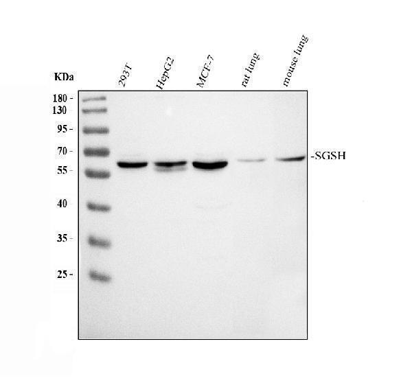
Anti-SGSH antibodies cover imaged human paraffin-section IHC (A04608-1 image caption) and listed human IF/ICC applications without an IF image (M04608-1 applications; IF image alts).
A04608-1 has an IHC image from a paraffin section of human appendiceal adenocarcinoma; its listed reactivity is human, mouse and rat (A04608-1 image caption; catalog reactivity). M04608-1 lists human IHC and IF/ICC applications, but supplies no IHC or IF image (M04608-1 applications; reactivity; image alts).
Which to pick: For tissue IHC, choose A04608-1 for its documented human paraffin-section staining; the fixative is unreported (A04608-1 image caption). For IF/ICC, choose the rabbit monoclonal M04608-1, which lists those applications for human samples (M04608-1 short description; applications; reactivity). For mouse or rat samples, A04608-1 lists reactivity in both species, though its IHC image documents human tissue only (A04608-1 catalog reactivity; image caption).