This website uses cookies to ensure you get the best experience on our website.


- Table of Contents
Real validated SHBG Western blot protocols, expected-band and isoform facts, troubleshooting for weak or shifted signal, and recommended anti-SHBG WB antibodies. Everything you need to plan the experiment before you commit precious samples.
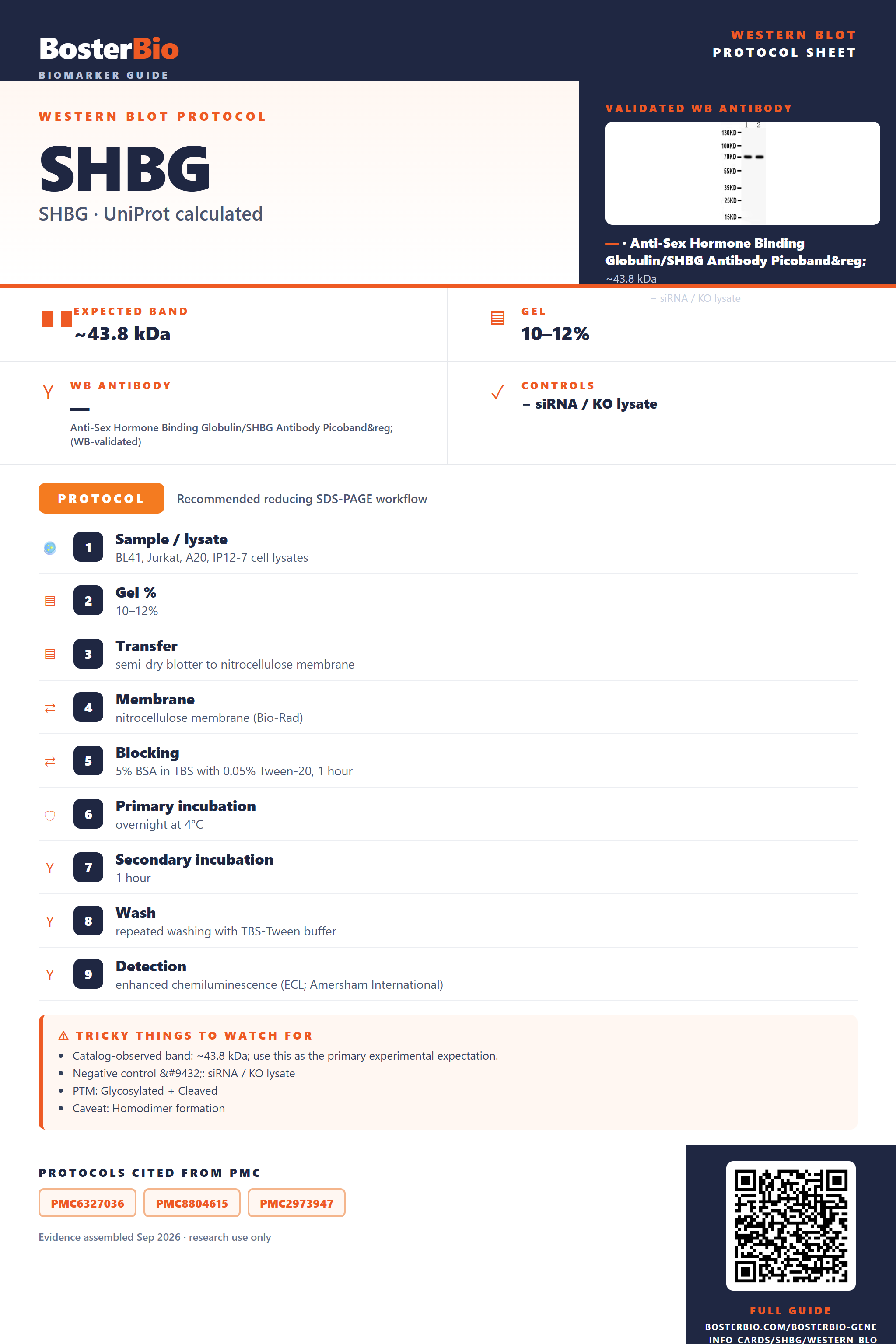
Expected bands, validated protocols, controls and antibodies — the at-a-glance facts below, then the full design guide.
| Expected band | ~43.8 kDa | |
| Observed band | 70 kDa | |
| Gel | 10–12% | |
| Negative control | siRNA / KO lysate |
| PTM | Glycosylated + Cleaved | |
| Caveat | Homodimer formation | |
| Regulation | LPS-induced | |
| Isoform | 5 isoform(s) |
Literature-validated Western blot parameters for SHBG — gel percentage, transfer, blocking, antibody incubation and detection, extracted from published methods.
| Sample / lysate | BL41, Jurkat, A20, IP12-7 cell lysates |
| Gel % | 10–12% |
| Transfer | semi-dry blotter to nitrocellulose membrane |
| Membrane | nitrocellulose membrane (Bio-Rad) |
| Blocking | 5% BSA in TBS with 0.05% Tween-20, 1 hour |
| Primary incubation | overnight at 4°C |
| Secondary incubation | 1 hour |
| Wash | repeated washing with TBS-Tween buffer |
| Detection | enhanced chemiluminescence (ECL; Amersham International) |
SHBG has a 43.8 kDa polypeptide backbone but runs at ~70 kDa on blots due to heavy N- and O-linked glycosylation, with a homodimer possible under non-reducing conditions.
| single band near 70 kDa | the mature, glycosylated SHBG monomer migrating well above its 43.8 kDa unmodified backbone |
| diffuse or smeared band around 65-75 kDa | heterogeneous glycoform occupancy across the three glycosylation sites (O-linked Thr36; N-linked Asn380, Asn396) |
| faint higher band near double the monomer mass under non-reducing conditions | incompletely reduced or intact SHBG homodimer stabilized by its disulfide bonds |
| little or no band in whole-cell lysate | SHBG is a secreted protein and accumulates in serum, conditioned media, or luminal fluid rather than inside cells |
| multiple closely spaced bands | detection of different SHBG splice isoforms rather than a single species |
| Predicted mass (UniProt) | 43.8 kDa unmodified polypeptide backbone serves as the baseline before any modification is added |
| O-linked glycosylation at Thr36 | adds mass and heterogeneity, shifting and broadening the band above the predicted backbone |
| N-linked glycosylation at Asn380 and Asn396 | further increases apparent mass and contributes to the diffuse band consistent with the ~70 kDa observed size |
| Disulfide bonds (Cys193-Cys217, Cys362-Cys390) and homodimer formation | stabilize folding and can support a dimer species running near double the monomer mass if reduction is incomplete |
| Signal peptide (residues 1-29) | cleaved during secretion, so the mature secreted protein runs slightly smaller than the unprocessed precursor |
| Splice isoforms (5 named) | can generate additional bands of slightly different sizes depending on which isoform(s) are present |
| Situation | Likely cause | Next action |
|---|---|---|
| No band in lysate | SHBG is secreted and does not accumulate intracellularly | test serum, plasma, or conditioned media instead of whole-cell lysate |
| Band higher than expected | heavy N- and O-glycosylation shifts the 43.8 kDa backbone up to the ~70 kDa observed monomer, and incomplete reduction can allow the disulfide-stabilized homodimer to run near double that mass | compare against the empirical ~70 kDa band rather than predicted mass and ensure complete reduction with fresh reducing agent and full boiling |
| Broad smear instead of sharp band | heterogeneous occupancy and composition across the three glycosylation sites | treat the sample with a deglycosylation enzyme to collapse glycoforms into a single sharper band |
| Multiple bands | co-detection of different SHBG splice isoforms | check which isoform(s) the antibody epitope covers before interpreting extra bands as nonspecific |
| Weak or no signal | low abundance of secreted protein in the sampled compartment or epitope masking by glycosylation | increase loading, sample serum or secreted media, or deglycosylate to expose masked epitopes |
| Fragments below expected size | normal cleavage of the signal peptide during secretion removes residues 1-29 from the precursor | confirm the antibody targets mature secreted SHBG rather than the signal-peptide-containing precursor region |

Comprehensive Human Protein Atlas IHC scoring per tissue. Rows are taken directly from the HPA tissue chart — click any row's HPA link to view the source.
| Tissue | Cell type | Level | Evidence | Source |
|---|
| Tissue | Cell type | Level | Evidence | Source |
|---|
Deeper troubleshooting and optimisation questions for SHBG, answered from its protein features.
BosterBio's SHBG antibodies are among the best-performing WB antibodies on the market — well cited, thoroughly validated, and orthogonally cross-validated against negative tissues and complementary methods.
Our recommended anti-SHBG antibody is a best-performing, extensively cited reagent, thoroughly validated by Western blot and orthogonally cross-validated against negative tissue and complementary detection methods to ensure specific, reproducible SHBG detection across sample types.
Which to pick: Only one Boster SHBG antibody is listed: PB9326 (Picoband), backed by an actual Western blot image showing a ~44 kDa band in HeLa and COLO320 whole-cell lysates at 0.5 ug/ml—making it the default, validated choice.