This website uses cookies to ensure you get the best experience on our website.
- Table of Contents
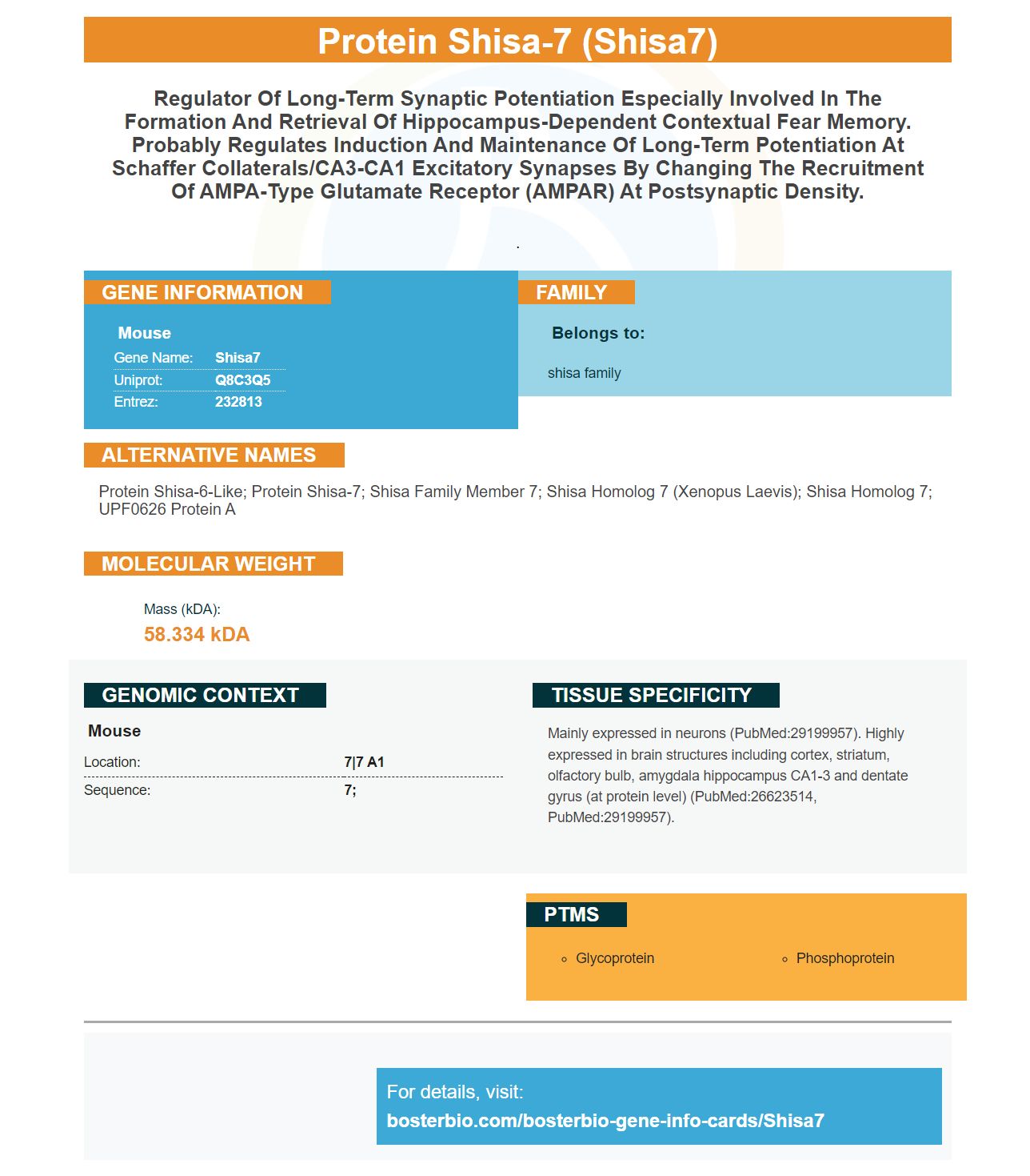
Facts about Protein shisa-7.
.
Mouse | |
---|---|
Gene Name: | Shisa7 |
Uniprot: | Q8C3Q5 |
Entrez: | 232813 |
Belongs to: |
---|
shisa family |
Protein Shisa-6-Like; Protein Shisa-7; Shisa Family Member 7; Shisa Homolog 7 (Xenopus Laevis); Shisa Homolog 7; UPF0626 Protein A
Mass (kDA):
58.334 kDA
Mouse | |
---|---|
Location: | 7|7 A1 |
Sequence: | 7; |
Mainly expressed in neurons (PubMed:29199957). Highly expressed in brain structures including cortex, striatum, olfactory bulb, amygdala hippocampus CA1-3 and dentate gyrus (at protein level) (PubMed:26623514, PubMed:29199957).
This article will discuss the Boster membrane, the SHISA7 marker and the AMPA-associated protein. It will also discuss recording autoradiography film results. This article was written by scientists who work with the Boster biosystem and have used rabbit and mouse for the development of antibodies against Shisa7. The SHISA7 marker, in addition to its excellent reactivity against rabbit and mouse samples, is useful in analyzing AMPA receptor recruitment.
The brain's inhibition of neurotransmission can be attributed to the Shisa7 protein. The HA–Shisa7 molecule correlated with vGAT, Gephyrin, GABAAR and GABAAR subunits within hippocampal cells. STED experiments revealed that Shisa7 colocalized in neurons and HEK cells with the GABAAR Subunits. The s prefix denotes that the molecule may be surface-bound. Single-cell KO of Shisa7 reduced the mIPSC frequency, but did not affect amplitude.
This antibody binds to an AMPAR homomeric peptide. Shisa7 decreases homomeric AMPARs' desensitization properties by decreasing the steady-state current and decreasing the desensitization period. Shisa7 also slows down the rectification process for GluA2-lacking AMPARs. These results are encouraging. They could have significant implications on future drug development.
A recent study in mice found that Shisa7 affects synaptic plasticity and is important for fear memory. It is important you understand that AMPARs and multiple AMPAR–associated proteins are involved within a single physiological phenotype. This new approach highlights how important it is to molecularly dissect highly composite AMPAR compounds.
This antibody targets Shisa7's N-terminus, which contains important restriction sites. The Shisa7 locus also contains an anti-FLAG M2 antibody. It has been affinity-purified and is suspended with 0.02% NaN3.
Boster Bio's AMPA receptor related protein Shisa7 belongs to the TARP family. It is also an auxiliary protein involved in AMPAR-mediated synaptic plasticity. Its absence has little effect on mEPSC amplifiers and is consistent to the Shisa6 result. This data supports the hypothesis that Shisa7 may contribute to synaptic plasticity even when present at basal levels.
Shisa7 KO mice showed reduced amplitudes NMDAR currents in the CA1 pyramidal neural neurons of their CA1 pyramidal mutants. In terms of their evoked NMDAR potentials, the Shisa7-KO mice were very similar to WT mice. Shisa7 has the potential to be a promising drug candidate that can be used to treat a variety of neurological conditions. Its target area is the AMPAR mediated amygdala channel, which is an important part of the nervous systems.
Shisa7, an innovative drug, may be able help prevent Parkinson’s disease. Shisa7 interacts directly with the protein machinery that is responsible for AMPAR exocytosis. Shisa7 also promotes diffusion trapping of PSD. This could be due in part to its C terminal type II PDZ ligand motif. These tests are preliminary and require rescue experiments with Shisa7 to confirm the hypothesis.
Membrane staining is a method that detects protein concentrations in cell cultures. Because membrane staining doesn't require any chemical compounds, it is more effective than other methods. In addition, it is much easier to clean than other methods. Here are some tips for membrane staining.
Different probes can be used. One example of this is the fluorescent styryl-pyridinium dyes. These dyes bind to the membrane’s negatively charged groups. Other reagents that can stain cells membranes include fluorescent styrylpyridinium stains and commercial lipophilic cell dye DiR. A cell membrane staining product can also be created by conjugating a quaternary amino group onto the hydrophobic Nile Red.
The most sensitive membrane stain is colloidal gold. Moeremans, et al. have written a paper on how to prepare colloidal-gold staining solutions. It is not yet widely available on the market. However, it is a highly sensitive membrane stain. This method can also be used in protein chemistry analysis, N terminal sequencing, and other types of protein chemistry. Because immunoreactivity correlates with specific stained bands, membrane staining can often be used in conjunction with immunoreactivity.
Swift(tm), Membrane Stain is a unique product that is faster than Ponceau S and has a higher level of sensitivity. This stain is more effective at binding proteins and provides better images than the Ponceau S stain. It is recommended for immunological research as it is reversible, has 500-fold greater sensitivity than Ponceau S, and is easy to use.
Autoradiography can be used to analyze cell binding patterns. This involves the detection of radiolabeled protein in SDS polyacrylamide gels or on blots and then exposing them for X-ray film. In addition to film exposure, autoradiography can also be enhanced with intensifying screens and organic scintillant-impregnated gels. Densitometry is used to quantify the image. Phosphorrimaging is an alternative method of film exposure, which is not necessary for autoradiography.
Autoradiography films are sensitive and should be treated with care. To prevent static artifacts, they should be removed from the sample immediately after exposure. To avoid static artifacts, the darkroom should be well-ventilated and equipped with the appropriate safelight conditions. Fog can occur when film is exposed to safelight for long periods. It is important to reduce exposure time when processing autoradiography films.
Autoradiography can also allow for brain imaging. It allows the detection of certain pharmacological targets both in diseased and healthy states. Recent advances in imaging technology allow for the production of autoradiograms that can be generated in real time. These films are made by gas detectors that measure a/b particle disintegrations and produce fast digital autoradiograms. To get the most exact results, blinded and randomized studies are necessary.
Preparation of tissues to be used in autoradiography plays a crucial role. Because it affects binding sites preservation, post-mortem prep is important. The radioligand must not be able to bind to any off-target sites. Moreover, the ligand-target complex must remain intact for the entire scope of the experiment. The kinetics and performance of the test can also be affected when radionuclides are used.
Autoradiography requires low radiation exposure. Before it can be activated, the radiated material must absorb at most 50 rad of radiation. The substance can also be activated with thermal neutrons. Thermal neutrons, however, are stronger than the radiation necessary to activate the substance. The autoradiography treatment is safe and efficient. A radiograph taken by autoradiography can help to determine the composition of a painting and the pigments used.
PMID: 26623514 by Farrow P., et al. Auxiliary subunits of the CKAMP family differentially modulate AMPA receptor properties.
PMID: 29199957 by Schmitz L.J.M., et al. The AMPA receptor-associated protein Shisa7 regulates hippocampal synaptic function and contextual memory.