SHMT1 / Serine hydroxymethyltransferase, cytosolic · IHC design guide

Design Immunohistochemistry for SHMT1

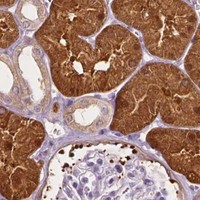
Plan SHMT1 chromogenic IHC in paraffin sections using cytoplasmic staining in hepatocytes and kidney tubule cells as positive references (HPA tissue IHC). Interpret nuclear signal in light of cell cycle dependent localization (UniProt).

Evidence assembled Oct 2026 · For research use; verify linked source records and product datasheet before use
Immunohistochemistry protocol sheet for SHMT1 (IHC for SHMT1): expected localisation Cytoplasmic tissue staining (HPA tissue IHC); nuclear location (UniProt), antibody M02944, validated IHC image, and IHC protocol steps
Printable SHMT1 IHC protocol sheet — expected localisation Cytoplasmic tissue staining (HPA tissue IHC); nuclear location (UniProt), antibody M02944, controls and protocol steps. Open the full SHMT1 IHC guide →

SHMT1 Immunohistochemistry Experimental Design Guide

Expected localisation, validated protocols, controls and antibodies — the at-a-glance facts below, then the full design guide.

Must know before staining
Expected localisation Cytoplasmic tissue staining (HPA tissue IHC); nuclear location (UniProt)
Staining pattern Cytoplasmic staining in hepatocytes and kidney tubule cells (HPA tissue IHC)
Antigen retrieval EDTA pH 8.0 HIER, heat-mediated (datasheet M02944)
Positive control ⓘ Appendix+4 more · see all
Negative control ⓘ Adipose tissue+4 more · see all
Important caveats
Reasons your staining may differ from the expected pattern.
Fixation Keep fixation consistent across paraffin sections. (standard IHC practice; not target-specific)
Caveat Cell cycle dependent nuclear localization may shift the pattern (UniProt)
Regulation Nuclear retention during S and G2/M phases (UniProt)
Isoform / epitope 4 isoforms; epitope differences are unspecified (UniProt)
Section 1

Recommended SHMT1 IHC & IF Protocols

The catalog antibody’s IHC-P protocol (datasheet: M02944) is followed by four published SHMT1 IHC protocols (PMC10728000; PMC12683878; PMC11724956; PMC6373090).

Recommended immunohistochemistry (IHC-P) protocol parameters
SampleParaffin-embedded human liver cancer tissue; fixative not specified (datasheet M02944)
FixationImage fixative and duration unreported (datasheet M02944); verify before use.
Sectioning4–5 µm sections on charged slides (standard)
DeparaffinisationXylene, graded ethanol series to water (standard)
Antigen retrievalHeat retrieval: EDTA pH 8.0 (datasheet M02944); 20 min, 95–100 °C (standard)
Peroxidase block3% H2O2, 10 min, room temperature (standard)
Blocking10% goat serum (datasheet M02944)
Primary antibodyMouse monoclonal (clone 9C7) anti-SHMT1, 2μg/ml (datasheet M02944)
Primary incubationOvernight at 4 °C (datasheet M02944)
DetectionStreptavidin-biotin complex (SABC), DAB chromogen (datasheet M02944)
CounterstainHematoxylin, blue, dehydrate and mount (standard)
Expected resultSHMT1-positive staining in glandular cells of appendix (HPA tissue IHC: High). HPA tissue profile: Cytoplasmic expression in several tissues. No signal in the no-primary control.
💡Decision noteStart with EDTA pH 8.0 heat retrieval for the catalog antibody (datasheet: M02944); citrate retrieval was reported for laryngeal carcinoma sections (PMC12683878).
Section 2

What Is the Expected SHMT1 Staining Pattern?

SHMT1 should stain mainly the cytoplasm of kidney tubular cells, hepatocytes and several glandular cell populations; spermatogonia also show high staining (HPA tissue IHC). Nuclear staining can occur, although cytosolic localization has stronger support in ICC-IF (UniProt P34896; HPA subcellular). SHMT1 has no transmembrane segment (UniProt P34896 topology). HPA rates tissue IHC reliability Enhanced, with medium consistency between antibody staining and RNA expression (HPA tissue IHC).

What am I looking at on my slide?
Clear cytoplasmic staining in kidney tubules or hepatocytes, with identifiable cell borders.This matches high staining in both cell populations (HPA tissue IHC) and SHMT1's cytoplasmic localization (UniProt P34896). Compare cells within the same section before judging intensity: HPA levels describe observed cell populations, not a required shade or staining threshold for every specimen (HPA tissue IHC).
Some nuclei stain along with the cytoplasm; or staining appears only as a crisp membrane outline.Nuclear signal can be plausible because SHMT1 enters the nucleus during the cell cycle (UniProt P34896); HPA reports additional nucleoplasmic ICC-IF localization as uncertain (HPA subcellular). A membrane-only pattern conflicts with the reported compartments and lack of a transmembrane segment (UniProt P34896 topology); investigate it as possible artefact.
Strong chromogen appears in adipocytes or cardiomyocytes while expected positive cells remain weak.HPA reports SHMT1 as not detected in those cell populations (HPA tissue IHC). The mismatch raises concern for nonspecific antibody binding or endogenous detection activity; it does not prove either cause. Review tissue morphology, antibody controls and detection-only controls before interpreting these cells as SHMT1 positive (general IHC practice).
Color covers stroma, blank spaces and many unrelated cells, obscuring cell boundaries.Diffuse deposition cannot establish the cell-restricted cytoplasmic pattern reported across several tissues (HPA tissue IHC). Assess background with a control lacking primary antibody, then review blocking, washes and detection chemistry (general IHC practice). A clean-looking field alone does not establish antibody specificity (general IHC practice).
Kidney tubules and hepatocytes show no convincing signal despite interpretable tissue morphology.Both are high-staining reference populations in HPA tissue IHC (HPA tissue IHC), so their simultaneous absence makes a technical failure plausible. Check the IHC-validated antibody's stated paraffin-section conditions and the run's positive control (general IHC practice). HPA's Enhanced rating supports the reported pattern but does not guarantee every specimen will stain (HPA tissue IHC).
💡Expected SHMT1 appearanceCall a section positive when cytoplasmic staining is clear in kidney tubular cells, hepatocytes or listed high-staining glandular cells (HPA tissue IHC), allowing plausible nuclear signal (UniProt P34896); isolated membrane outlines or widespread cell-free deposit are suspect (UniProt P34896 topology; general IHC practice).
How each factor affects the staining
Choice of reference tissueKidney tubules and hepatocytes are high-staining examples; adipocytes and cardiomyocytes are reported not detected (HPA tissue IHC). Use the named cell populations when comparing sections, since a tissue contains more than one cell type (general IHC practice).
Compartment and cell-cycle contextSHMT1 is cytoplasmic and nuclear; nuclear import and enrichment during S and G2/M phases are reported (UniProt P34896). Nuclear staining therefore needs cell context and cannot be rejected solely for being nuclear (UniProt P34896).
Antibody evidenceHPA023314 and HPA078682 each have Enhanced IHC validation (HPA antibodies). Tissue reliability is also Enhanced, but staining and RNA show medium consistency (HPA tissue IHC). Treat an unexpected pattern as a finding to verify, not as automatic proof of expression.
Isoforms and epitope coverageFour SHMT1 isoforms are listed (UniProt P34896). The supplied record gives no antibody epitope or isoform coverage, so it cannot predict whether the catalog antibody detects every isoform or explain an apparent negative by isoform choice.
IF/ICC Q: Where should fluorescence appear?A: Mainly in cytosol; additional nucleoplasm is reported with uncertain support (HPA subcellular). This informs localization checks only: ICC-IF images do not supply an IHC-P protocol or establish the expected chromogenic intensity in tissue sections (HPA subcellular; HPA tissue IHC).
Why is my staining missing, weak or wrong?
SituationLikely causeNext action
No signal in a kidney section.The sampled cells may not include intact tubules, or the IHC run may have failed; tubule cells are high staining in HPA (HPA tissue IHC).Confirm tubule morphology and inspect a concurrently processed positive control; then check the catalog antibody's IHC-P instructions and detection reagents (general IHC practice).
Nuclei stain more strongly than cytoplasm.Nuclear SHMT1 is biologically plausible, with cell-cycle-linked localization reported (UniProt P34896), but nonspecific nuclear signal remains possible (general IHC practice).Compare the pattern across cells and sections, and inspect controls before scoring it; HPA's additional nucleoplasmic ICC-IF location has uncertain support (HPA subcellular).
Only cell membranes have sharp staining.That distribution conflicts with cytoplasm/nucleus localization and the absence of a transmembrane segment (UniProt P34896 topology).Review morphology and detection-only controls; repeat with an independently validated IHC antibody if available (general IHC practice; HPA antibodies: two Enhanced IHC antibodies).
Brown deposit appears without the primary antibody.The primary antibody cannot account for that control's deposit; endogenous detection activity or detection-reagent background is possible (general IHC practice).Check the enzyme and chromogen system, its appropriate endogenous-activity control and blocking steps, then repeat the control alongside the specimen (general IHC practice).
Diffuse staining hides differences between listed high and not-detected cells.Background or excessive detection signal may obscure the cell-specific pattern reported by HPA (HPA tissue IHC; general IHC practice).Compare a primary-omission control, review washes and blocking, and adjust the assay using the catalog antibody's IHC-P instructions (general IHC practice).
An HPA not-detected population stains reproducibly.HPA's designation describes its observed assay result, not an absolute absence of protein in every specimen (HPA tissue IHC). The new staining could also be nonspecific (general IHC practice).Record the exact cell type and compartment, compare a known high-staining population, and seek independent antibody or orthogonal evidence before assigning SHMT1 expression (HPA tissue IHC; general IHC practice).

Sample controls for SHMT1 IHC & IF

🧪Run liver first: hepatocytes should stain strongly (HPA: High in liver hepatocytes). Run adipose tissue as a negative comparator: adipocytes should lack specific staining (HPA: Not detected in adipocytes); within the liver slide, any unstained non-hepatocyte cells can serve as a background reference, but their SHMT1-negative status is unestablished by the supplied HPA rows.
Positive control tissue: Appendix (Glandular cells, HPA High)
Negative control tissue: Adipose tissue (HPA Not detected)
ICC-IF cell lines (HPA subcellular resource): HPA ICC-IF images show SHMT1 in A-431, U-251MG, U2OS, KOLF2.1J, with annotated localisation: Cytosol (supported) (HPA subcellular).
Technical controls: Include a no-primary, secondary-only control and a mouse isotype control matched to the primary antibody’s isotype and clonality when known (catalog IHC caption: mouse primary); use SHMT1 knockout material, if available, as a biological specificity control. Block endogenous peroxidase and assess endogenous biotin background in liver sections because the reported detection uses a biotin-based complex and DAB (catalog IHC caption: SABC/DAB).
⚠️Feasibility: A target-specific fixation window and fixation effect are unreported, and the selected M02944 paraffin-section caption does not state the fixative (catalog IHC caption: fixative unreported). The caption reports heat retrieval in EDTA at pH 8.0, but does not establish that retrieval is required; no supplied matched evidence shows that frozen sections or IF/ICC are easier (catalog IHC caption: EDTA retrieval). For liver, check peroxidase and biotin background when interpreting the reported SABC/DAB staining (catalog IHC caption: SABC/DAB).

HPA tissue IHC evidence for SHMT1

Comprehensive Human Protein Atlas IHC scoring per tissue (reliability: Enhanced — Medium consistency between antibody staining and RNA expression data.). Rows are taken directly from the HPA tissue chart — click any row's HPA link to view the source.

Positive expression · recommended positive controls

TissueCell typeLevelEvidenceSource
Appendix Glandular cells High Protein (IHC) HPA →
Duodenum Glandular cells High Protein (IHC) HPA →
Epididymis Glandular cells High Protein (IHC) HPA →
Kidney Cells in tubules High Protein (IHC) HPA →
Liver Hepatocytes High Protein (IHC) HPA →

Undetected expression · recommended negative controls

TissueCell typeLevelEvidenceSource
Adipose tissue Adipocytes Not detected Protein (IHC) HPA →
Adrenal gland Glandular cells Not detected Protein (IHC) HPA →
Caudate Glial cells Not detected Protein (IHC) HPA →
Cerebellum Cells in granular layer Not detected Protein (IHC) HPA →
Heart muscle Cardiomyocytes Not detected Protein (IHC) HPA →
Section 3

Advanced SHMT1 IHC Tips

Troubleshoot SHMT1 staining in paraffin sections by checking retrieval, compartment, cell type, detection background and scoring against the supplied IHC evidence.

Which retrieval conditions should I start with for SHMT1 paraffin sections?
Start with heat-mediated retrieval in EDTA at pH 8.0 (datasheet M02944). The selected paraffin-section image used this retrieval before overnight incubation with 2 µg/mL catalog antibody at 4°C (caption M02944). If staining is weak, check section adherence, heating consistency and antibody coverage before changing retrieval conditions (standard IHC practice). Compare any retrieval adjustment on adjacent sections, keeping antibody concentration, detection and DAB development constant (standard IHC practice). Judge the result by staining in hepatocytes or kidney tubule cells and by background in an adipocyte-rich control area, where SHMT1 was not detected (HPA: high in hepatocytes and kidney tubule cells; not detected in adipocytes).
Could fixation explain weak or uneven SHMT1 staining?
Target-specific fixation sensitivity is unknown: the selected SHMT1 paraffin-section caption does not state a fixative (caption M02944). Record the specimen's actual fixation and processing history, then compare sections processed under documented conditions when investigating a weak result (standard IHC practice). Apply the stated EDTA pH 8.0 retrieval and 2 µg/mL antibody conditions consistently during that comparison (caption M02944). Check whether weak staining follows fixation or processing batches, section edges, or particular tissue regions before changing antibody concentration (standard IHC practice). High SHMT1 staining in hepatocytes and kidney tubule cells provides a pattern to assess, but does not establish fixation tolerance (HPA: high in hepatocytes and kidney tubule cells).
Should SHMT1 stain nuclei as well as cytoplasm in IHC?
Expect cytoplasmic staining across several tissues, while assessing nuclear staining separately (HPA tissue IHC: cytoplasmic expression in several tissues; UniProt P34896: cytoplasm and nucleus). Cytosol is the supported main location in cell imaging, whereas nucleoplasm is listed as an uncertain additional location (HPA subcellular: cytosol supported; nucleoplasm uncertain). Nuclear SHMT1 is biologically plausible because its nuclear translocation is RAN-dependent and its localization changes with the cell cycle (UniProt P34896: PubMed:30035852; PubMed:17446168). Score cytoplasmic and nuclear staining independently, using the same counterstain and assessment rules across sections (standard IHC practice). Treat isolated nuclear DAB signal cautiously when comparable cytoplasmic staining and appropriate controls are absent (HPA tissue IHC: cytoplasmic profile; standard IHC practice).
Could isoforms or epitope access alter the SHMT1 IHC pattern?
SHMT1 has 4 listed isoforms, and the supplied caption does not identify which isoforms the catalog antibody recognizes (UniProt P34896: isoforms 1–4; caption M02944). Its annotated topology has no transmembrane segment, so compartment-dependent staining should be evaluated against its cytoplasmic and nuclear localization (UniProt P34896: topology and subcellular location). UniProt lists a pyridoxal-phosphate lysine at residue 257 and an acetyllysine at 271, but the supplied antibody evidence does not locate its epitope (UniProt P34896: modified residues; caption M02944). If two specimens differ, compare matched retrieval and detection before attributing the difference to isoforms or modification (standard IHC practice). An isoform-specific conclusion requires antibody epitope information or independent validation (standard IHC practice).
How should I assess SHMT1 if I also examine sections by IF?
Keep the chromogenic paraffin-section result as the IHC reference; the selected antibody caption supplies no IF protocol or IF validation (caption M02944). For a separate IF assessment, multiplex SHMT1 with a marker identifying the cell type being assessed, such as hepatocytes, which show high tissue staining (HPA: high in hepatocytes; standard IF practice). Select a fluorophore and detection channel after checking tissue autofluorescence, especially in liver sections (standard IF practice). SHMT1 has no transmembrane segment and is reported in cytoplasm and nucleus, so assess permeabilisation for access to cytosolic and nuclear epitopes rather than assuming a surface epitope (UniProt P34896: topology and location; standard IF practice). Evaluate each channel's controls before interpreting overlap (standard IF practice).
What should I check when SHMT1 DAB staining is diffuse?
First compare the stained section with a no-primary control and inspect whether color follows tissue edges, folds or endogenous pigment (standard IHC practice). The selected workflow used 10% goat serum, a biotinylated secondary, a streptavidin–biotin complex and DAB (caption M02944). With that detection system, assess endogenous peroxidase and endogenous biotin as possible sources of signal, using appropriate controls and a peroxidase block as general IHC practice (standard IHC practice). Keep DAB development and counterstaining comparable across controls and specimens before adjusting the antibody incubation (standard IHC practice). A diffuse result is less convincing when it lacks the cytoplasmic distribution reported across several tissues (HPA tissue IHC: cytoplasmic expression).
How can I quantify SHMT1 staining across tissue sections? ⚠ ANSWER MARKED FOR VERIFICATION
Define the cell population and compartment before scoring, because HPA reports cytoplasmic tissue staining while UniProt also lists nuclear localization (HPA tissue IHC: cytoplasmic profile; UniProt P34896: subcellular location). For chromogenic sections, report the percentage of positive cells and an H-score based on staining intensity, with separate nuclear and cytoplasmic values when both are present (standard IHC practice). Normalize cell-based scores to the number of evaluable cells of the same type, or report positive-cell density per mm² of evaluable tissue area (standard IHC practice). Apply consistent thresholds, counterstaining and exclusion rules for folds and necrosis across specimens (standard IHC practice). Identify the sampled cell type because HPA staining differs across tissues and cells (HPA tissue IHC).
How do I distinguish true SHMT1 staining from artefact?
A credible pattern should fit the sampled cell type: HPA reports high SHMT1 staining in hepatocytes and kidney tubule cells, but no detection in adipocytes (HPA tissue IHC). Predominantly cytoplasmic staining fits the tissue profile; nuclear staining can be plausible, but merits separate assessment because nucleoplasmic localization is uncertain in cell imaging (HPA tissue IHC; HPA subcellular; UniProt P34896: nucleus). Suspect artefact when color concentrates at cut edges or necrotic areas, appears in the no-primary control, or tracks endogenous enzyme activity (standard IHC practice). Check those features alongside a matched positive tissue area and the specimen's morphology before calling a weak field positive (HPA: high in hepatocytes and kidney tubule cells; standard IHC practice). The selected liver-cancer image demonstrates staining under its stated protocol, not specificity in every specimen (caption M02944; standard IHC practice).
Boster reagents

Best SHMT1 / Serine hydroxymethyltransferase, cytosolic IHC Antibodies

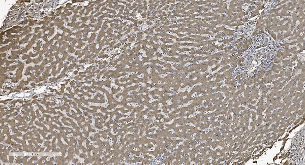
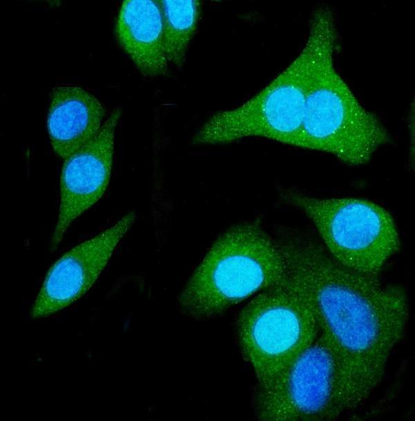
The catalog shows SHMT1 IHC in a human paraffin-embedded liver cancer section (M02944 image caption) and IF/ICC in A431 cells (A02944-1 image caption); both antibodies list Human, Monkey, Mouse and Rat reactivity (catalog).

Real IHC data IHC analysis of SHMT1 using anti-SHMT1 antibody (M02944). SHMT1 was detected in paraffin-embedded section of human liver cancer tissue. Heat mediated antigen retrieval was performed in EDTA buffer (pH8.0, epitope retrieval solution). The tissue section was blocked with 10% goat serum. The tissue section was then incubated with 2μg/ml mouse anti-SHMT1 Antibody (M02944) overnight at 4°C. Biotinylated goat anti-mouse IgG was used as secondary antibody and incubated for 30 minutes at 37°C. The tissue section was developed using Strepavidin-Biotin-Complex (SABC) (Catalog # SA1021) with DAB as the chromogen.
Anti-SHMT1 Antibody ® (monoclonal, 9C7)
Cat # M02944
Real IF data IF analysis of SHMT1 using anti-SHMT1 antibody (A02944-1). SHMT1 was detected in immunocytochemical section of A431 cells. Enzyme antigen retrieval was performed using IHC enzyme antigen retrieval reagent (AR0022) for 15 mins. The cells were blocked with 10% goat serum. And then incubated with 5μg/mL rabbit anti-SHMT1 Antibody (A02944-1) overnight at 4°C. DyLight®594 Conjugated Goat Anti-Rabbit IgG (BA1142) was used as secondary antibody at 1:100 dilution and incubated for 30 minutes at 37°C. The section was counterstained with DAPI. Visualize using a fluorescence microscope and filter sets appropriate for the label used.
Anti-SHMT1 Antibody ®
Cat # A02944-1

M02944 is listed for IHC and IF/ICC, with its own IHC figure showing a human paraffin-embedded liver cancer section (catalog; M02944 image caption). A02944-1 is listed for IHC and IF/ICC, with its own IF figure showing A431 cells (catalog; A02944-1 image caption).

Which to pick: For tissue IHC, choose the mouse monoclonal M02944 (clone 9C7): its own caption documents chromogenic staining of a paraffin-embedded human liver cancer section; the fixative is unreported (catalog; M02944 image caption). For IF/ICC, choose the rabbit A02944-1, whose own figure documents staining in A431 cells at 5 μg/mL (catalog; A02944-1 image caption). For cross-species work, both list Human, Monkey, Mouse and Rat reactivity, while the cited figures show human samples (catalog; M02944 image caption; A02944-1 image caption).

Each figure is that product's own IHC / IF validation image from its datasheet.

References

  1. UniProt Consortium. UniProt entry P34896 (GLYC_HUMAN, Serine hydroxymethyltransferase, cytosolic).
  2. Human Protein Atlas. SHMT1 tissue IHC expression (reliability: Enhanced).
  3. Human Protein Atlas. SHMT1 subcellular location (ICC-IF): Mainly localized to the cytosol. In addition localized to the nucleoplasm..
  4. Human Protein Atlas. SHMT1 antibody validation summary (2 antibodies).
  5. HOXD8 suppresses renal cell carcinoma growth by upregulating SHMT1 expression. Cancer science 2023 — PMC10728000.
  6. SHMT1, an amino acid metabolism-related gene, is an independent prognostic biomarker in laryngeal squamous cell carcinoma. BMC cancer 2025 — PMC12683878.
  7. Integrative bioinformatics approach identifies novel drug targets for hyperaldosteronism, with a focus on SHMT1 as a promising therapeutic candidate. Scientific reports 2025 — PMC11724956.
  8. SHMT1 inhibits the metastasis of HCC by repressing NOX1-mediated ROS production. Journal of experimental & clinical cancer research : CR 2019 — PMC6373090.
  9. PubMed PMID:8505317 — UniProt-cited evidence.
  10. PubMed PMID:9056951 — UniProt-cited evidence.
  11. PubMed PMID:14702039 — UniProt-cited evidence.