This website uses cookies to ensure you get the best experience on our website.


- Table of Contents
Plan chromogenic SHOC2 IHC on paraffin sections using cytoplasmic staining as the tissue reference pattern (HPA tissue IHC). This guide covers control selection, fixation consistency and the catalog antibody A07214-1 starting range of 2–5 μg/mL (datasheet).

Expected localisation, validated protocols, controls and antibodies — the at-a-glance facts below, then the full design guide.
| Expected localisation | Cytoplasmic tissue staining (HPA tissue IHC); nuclear entry after growth factor stimulation (UniProt) | |
| Staining pattern | Cytoplasmic; some glandular and neuronal cells stain strongly (HPA tissue IHC) | |
| Antigen retrieval | Tris-EDTA pH 9.0 HIER, 95–98 °C, 20 min (rule: nuclear antigen) | |
| Positive control | Appendix+4 more · see all | |
| Negative control | Adipose tissue+4 more · see all |
| Fixation | Keep fixation consistent across sections. (standard IHC practice; not target-specific) | |
| Caveat | Tissue staining has medium consistency with RNA data (HPA tissue IHC) | |
| Regulation | Growth factors shift nuclear signal (UniProt) | |
| Isoform / epitope | 2 isoforms; no extracellular segment; epitope map unknown (UniProt) |
The catalog antibody protocol is accompanied by published SHOC2 IHC methods for breast and pancreatic cancer tissue (PMC6947812; PMC6503676).
| Sample | Tissue sections; selected-image fixative not specified (standard IHC workflow) |
| Fixation | Image fixative and duration unreported (datasheet A07214); verify before use. |
| Sectioning | 4–5 µm sections on charged slides (standard) |
| Deparaffinisation | Xylene, graded ethanol series to water (standard) |
| Antigen retrieval | Heat-induced epitope retrieval in Tris-EDTA buffer, pH 9.0, 20 min at 95–98 °C (standard rule: nuclear antigen) |
| Peroxidase block | 3% H2O2, 10 min, room temperature (standard) |
| Blocking | 10% normal serum of the secondary host, 30 min, room temperature (standard) |
| Primary antibody | Rabbit anti-SHOC2, 5 μg/mL (datasheet A07214) |
| Primary incubation | Overnight at 4 °C (standard) |
| Detection | HRP-polymer secondary, DAB chromogen 5–10 min (standard) |
| Counterstain | Hematoxylin, blue, dehydrate and mount (standard) |
| Expected result | SHOC2-positive staining in glandular cells of appendix (HPA tissue IHC: High). HPA tissue profile: Cytoplasmic expression in a majority of tissues. No signal in the no-primary control. |
SHOC2 should appear mainly in the cytoplasm of many tissue cell types, with nuclear staining also plausible because it can move into the nucleus after growth factor stimulation (HPA: tissue IHC profile; UniProt Q9UQ13: subcellular location). High staining is reported in several glandular, neuronal and squamous epithelial cell populations (HPA: tissue IHC). SHOC2 has no transmembrane segment (UniProt Q9UQ13: topology). HPA rates its tissue staining Approved, with medium consistency against RNA data (HPA: tissue IHC reliability).
| Cytoplasmic chromogenic staining in colon glandular cells or cerebral cortex neurons. | This matches cell populations scored High in those tissues and the predominant cytoplasmic tissue pattern (HPA: tissue IHC). Judge the stained cells and their compartment together; broad tissue expression alone does not make every stained cell a convincing positive (HPA: low RNA tissue specificity; general IHC practice). |
| Nuclear staining accompanies a cytoplasmic signal. | Nuclear localization is biologically plausible: SHOC2 translocates from cytoplasm to nucleus after growth factor stimulation (UniProt Q9UQ13: subcellular location). HPA ICC-IF also places it in the nucleoplasm, but that observation does not establish an expected nuclear intensity for paraffin IHC (HPA: subcellular ICC-IF). |
| A sharp membrane-only rim or extracellular deposit dominates the slide. | Treat this as a compartment mismatch requiring investigation, rather than a confident SHOC2 pattern: the reported locations are cytoplasm and nucleus, and SHOC2 lacks a transmembrane segment (UniProt Q9UQ13: subcellular location and topology). Compare a positive tissue and detection controls before assigning cause (general IHC practice). |
| Strong staining appears in adipocytes or bronchial respiratory epithelial cells. | These cell populations were Not detected in the cited HPA tissue images (HPA: adipose tissue adipocytes; bronchus respiratory epithelial cells). Consider cross-reactivity or endogenous detection activity, while remembering that an HPA nondetection is an observation under its assay conditions, not proof that expression is impossible (general IHC practice; HPA: tissue IHC). |
| Uniform haze covers cells and stroma, or a known-positive tissue has no visible signal. | Haze without cell-specific contrast is background; absence from a High population such as colon glandular cells suggests an assay failure or sample-dependent issue (general IHC practice; HPA: colon glandular cells High). Neither observation alone establishes whether SHOC2 is absent from the specimen (general IHC practice). |
| Cell population and tissue context | HPA scores appendix, colon, duodenum and gallbladder glandular cells, esophageal squamous epithelial cells, and caudate, cerebral cortex and hippocampal neurons High (HPA: tissue IHC). Its low RNA tissue specificity and cytoplasmic staining in a majority of tissues make cell-level interpretation more useful than a simple positive or negative tissue label (HPA: tissue IHC profile and RNA specificity). |
| Growth factor state and compartment | SHOC2 can move from cytoplasm to nucleus after growth factor stimulation (UniProt Q9UQ13: subcellular location). Do not reject a nuclear component solely because the tissue IHC profile emphasizes cytoplasm; do investigate a membrane-only pattern (HPA: tissue IHC profile; UniProt Q9UQ13: topology). |
| Antibody evidence | HPA048134 is Approved for IHC, while HPA009164 is Supported for ICC and has no listed IHC status (HPA: antibody validation). Those labels refer to different applications; ICC support alone does not validate an antibody for paraffin-section interpretation (HPA: antibody validation; general IHC practice). |
| Isoforms and processing | UniProt lists two SHOC2 isoforms, a full-length chain spanning residues 1–582, no signal peptide or propeptide, and no annotated glycosylation sites (UniProt Q9UQ13: isoforms and processing). The supplied evidence gives no epitope map, so it cannot establish whether a given antibody detects both isoforms (UniProt Q9UQ13: isoforms; HPA: antibody validation). |
| IF/ICC Q&A | Q: Can an IF/ICC nuclear signal be expected? A: Yes; HPA reports approved nucleoplasm and supported cytosol localization, and UniProt reports stimulus-dependent nuclear translocation (HPA: subcellular ICC-IF; UniProt Q9UQ13: subcellular location). These observations inform localization, without specifying a paraffin IHC detection condition. |
| Situation | Likely cause | Next action |
|---|---|---|
| No signal in colon glandular cells or hippocampal neurons. | Both are High in HPA tissue IHC; a blank result may reflect a failed staining run or specimen-specific conditions (HPA: colon glandular cells and hippocampal neurons High; general IHC practice). | Confirm that a concurrently processed positive section stains, then check the primary-antibody step, detection reagents and the assay's validated retrieval settings (general IHC practice). No SHOC2-specific retrieval or fixation sensitivity is supplied. |
| Diffuse brown haze obscures cell boundaries. | Background can arise from nonspecific binding or endogenous chromogenic detection activity (general IHC practice). HPA's cytoplasmic tissue profile does not support interpreting uniform haze as SHOC2 (HPA: tissue IHC profile). | Inspect a no-primary control and the detection system; adjust blocking, washing or detection conditions according to the validated assay (general IHC practice). |
| Signal is restricted to a crisp cell-surface outline. | A membrane-only pattern conflicts with SHOC2's reported cytoplasmic and nuclear locations and lack of a transmembrane segment (UniProt Q9UQ13: subcellular location and topology). | Compare cell-level staining in an HPA High reference tissue and a no-primary control; review nonspecific signal before calling the rim positive (HPA: tissue IHC; general IHC practice). |
| Adipocytes or bronchial respiratory epithelial cells stain strongly. | HPA reports Not detected for those populations under its tissue IHC conditions (HPA: adipose tissue adipocytes; bronchus respiratory epithelial cells). Cross-reactivity or detection-system activity is possible (general IHC practice). | Check whether the signal persists in a no-primary control and whether an HPA High cell population stains with the expected compartment pattern (general IHC practice; HPA: tissue IHC profile). |
| A nuclear component appears alongside cytoplasmic staining. | Nuclear translocation after growth factor stimulation is documented, and HPA ICC-IF reports nucleoplasmic localization (UniProt Q9UQ13: subcellular location; HPA: subcellular ICC-IF). | Score nuclear and cytoplasmic compartments separately and compare the cell-specific pattern with controls; do not classify a nuclear component alone as artefact (general IHC practice; UniProt Q9UQ13: subcellular location). |
| An ICC-supported antibody gives uncertain paraffin IHC staining. | HPA009164 is Supported for ICC but has no listed IHC status; HPA048134 is Approved for IHC (HPA: antibody validation). | Base paraffin IHC interpretation on IHC-specific validation and an appropriate positive section; do not transfer the ICC validation label to IHC (HPA: antibody validation; general IHC practice). |
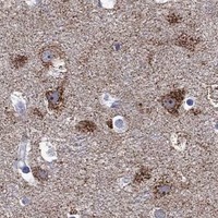


Comprehensive Human Protein Atlas IHC scoring per tissue (reliability: Approved — Medium consistency between antibody staining and RNA expression data.). Rows are taken directly from the HPA tissue chart — click any row's HPA link to view the source.
| Tissue | Cell type | Level | Evidence | Source |
|---|---|---|---|---|
| Appendix | Glandular cells | High | Protein (IHC) | HPA → |
| Caudate | Neuronal cells | High | Protein (IHC) | HPA → |
| Cerebral cortex | Neuronal cells | High | Protein (IHC) | HPA → |
| Colon | Glandular cells | High | Protein (IHC) | HPA → |
| Duodenum | Glandular cells | High | Protein (IHC) | HPA → |
| Tissue | Cell type | Level | Evidence | Source |
|---|---|---|---|---|
| Adipose tissue | Adipocytes | Not detected | Protein (IHC) | HPA → |
| Bone marrow | Hematopoietic cells | Not detected | Protein (IHC) | HPA → |
| Breast | Adipocytes | Not detected | Protein (IHC) | HPA → |
| Bronchus | Respiratory epithelial cells | Not detected | Protein (IHC) | HPA → |
| Cervix | Glandular cells | Not detected | Protein (IHC) | HPA → |
Troubleshoot SHOC2 staining in paraffin section IHC by checking retrieval, cellular localisation, controls and scoring before interpreting signal.
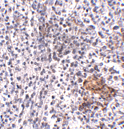
IHC images show human spleen (A07214 image caption), human liver and liver cancer, and mouse and rat brain (A07214-1 image captions). No IF images are supplied (catalog IF image alts).
A07214 is the sole rendered SKU; its IHC image shows human spleen at 5 μg/mL (A07214 image caption). It is listed for IHC-P and human, mouse and rat reactivity, with IHC validation specified for human samples (A07214 catalog).
Which to pick: For human tissue IHC-P, choose A07214 based on its human spleen image; its caption does not report the fixative (A07214 application list and image caption). For paraffin sections across species, A07214-1 has IHC images of mouse and rat brain and human liver and liver cancer; its captions report EDTA retrieval at pH 8.0 and primary antibody at 2 μg/mL, but do not report the fixative, and neither rabbit antibody has a clone designation supplied (A07214-1 image captions; catalog host and clone fields). Neither SKU has IF/ICC listed or an IF image, so there is no catalog-supported IF/ICC choice (catalog applications and IF image alts).