This website uses cookies to ensure you get the best experience on our website.


- Table of Contents
Plan SHQ1 chromogenic IHC on paraffin sections using the IHC-validated antibody A10185-1 (datasheet A10185-1). Compare cytoplasmic staining in reported positive cells with controls, and interpret the tissue profile cautiously because its reliability is uncertain (HPA tissue IHC).

Expected localisation, validated protocols, controls and antibodies — the at-a-glance facts below, then the full design guide.
| Expected localisation | Cytoplasmic in tissue (HPA tissue IHC); nucleoplasmic localization is possible (UniProt) | |
| Staining pattern | Cytoplasmic staining in selected squamous, exocrine and Leydig cells (HPA tissue IHC) | |
| Antigen retrieval | EDTA pH 8.0 HIER, heat-mediated (datasheet A10185-1) | |
| Positive control | Oral mucosa+4 more · see all | |
| Negative control | Adipose tissue+4 more · see all |
| Fixation | Keep fixation consistent across sections (standard IHC practice; not target-specific); Matched tissue-IHC evidence does not establish the fixation claim. Validate the specimen-specific method before use. (selected-SKU IHC image A10185-1) | |
| Caveat | Staining has low consistency with RNA expression (HPA tissue IHC) | |
| Regulation | Expression regulation is not annotated (UniProt) | |
| Isoform / epitope | 2 isoforms; epitope differences are unspecified (UniProt) |
The catalog antibody has an IHC-P protocol (datasheet A10185-1). The two published SHQ1 IHC protocols below cover mouse spleen sections (PMC6189109: Immunohistochemistry) and tumor/tissue sections (PMC7286909: Immunohistochemistry).
| Sample | Paraffin-embedded human breast cancer tissue; fixative not specified (datasheet A10185-1) |
| Fixation | Image fixative and duration unreported (datasheet A10185-1); verify before use. |
| Sectioning | 4–5 µm sections on charged slides (standard) |
| Deparaffinisation | Xylene, graded ethanol series to water (standard) |
| Antigen retrieval | Heat retrieval: EDTA pH 8.0 (datasheet A10185-1); 20 min, 95–100 °C (standard) |
| Peroxidase block | 3% H2O2, 10 min, room temperature (standard) |
| Blocking | 10% goat serum (datasheet A10185-1) |
| Primary antibody | Rabbit anti-SHQ1, 2-5 μg/ml (datasheet A10185-1) |
| Primary incubation | Overnight at 4 °C (datasheet A10185-1) |
| Detection | HRP-conjugated secondary, DAB chromogen (datasheet A10185-1) |
| Counterstain | Hematoxylin, blue, dehydrate and mount (standard) |
| Expected result | SHQ1-positive staining in squamous epithelial cells of oral mucosa (HPA tissue IHC: High). HPA tissue profile: Cytoplasmic expression in a few tissues. No signal in the no-primary control. |
SHQ1 is a soluble protein found in the cytosol and nucleoplasm; it has no transmembrane segment (UniProt Q6PI26). In tissue IHC, expect mainly cytoplasmic staining, strongest in oral squamous epithelial cells, pancreatic exocrine glandular cells, and testicular Leydig cells (HPA: High; cytoplasmic expression in a few tissues). Treat the tissue pattern as provisional: HPA rates its IHC evidence Uncertain because staining and RNA expression have low consistency (HPA: Uncertain).
| Distinct cytoplasmic staining in oral squamous epithelium, pancreatic exocrine cells, or Leydig cells. | This matches HPA's reported High staining in those cell types and its mainly cytoplasmic tissue profile (HPA: tissue IHC). Score the relevant cells and compartment; HPA's Uncertain reliability means a matching pattern alone does not establish antibody specificity (HPA: Uncertain). |
| Nucleoplasmic staining, or staining confined to nucleoli, Cajal bodies, or cell membranes. | Nucleoplasmic signal can be plausible: it is supported in ICC-IF and described by UniProt (HPA: subcellular; UniProt Q6PI26). UniProt reports nucleolar and Cajal body exclusion; membrane staining conflicts with its soluble localization and lack of a transmembrane segment (UniProt Q6PI26). Investigate those discordant patterns before scoring them as SHQ1. |
| Strong staining in adipocytes or bone marrow hematopoietic cells while expected positive cells are weak. | HPA reports SHQ1 as Not detected in those cells (HPA: tissue IHC). Cross-reactivity or endogenous detection activity is possible in chromogenic IHC (general IHC practice). Because HPA rates its tissue IHC Uncertain, use these cells as comparison points, not definitive biological negatives (HPA: Uncertain). |
| Diffuse color across several cell types, tissue spaces, or the whole section, without a clear cellular compartment. | That distribution does not resemble HPA's cytoplasmic tissue profile or its supported nucleoplasmic ICC-IF localization (HPA: tissue IHC; HPA: subcellular). Nonspecific binding, detection background, or excessive chromogen development are possible causes (general IHC practice). Compare with a no-primary control. |
| No discernible signal in oral squamous epithelium, pancreatic exocrine cells, or Leydig cells. | Those are HPA's High-staining examples (HPA: tissue IHC). Check whether the expected cells are present, then review retrieval, primary antibody application, and detection controls (general IHC practice). An absent signal does not by itself resolve SHQ1 expression because the reference IHC pattern is rated Uncertain (HPA: Uncertain). |
| Tissue reference strength | HPA calls SHQ1 tissue IHC Uncertain because antibody staining has low consistency with RNA expression; its High, Medium, Low, and Not detected entries are observed patterns, not validated expression thresholds (HPA: tissue IHC). Compare cell types within the section and seek an independent specificity check before assigning biological meaning. |
| Compartment depends on assay | HPA tissue IHC describes cytoplasmic expression, while HPA ICC-IF supports nucleoplasm as the main location (HPA: tissue IHC; HPA: subcellular). UniProt describes cytosol and nucleoplasm, possible shuttling, and granular nucleoplasmic distribution (UniProt Q6PI26). Interpret a nuclear component in light of the assay and antibody controls. |
| Topology and processing | SHQ1 has no transmembrane segment, signal peptide, propeptide, or annotated glycosylation sites; its annotated chain spans residues 1–577 (UniProt Q6PI26). These annotations support evaluating intracellular staining and offer no basis for expecting a membrane rim or secreted deposit. |
| Isoforms and antibody evidence | UniProt lists two SHQ1 isoforms; the supplied record gives no antibody epitope or isoform coverage (UniProt Q6PI26). HPA lists one rabbit polyclonal antibody, HPA042792, as IHC Uncertain and ICC Supported (HPA: antibodies). Do not infer that an IHC pattern detects both isoforms or that ICC support validates tissue IHC. |
| IF/ICC Q&A: what should I expect? | Mainly nucleoplasmic signal is supported in the HPA ICC-IF record, with images listed for A-431, U-251MG, and U2OS (HPA: subcellular). UniProt also allows cytosolic localization and describes nucleolar and Cajal body exclusion (UniProt Q6PI26). This ICC-IF observation does not set an IHC protocol or an expected IHC nuclear intensity. |
| Situation | Likely cause | Next action |
|---|---|---|
| Positive reference tissue is blank. | The expected cells may be absent, or the IHC detection workflow may have failed (general IHC practice); HPA reports High staining in the specified cells but rates tissue IHC Uncertain (HPA: tissue IHC). | Confirm the cell type on the counterstained section; review retrieval, primary application, and detection controls using the IHC-validated antibody's instructions (general IHC practice). |
| The entire section has weak, flat color. | Background may obscure compartment-specific signal, or chromogen development may be excessive (general IHC practice). This differs from HPA's cytoplasmic tissue profile (HPA: tissue IHC). | Compare a no-primary control; review blocking, washing, primary dilution, and detection timing within the established IHC workflow (general IHC practice). |
| Color appears mainly at cell membranes or outside cells. | That pattern conflicts with SHQ1's cytosolic and nucleoplasmic localization and lack of a transmembrane segment (UniProt Q6PI26). Nonspecific or detection-related staining is possible (general IHC practice). | Inspect cell boundaries with the counterstain and compare the no-primary control; require a reproducible intracellular pattern before assigning SHQ1 staining (general IHC practice; UniProt Q6PI26). |
| Nuclear color is present, but cytoplasmic IHC color is faint. | A nuclear component is biologically plausible, although HPA's tissue IHC profile is cytoplasmic and its supported nucleoplasmic evidence comes from ICC-IF (UniProt Q6PI26; HPA: tissue IHC; HPA: subcellular). | Record nuclear and cytoplasmic scores separately; check whether color is nucleoplasmic rather than nucleolar, and interpret tissue IHC conservatively given its Uncertain rating (UniProt Q6PI26; HPA: Uncertain). |
| Cells listed as Not detected stain strongly. | Unexpected staining could reflect cross-reactivity or endogenous detection activity (general IHC practice). HPA lists adipocytes and bone marrow hematopoietic cells as Not detected, with Uncertain overall tissue IHC reliability (HPA: tissue IHC). | Compare a no-primary control and a High-staining reference cell type; investigate persistent discordance before calling the cells SHQ1-positive (general IHC practice; HPA: tissue IHC). |
| Runs give different intensity in the same reference cell type. | Variation in retrieval, antibody dilution, or chromogen development can change apparent IHC intensity (general IHC practice). HPA's High designation is an observed tissue category, not a quantitative acceptance limit (HPA: tissue IHC). | Keep the IHC workflow and scoring criteria consistent across runs, include the same reference cell type, and compare compartment and background as well as intensity (general IHC practice; HPA: tissue IHC). |




Comprehensive Human Protein Atlas IHC scoring per tissue (reliability: Uncertain — Low consistency between antibody staining and RNA expression data. Pending external verification.). Rows are taken directly from the HPA tissue chart — click any row's HPA link to view the source.
| Tissue | Cell type | Level | Evidence | Source |
|---|---|---|---|---|
| Oral mucosa | Squamous epithelial cells | High | Protein (IHC) | HPA → |
| Pancreas | Exocrine glandular cells | High | Protein (IHC) | HPA → |
| Testis | Leydig cells | High | Protein (IHC) | HPA → |
| Cervix | Squamous epithelial cells | Medium | Protein (IHC) | HPA → |
| Colon | Glandular cells | Medium | Protein (IHC) | HPA → |
| Tissue | Cell type | Level | Evidence | Source |
|---|---|---|---|---|
| Adipose tissue | Adipocytes | Not detected | Protein (IHC) | HPA → |
| Adrenal gland | Glandular cells | Not detected | Protein (IHC) | HPA → |
| Appendix | Glandular cells | Not detected | Protein (IHC) | HPA → |
| Bone marrow | Hematopoietic cells | Not detected | Protein (IHC) | HPA → |
| Bronchus | Respiratory epithelial cells | Not detected | Protein (IHC) | HPA → |
Use the catalog antibody’s paraffin-section IHC conditions as a starting point, then assess compartment, cell type, and controls before scoring SHQ1 staining.
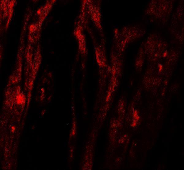
Anti-SHQ1 antibodies have IHC images from human breast cancer, renal cancer and spleen, and mouse kidney paraffin sections (A10185-1 image captions); IF images show human heart tissue and A549 cells (A10185 and A10185-1 image captions).
A10185-1 lists IHC and IF/ICC applications for human, mouse and rat samples, with IHC images from human breast cancer, renal cancer and spleen, and mouse kidney paraffin sections, plus an IF image from A549 cells (catalog applications and reactivity; A10185-1 image captions). A10185 lists human IF and shows an IF image from human heart tissue; IHC is not listed for this SKU (catalog applications and reactivity; A10185 image caption).
Which to pick: Choose A10185-1 for tissue IHC: its paraffin-section images use heat retrieval in EDTA at pH 8.0 and 2 μg/ml primary antibody; the fixative is unreported (A10185-1 IHC image captions). For IF/ICC, choose A10185-1 when ICC is needed (catalog applications; A549 IF image caption), or A10185 for human tissue IF (catalog reactivity; human heart IF image caption). For work across species, A10185-1 lists human, mouse and rat reactivity, with a mouse kidney paraffin-section IHC image; A10185 lists human reactivity (catalog reactivity; A10185-1 IHC image caption).