This website uses cookies to ensure you get the best experience on our website.
- Table of Contents
3 Citations 11 Q&As
Facts about NAD-dependent protein deacetylase sirtuin-2.

Plays a major role in the control of cell cycle progression and genomic stability. Functions in the antephase checkpoint preventing precocious mitotic entry in response to microtubule anxiety agents, and hence allowing appropriate inheritance of chromosomes.
| Human | |
|---|---|
| Gene Name: | SIRT2 |
| Uniprot: | Q8IXJ6 |
| Entrez: | 22933 |

| Belongs to: |
|---|
| sirtuin family |

EC 3.5.1; EC 3.5.1.-; FLJ35621; FLJ37491; S.cerevisiae, homolog) 2; sirtuin 2; sirtuin type 2
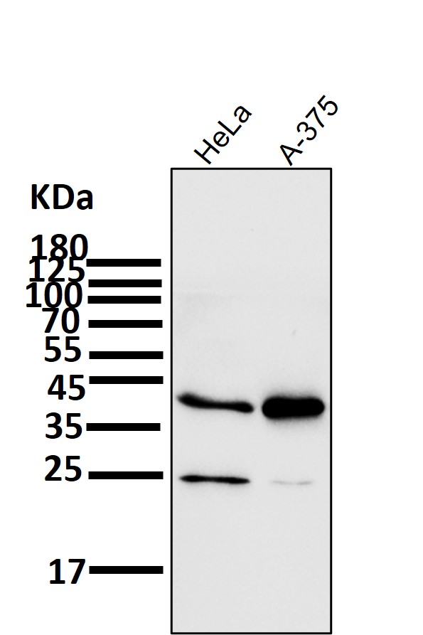
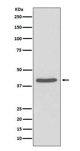
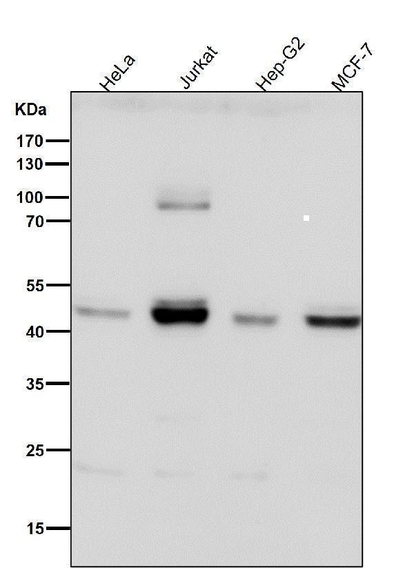
Mass (kDA):
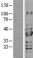
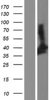
43.182 kDA

| Human | |
|---|---|
| Location: | 19q13.2 |
| Sequence: | 19; NC_000019.10 (38878555..38899862, complement) |
Isoform 1 is expressed in heart, liver and skeletal muscle, weakly expressed in the cortex. Isoform 2 is strongly expressed in the cortex, weakly expressed in heart and liver. Weakly expressed in several malignancies including breast, liver, brain, kidney and prostate cancers compared to normal tissues. Weakly expressed in glioma cell lines compared to normal brain tissues (at protein level). Widely expressed. Highly expressed in heart, brain and skeletal muscle, while it is weakly expressed in placenta and lung. Down-regulated in many gliomas suggesting that it may act as a tumor suppressor gene in human gliomas possibly through the regulation of microtubule network.
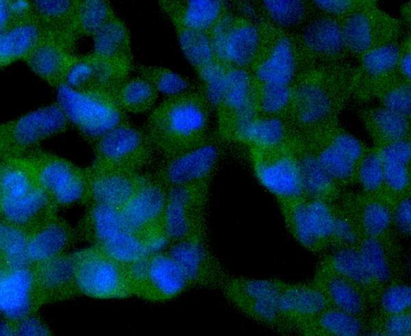
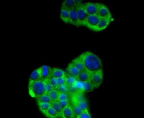
Nucleus. Cytoplasm, perinuclear region. Cytoplasm. Cytoplasm, cytoskeleton. Cytoplasm, cytoskeleton, microtubule organizing center, centrosome. Cytoplasm, cytoskeleton, microtubule organizing center, centrosome, centriole. Cytoplasm, cytoskeleton, spindle. Midbody. Chromosome. Perikaryon. Cell projection. Cell projection, growth cone. Myelin membrane. Deacetylates FOXO3 in the cytoplasm. Colocalizes with PLP1 in internodal regions, at paranodal axoglial junction and Schmidt-Lanterman incisures of myelin sheat. Colocalizes with CDK5R1 in the perikaryon, neurites and growth cone of hippocampal n


PMID: 10381378 by Frye R.A.; Characterization of five human cDNAs with homology to the yeast SIR2 gene: Sir2-like proteins (sirtuins) metabolize NAD and may have protein ADP-ribosyltransferase activity.
PMID: 10393250 by Afshar G., et al. Characterization of a human gene with sequence homology to Saccharomyces cerevisiae SIR2.