This website uses cookies to ensure you get the best experience on our website.


- Table of Contents
Plan SIRT4 paraffin-section IHC using placental trophoblasts as a high-staining reference (HPA tissue IHC). Score cytoplasmic signal by cell type, with lymph-node germinal-center cells as a reported unstained comparison (HPA tissue IHC).

Expected localisation, validated protocols, controls and antibodies — the at-a-glance facts below, then the full design guide.
| Expected localisation | Cytoplasmic in tissue IHC (HPA tissue IHC); mitochondrial matrix (UniProt) | |
| Staining pattern | Strong cytoplasmic staining in placental trophoblasts (HPA tissue IHC) | |
| Antigen retrieval | Citrate pH 6 HIER, heat-mediated (datasheet A03764-1) | |
| Positive control | Placenta+4 more · see all | |
| Negative control | Lymph node+4 more · see all |
| Fixation | Matched tissue-IHC evidence does not establish the fixation claim. Validate the specimen-specific method before use. (selected-SKU IHC image A03764-1); keep fixation consistent. (standard IHC practice; not target-specific) | |
| Caveat | Antibody staining and RNA show medium consistency (HPA tissue IHC) | |
| Regulation | mTORC1 represses SIRT4 expression (UniProt) | |
| Isoform / epitope | No listed isoforms; residues 1–28 precede the mature chain (UniProt) |
The catalog antibody protocol is paired with 4 published SIRT4 IHC protocols covering bladder, prostate, colorectal, and neuroblastoma tissue (PMC10257844; PMC9288510; PMC4522635; PMC6234993).
| Sample | Paraffin-embedded human ovary cancer tissue; fixative not specified (datasheet A03764-1) |
| Fixation | Image fixative and duration unreported (datasheet A03764-1); verify before use. |
| Sectioning | 4–5 µm sections on charged slides (standard) |
| Deparaffinisation | Xylene, graded ethanol series to water (standard) |
| Antigen retrieval | Heat retrieval: Citrate pH 6, 20 min (datasheet A03764-1) |
| Peroxidase block | 3% H2O2, 10 min, room temperature (standard) |
| Blocking | 10% goat serum (datasheet A03764-1) |
| Primary antibody | Rabbit anti-SIRT4, 1μg/ml (datasheet A03764-1) |
| Primary incubation | Overnight at 4 °C (datasheet A03764-1) |
| Detection | Streptavidin-biotin complex (SABC), DAB chromogen (datasheet A03764-1) |
| Counterstain | Hematoxylin, blue, dehydrate and mount (standard) |
| Expected result | SIRT4-positive staining in trophoblastic cells of placenta (HPA tissue IHC: High). HPA tissue profile: Cytoplasmic expression in several cell types, most abundant in placenta. No signal in the no-primary control. |
SIRT4 resides in the mitochondrial matrix and has no transmembrane segment (UniProt Q9Y6E7). In paraffin IHC, expect cytoplasmic staining, strongest in placental trophoblastic cells and round or early spermatids (HPA: High; cytoplasmic profile). HPA rates its tissue IHC evidence Enhanced, with medium consistency between staining and RNA data (HPA: tissue IHC).
| Cytoplasmic stain in placental trophoblastic cells or round or early spermatids. | This matches HPA's High staining in those cells and its cytoplasmic profile (HPA: tissue IHC). Mitochondrial matrix residence supports a cytoplasmic location, though chromogenic IHC may not resolve individual mitochondria (UniProt Q9Y6E7; general IHC practice). |
| Predominantly nuclear or surface outlining stain, with little cytoplasmic signal. | This conflicts with the expected mitochondrial matrix location and HPA cytoplasmic profile (UniProt Q9Y6E7; HPA: tissue IHC). Treat the pattern as suspect and review controls and morphology before assigning it to SIRT4 (general IHC practice). |
| Strong stain in an HPA-listed undetected cell population, such as lymph-node germinal center cells. | HPA reports SIRT4 not detected in those cells (HPA: tissue IHC). Assess antibody specificity or endogenous chromogen activity before interpreting the signal; a single discordant site does not prove cross-reactivity (general IHC practice). |
| Diffuse color across cells, stroma, or the whole section, obscuring cell boundaries. | This is difficult to reconcile with HPA's cell-dependent cytoplasmic pattern (HPA: tissue IHC). Background from detection reagents or inadequate washing is possible; compare a negative reagent control before scoring (general IHC practice). |
| No stain in placental trophoblastic cells or round or early spermatids. | These are HPA High reference populations, so their absence raises a run-performance or specimen concern (HPA: tissue IHC). Check section integrity and controls before concluding the sample lacks SIRT4 (general IHC practice). |
| Cell type and reference tissue | Use placental trophoblastic cells or round or early spermatids as high-staining references (HPA: tissue IHC). Medium glandular staining in appendix, breast, cervix, and colon offers a less intense comparison (HPA: tissue IHC). Compare the named cells, not every cell in the tissue. |
| Subcellular resolution | SIRT4 is a mitochondrial matrix protein without a transmembrane segment (UniProt Q9Y6E7). HPA describes tissue staining as cytoplasmic (HPA: tissue IHC). A resolved mitochondrial pattern can support localisation, but routine chromogenic IHC need not show distinct puncta (general IHC practice). |
| Protein processing and epitope choice | UniProt annotates a mature chain at residues 29–314 and a sirtuin-type domain at 37–314 (UniProt Q9Y6E7). If an antibody epitope is known, check whether it lies in the retained chain; no epitope is supplied here, so no antibody-specific effect can be predicted. |
| Antibody evidence and limits | HPA lists HPA029692 as IHC Enhanced and HPA029691 as IHC Approved (HPA: antibodies). The tissue profile's Enhanced rating has medium staining-to-RNA consistency (HPA: tissue IHC); neither rating guarantees specificity in a new specimen or detection setup (general IHC practice). |
| IF/ICC expectation? | UniProt's mitochondrial matrix annotation suggests intracellular, potentially mitochondrial fluorescence after appropriate cellular access (UniProt Q9Y6E7; general IF practice). HPA supplies no main IF location or ICC-IF images for SIRT4, so that appearance remains a prediction rather than an HPA-observed IF pattern (HPA: subcellular). |
| Situation | Likely cause | Next action |
|---|---|---|
| A high-reference tissue has no detectable stain. | A failed detection run, unsuitable antibody conditions, or compromised section is possible (general IHC practice); HPA reports High staining in trophoblastic cells and round or early spermatids (HPA: tissue IHC). | Inspect the named cell population and run controls, then verify the catalog antibody's IHC-P instructions and detection reagents before interpreting a biological negative (general IHC practice). |
| Only nuclei or cell borders stain strongly. | The compartment conflicts with mitochondrial matrix localisation and the HPA cytoplasmic tissue profile (UniProt Q9Y6E7; HPA: tissue IHC). | Review morphology and reagent controls; repeat with an independently validated IHC antibody if available before accepting the compartment assignment (general IHC practice; HPA: antibodies). |
| A purported negative population stains strongly. | Cross-reactivity or endogenous detection activity is possible (general IHC practice). HPA lists germinal center cells, follicle cells, and skin fibroblasts as not detected, but its survey does not establish universal biological absence (HPA: tissue IHC). | Check the exact cell type against HPA, assess a negative reagent control, and compare staining with a high-reference population in the same run (HPA: tissue IHC; general IHC practice). |
| The section shows widespread haze or precipitate. | Non-specific detection, insufficient washing, or deposited chromogen can obscure the cell-level pattern (general IHC practice). HPA's reported pattern is cytoplasmic in several cell types (HPA: tissue IHC). | Examine a negative reagent control, inspect chromogen deposits, and adjust general blocking, washing, or detection conditions as indicated by the controls (general IHC practice). |
| A medium or low reference looks weaker than placenta. | Different intensity is expected: HPA reports High trophoblastic staining, Medium glandular staining in several tissues, and Low signal in selected cells (HPA: tissue IHC). | Score the specified cells against tissue-appropriate expectations and the run's positive control; avoid requiring placental intensity in every tissue (HPA: tissue IHC; general IHC practice). |
| The proposed IF pattern is used to validate the IHC result. | HPA supplies no SIRT4 ICC-IF images or main IF location; its cytoplasmic tissue profile comes from IHC (HPA: subcellular; HPA: tissue IHC). | Treat mitochondrial fluorescence as a localisation hypothesis from UniProt, and judge the paraffin IHC slide against HPA's cell-specific tissue pattern and IHC controls (UniProt Q9Y6E7; HPA: tissue IHC; general IHC practice). |


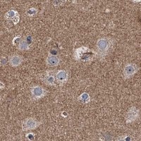


Comprehensive Human Protein Atlas IHC scoring per tissue (reliability: Enhanced — Medium consistency between antibody staining and RNA expression data.). Rows are taken directly from the HPA tissue chart — click any row's HPA link to view the source.
| Tissue | Cell type | Level | Evidence | Source |
|---|---|---|---|---|
| Placenta | Trophoblastic cells | High | Protein (IHC) | HPA → |
| Testis | Round or early spermatids | High | Protein (IHC) | HPA → |
| Appendix | Glandular cells | Medium | Protein (IHC) | HPA → |
| Breast | Glandular cells | Medium | Protein (IHC) | HPA → |
| Caudate | Neuronal cells | Medium | Protein (IHC) | HPA → |
| Tissue | Cell type | Level | Evidence | Source |
|---|---|---|---|---|
| Lymph node | Germinal center cells | Not detected | Protein (IHC) | HPA → |
| Ovary | Follicle cells | Not detected | Protein (IHC) | HPA → |
| Skin | Fibroblasts | Not detected | Protein (IHC) | HPA → |
| Soft tissue | Chondrocytes | Not detected | Protein (IHC) | HPA → |
| Spleen | Cells in red pulp | Not detected | Protein (IHC) | HPA → |
Troubleshoot SIRT4 staining in paraffin section IHC using the catalog antibody’s tissue example, reported expression patterns, and its mitochondrial matrix localisation.
One anti-SIRT4 antibody has IHC images from human paraffin sections (A03764-1 IHC captions); the catalog lists Human, Mouse and Rat reactivity (A03764-1 catalog).
A03764-1 is the only SKU listed for IHC, with images from human ovary cancer, placenta, gastric cancer and glioma paraffin sections (A03764-1 applications and IHC captions). Its listed reactivity is Human, Mouse and Rat; no IF images are supplied (A03764-1 catalog).
Which to pick: For tissue IHC, choose A03764-1: its captions document staining in human paraffin sections, but do not report the fixative (A03764-1 IHC captions). For IF/ICC, there is no supported SKU to recommend here because A03764-1 does not list IF/ICC or provide IF images (A03764-1 applications and image catalog). For mouse or rat IHC, A03764-1 lists those species and IHC, although its illustrated IHC examples are human only (A03764-1 reactivity, applications and IHC captions).