This website uses cookies to ensure you get the best experience on our website.


- Table of Contents
Plan chromogenic IHC for SKA3 in paraffin sections using the catalog antibody's 2–5 μg/ml range (datasheet A07561-1). Assess cytoplasmic staining in testis and epididymis, interpreting intensity cautiously because staining and RNA show low concordance (HPA tissue IHC).

Expected localisation, validated protocols, controls and antibodies — the at-a-glance facts below, then the full design guide.
| Expected localisation | Cytoplasmic staining in tissue (HPA tissue IHC) | |
| Staining pattern | Cytoplasmic in testis and epididymal glandular cells (HPA tissue IHC) | |
| Antigen retrieval | EDTA pH 8.0 HIER, heat-mediated (datasheet A07561-1) | |
| Positive control | Epididymis+1 more · see all | |
| Negative control | Adipose tissue+4 more · see all |
| Fixation | Keep fixation consistent across sections (standard IHC practice; not target-specific) | |
| Caveat | Staining and RNA show low concordance (HPA tissue IHC) | |
| Regulation | Expression regulation unreported (UniProt) | |
| Isoform / epitope | 3 isoforms; epitope coverage unknown (UniProt) |
The catalog antibody’s IHC-P protocol (datasheet: A07561-1) is accompanied by published SKA3 protocols for pancreatic adenocarcinoma (PMC11120893) and hepatocellular carcinoma (PMC6895034).
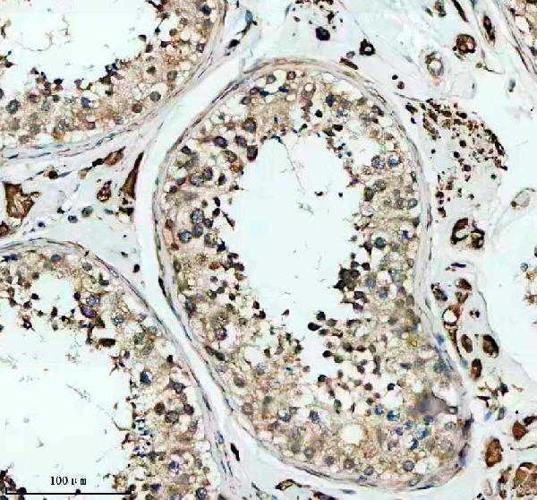
| Sample | Paraffin-embedded human testis tissue; fixative not specified (datasheet A07561-1) |
| Fixation | Image fixative and duration unreported (datasheet A07561-1); verify before use. |
| Sectioning | 4–5 µm sections on charged slides (standard) |
| Deparaffinisation | Xylene, graded ethanol series to water (standard) |
| Antigen retrieval | Heat retrieval: EDTA pH 8.0 (datasheet A07561-1); 20 min, 95–100 °C (standard) |
| Peroxidase block | 3% H2O2, 10 min, room temperature (standard) |
| Blocking | 10% goat serum (datasheet A07561-1) |
| Primary antibody | Rabbit anti-SKA3, 2-5 μg/ml (datasheet A07561-1) |
| Primary incubation | Overnight at 4 °C (datasheet A07561-1) |
| Detection | HRP-conjugated secondary, DAB chromogen (datasheet A07561-1) |
| Counterstain | Hematoxylin, blue, dehydrate and mount (standard) |
| Expected result | SKA3-positive staining in glandular cells of epididymis (HPA tissue IHC: High). HPA tissue profile: Cytoplasmic expression in testis and epididymis. No signal in the no-primary control. |
SKA3 is a non-membrane protein associated with the outer kinetochore and spindle microtubules during mitosis (UniProt Q8IX90: topology and subcellular location). In tissue IHC, expect cytoplasmic staining in testicular pachytene spermatocytes and epididymal glandular cells (HPA: High in both cell types; cytoplasmic profile). Treat this as a provisional reference pattern: HPA rates the IHC staining Approved but reports low consistency with RNA expression and pending external verification (HPA: reliability description).
| Strong cytoplasmic chromogen in pachytene spermatocytes or epididymal glandular cells, with recognizable tissue structure. | This matches the reported tissue pattern (HPA: High in both cell types; cytoplasmic profile). Score the named cells and their compartment, rather than calling an entire section positive; HPA's Approved rating carries a low RNA-consistency caveat and pending external verification (HPA: reliability description). |
| Signal concentrated at a cell-surface rim, in nuclei throughout the section, or in another compartment without the expected cytoplasmic pattern. | Reassess specificity and slide artefact before scoring it as SKA3 (HPA: cytoplasmic tissue profile; UniProt Q8IX90: no transmembrane segment). Mitotic kinetochore or spindle association is biologically plausible (UniProt Q8IX90: subcellular location), so assess compartment together with cell morphology and mitotic state. |
| Strong staining in a cell population reported as undetected, such as adipocytes in adipose tissue. | Consider cross-reactivity or endogenous detection activity and check controls before interpreting it as SKA3 (HPA: adipose adipocytes Not detected; standard IHC practice). An HPA negative is an observed staining result for its tested material, not proof that every specimen must remain negative (HPA: tissue IHC profile). |
| Diffuse color covers stroma, empty spaces, or many unrelated cell types and obscures boundaries. | Treat this as background until controls support a cell-specific pattern (standard IHC practice). Review blocking, washes, chromogen development, and detection controls (standard IHC practice); the reported SKA3 tissue pattern identifies particular cells, not uniform section-wide staining (HPA: tissue IHC profile). |
| No signal in testicular pachytene spermatocytes or epididymal glandular cells. | First verify the named cells are present and the assay's positive control develops (HPA: High in both cell types; standard IHC practice). Then review the catalog antibody's IHC-P instructions and reagent performance (standard IHC practice). HPA supplies no target-specific fixation sensitivity for this result (HPA: tissue IHC profile). |
| Cell cycle and compartment | SKA3 reaches the outer kinetochore and spindle microtubules during mitosis (UniProt Q8IX90: subcellular location). Interpret a mitotic focus in its cellular context; the reported paraffin-tissue pattern is cytoplasmic in testis and epididymis (HPA: tissue IHC profile). |
| Tissue selection and confidence | Use testis or epididymis as pattern references (HPA: High in specified cells), while recording HPA's low staining–RNA consistency and pending external verification (HPA: reliability description). Bone-marrow hematopoietic cells were Not detected by IHC despite tissue-enhanced bone-marrow RNA (HPA: tissue IHC and RNA specificity). |
| Antibody validation | HPA039272 is Approved for IHC; that rating does not remove the tissue-profile caveat (HPA: antibody validation; reliability description). Match the interpretation to the antibody and application used, and use appropriate assay controls (standard IHC practice). |
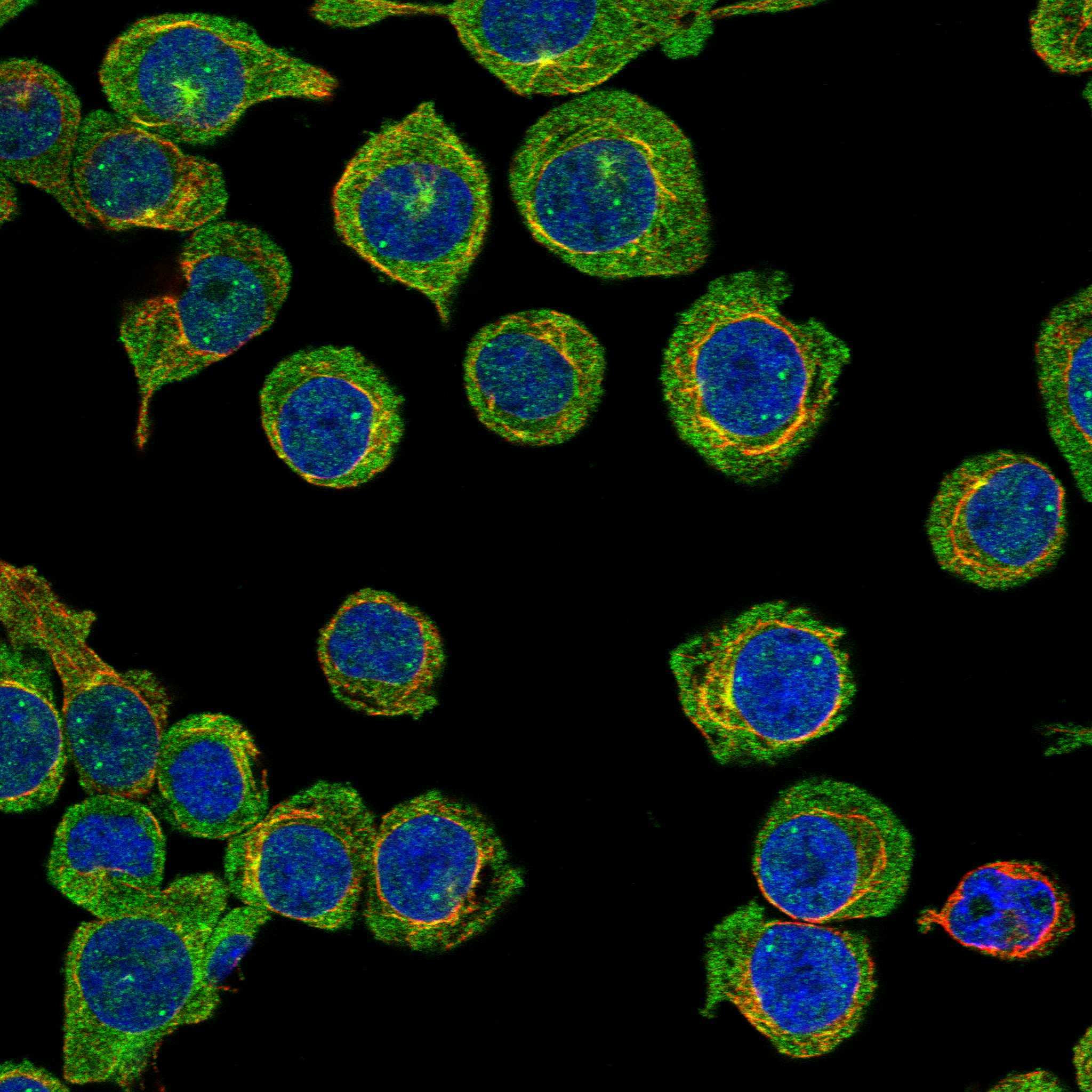
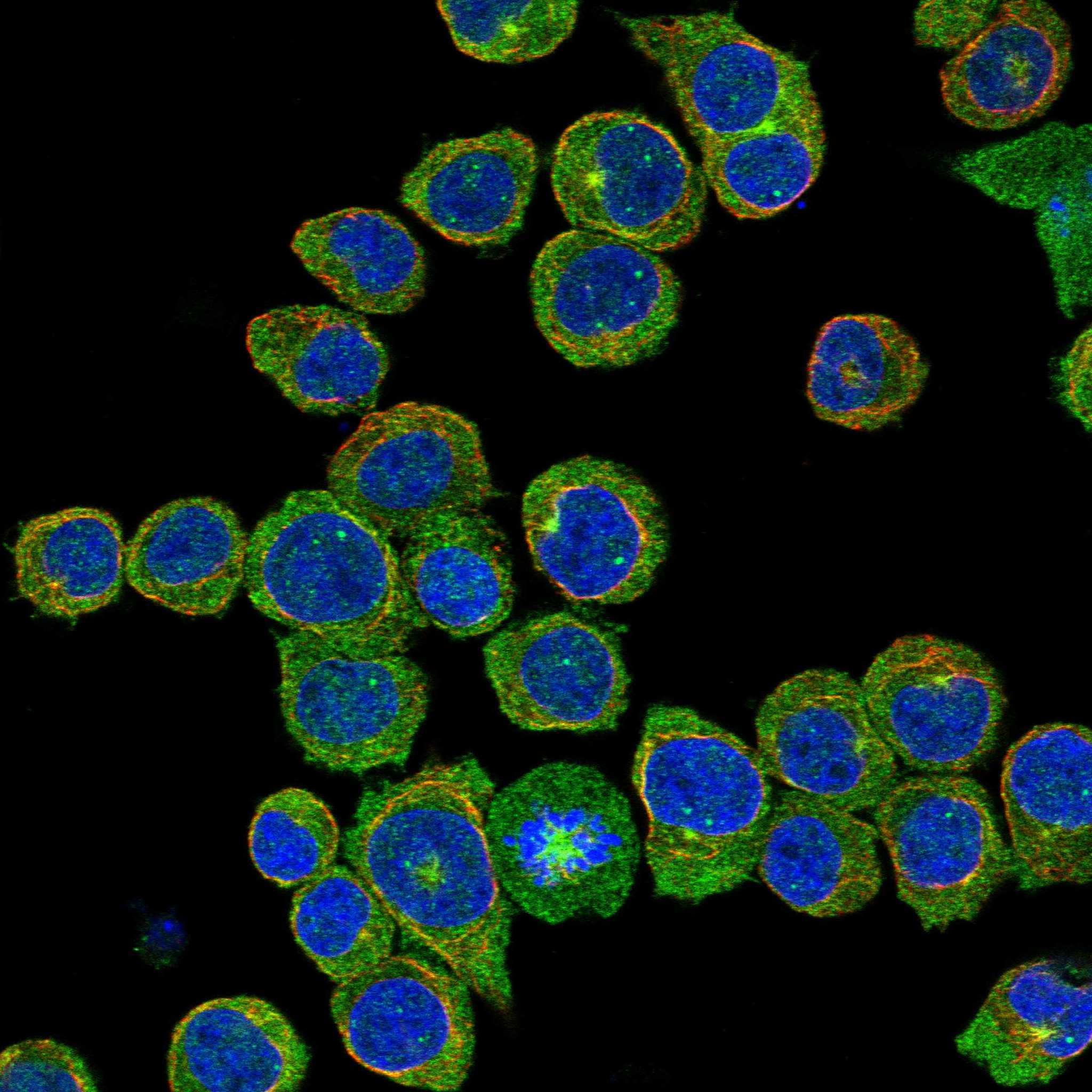
| IF/ICC Q&A: What pattern should be checked? | Check centrosome and mitotic-spindle signal, with possible cytosolic signal (HPA: subcellular summary). HPA marks those locations uncertain; HPA061534 has Supported ICC validation, whereas HPA039356 is Uncertain (HPA: subcellular locations; antibody validation). This IF/ICC observation does not establish a paraffin-IHC protocol (HPA: application-specific validation). |
| Isoforms and antibody epitope | Three isoforms are listed (UniProt Q8IX90: isoforms 1–3). Without an epitope map in the supplied evidence, do not assume that a given antibody recognizes all three or that isoform choice explains an absent stain (UniProt Q8IX90: isoform record). |
| Situation | Likely cause | Next action |
|---|---|---|
| Expected positive cells are blank. | The named cells may be absent from the section, or the IHC run may have failed (HPA: High in specified cells; standard IHC practice). | Confirm cell identity and inspect the run's positive control; then check the catalog antibody's IHC-P instructions, retrieval conditions, detection reagents, and counterstain (standard IHC practice). |
| All tissue compartments show diffuse color. | Nonspecific reagent binding, insufficient washing, or excessive detection development can create background (standard IHC practice). | Review blocking, washes, antibody concentration, and chromogen timing; compare a detection control with the stained slide (standard IHC practice). |
| An HPA-undetected cell type stains strongly. | Cross-reactivity or endogenous detection activity is possible (standard IHC practice); the discordance is clearest when cell identity is secure (HPA: negative tissue entries). | Verify the cell type, examine a control without primary antibody, and repeat with an independently validated antibody if available (standard IHC practice). |
| The dominant result is a membrane rim or broad nuclear staining. | That distribution conflicts with the reported cytoplasmic tissue pattern and SKA3's lack of a transmembrane segment (HPA: tissue IHC profile; UniProt Q8IX90: topology). | Check morphology, controls, and antibody identity before scoring; distinguish a mitotic kinetochore-associated focus from staining across ordinary nuclei (UniProt Q8IX90: mitotic localization; standard IHC practice). |
| Bone marrow is blank despite an expectation from RNA data. | HPA reports tissue-enhanced bone-marrow RNA but Not detected staining in hematopoietic cells (HPA: RNA specificity; tissue IHC). | Use the observed IHC result when choosing a staining reference; check testicular pachytene spermatocytes or epididymal glandular cells and document the RNA–IHC discrepancy (HPA: tissue IHC profile; reliability description). |
| A bright IF/ICC spindle result is used to justify a tissue-IHC call. | The subcellular locations are marked uncertain, and HPA lists validation separately by application (HPA: subcellular locations; antibody validation). | Interpret paraffin IHC against the reported tissue-cell pattern and its reliability caveat; evaluate IF/ICC localization on its own application-specific evidence (HPA: tissue IHC profile; reliability description; subcellular summary). |
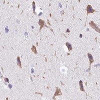


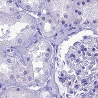
Comprehensive Human Protein Atlas IHC scoring per tissue (reliability: Approved — Low consistency between antibody staining and RNA expression data. Pending external verification.). Rows are taken directly from the HPA tissue chart — click any row's HPA link to view the source.
| Tissue | Cell type | Level | Evidence | Source |
|---|---|---|---|---|
| Epididymis | Glandular cells | High | Protein (IHC) | HPA → |
| Testis | Pachytene spermatocytes | High | Protein (IHC) | HPA → |
| Tissue | Cell type | Level | Evidence | Source |
|---|---|---|---|---|
| Adipose tissue | Adipocytes | Not detected | Protein (IHC) | HPA → |
| Adrenal gland | Glandular cells | Not detected | Protein (IHC) | HPA → |
| Appendix | Glandular cells | Not detected | Protein (IHC) | HPA → |
| Bone marrow | Hematopoietic cells | Not detected | Protein (IHC) | HPA → |
| Breast | Adipocytes | Not detected | Protein (IHC) | HPA → |
These questions focus on interpreting and troubleshooting SKA3 staining in paraffin sections, with one entry for companion IF experiments.
One human-reactive anti-SKA3 antibody has real IHC data from a paraffin-embedded human testis section (catalog; IHC image caption). No IF image is supplied (catalog).
A07561-1 is the only SKU that will render; its applications include IHC, and its image shows staining in a paraffin-embedded human testis section (catalog; IHC image caption). The catalog lists human reactivity and an IHC concentration of 2–5 μg/ml for A07561-1 (catalog).
Which to pick: Choose A07561-1 for human paraffin-section IHC: it is a rabbit polyclonal antibody with IHC listed, and its own image caption documents staining in human testis after heat retrieval in EDTA at pH 8.0 (catalog; IHC image caption). The caption does not report the fixative (IHC image caption). IF/ICC and cross-species use have no supporting application or reactivity listing for A07561-1 (catalog).