SKIL / Ski-like protein · IHC design guide

Design Immunohistochemistry for SKIL

Plan SKIL staining in paraffin sections using the reported cytoplasmic and membranous tissue pattern as a starting point (HPA tissue IHC). This guide covers the catalog antibody’s IHC protocol and tissue controls, with caution because the reported staining has low concordance with RNA expression (datasheet A04131-2; HPA tissue IHC).

Evidence assembled Oct 2026 · For research use; verify linked source records and product datasheet before use
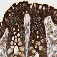
Immunohistochemistry protocol sheet for SKIL (IHC for SKIL): expected localisation Reported cytoplasmic and membranous staining; uncertain (HPA tissue IHC), antibody A04131-2, validated IHC image, and IHC protocol steps
Printable SKIL IHC protocol sheet — expected localisation Reported cytoplasmic and membranous staining; uncertain (HPA tissue IHC), antibody A04131-2, controls and protocol steps. Open the full SKIL IHC guide →

SKIL Immunohistochemistry Experimental Design Guide

Expected localisation, validated protocols, controls and antibodies — the at-a-glance facts below, then the full design guide.

Must know before staining
Expected localisation Reported cytoplasmic and membranous staining; uncertain (HPA tissue IHC)
Staining pattern Most cells show cytoplasmic and membranous staining, except in lymphoid tissue and CNS (HPA tissue IHC)
Antigen retrieval EDTA pH 8.0 HIER, heat-mediated (datasheet A04131-2)
Positive control ⓘ Appendix+4 more · see all
Negative control ⓘ Cerebellum+4 more · see all
Important caveats
Reasons your staining may differ from the expected pattern.
Fixation Keep fixation consistent across sections (standard IHC practice; not target-specific)
Caveat Staining has low concordance with RNA expression (HPA tissue IHC)
Regulation Isoform expression varies by tissue (UniProt)
Isoform / epitope 5 isoforms; confirm epitope coverage across variants (UniProt)
Section 1

Recommended SKIL IHC & IF Protocols

The catalog antibody has a datasheet IHC-P protocol (datasheet A04131-2). These four published SKIL IHC protocols provide additional conditions for paraffin sections (PMC5047385; PMC4467434; PMC10928349; PMC7710697).

Recommended immunohistochemistry (IHC-P) protocol parameters
SampleParaffin-embedded human esophageal squamous carcinoma tissue; fixative not specified (datasheet A04131-2)
FixationImage fixative and duration unreported (datasheet A04131-2); verify before use.
Sectioning4–5 µm sections on charged slides (standard)
DeparaffinisationXylene, graded ethanol series to water (standard)
Antigen retrievalHeat retrieval: EDTA pH 8.0 (datasheet A04131-2); 20 min, 95–100 °C (standard)
Peroxidase block3% H2O2, 10 min, room temperature (standard)
Blocking10% goat serum (datasheet A04131-2)
Primary antibodyRabbit anti-SKIL, 2-5 μg/ml (datasheet A04131-2)
Primary incubationOvernight at 4 °C (datasheet A04131-2)
DetectionStreptavidin-biotin complex (SABC), DAB chromogen (datasheet A04131-2)
CounterstainHematoxylin, blue, dehydrate and mount (standard)
Expected resultSKIL-positive staining in glandular cells of appendix (HPA tissue IHC: High). HPA tissue profile: General cytoplasmic and membranous expression in essentially all cell types, except lymphoid tissue and CNS. No signal in the no-primary control.
💡Decision noteStart with heat-mediated EDTA pH 8.0 retrieval for the catalog antibody (datasheet A04131-2). If optimizing, the cited studies report citrate or Tris-EDTA retrieval (PMC5047385; PMC4467434; PMC7710697).
Section 2

What Is the Expected SKIL Staining Pattern?

In paraffin-section IHC, expect mainly cytoplasmic and membranous SKIL staining in glandular and respiratory epithelial cells (HPA tissue IHC). Interpret this cautiously: the tissue profile is Uncertain because antibody staining has low consistency with RNA data (HPA tissue IHC). SKIL has no annotated transmembrane segment, so membranous staining is an observed IHC pattern, not evidence that SKIL spans the membrane (UniProt P12757 topology; HPA tissue IHC).

What am I looking at on my slide?
Strong cytoplasmic or membranous staining in glandular cells of appendix, breast or colon.These are observed high-staining cell populations (HPA tissue IHC). Compare signal with adjacent cells; intensity alone cannot confirm specificity while tissue IHC reliability is Uncertain (HPA tissue IHC).
Predominantly nuclear staining in a paraffin section, with little cytoplasmic signal.This differs from the general tissue IHC profile and needs investigation (HPA tissue IHC). Nuclear SKIL cannot simply be dismissed as artefact: nucleoplasm is the supported main location in ICC-IF (HPA subcellular ICC-IF).
Strong staining in lymph-node germinal center cells or cerebellar granular-layer cells.Both populations were not detected in the supplied tissue IHC profile (HPA tissue IHC). Check for cross-reactivity or endogenous detection activity before interpreting the signal as SKIL (general IHC practice).
Weak, diffuse colour across cells, stroma and blank areas.This lacks the cell-associated pattern needed to interpret SKIL staining (HPA tissue IHC). Uneven blocking, excess detection reagent or inadequate washing can produce background (general IHC practice).
No signal in appendix glandular cells despite a visible counterstain.Appendix glandular cells were rated High (HPA tissue IHC). A blank result raises a technical question, although the Uncertain tissue-IHC reliability means one specimen cannot establish assay failure (HPA tissue IHC).
💡Expected SKIL appearanceA plausible positive is distinct cytoplasmic or membranous staining in glandular or respiratory epithelial cells rated High (HPA tissue IHC); uniform tissue-wide colour or equally strong staining in HPA-undetected cells warrants investigation (HPA tissue IHC; general IHC practice).
How each factor affects the staining
Tissue and cell contextSeveral glandular populations and bronchial respiratory epithelium are rated High, while lymph-node germinal center cells and pancreatic exocrine cells were not detected (HPA tissue IHC). Choose controls by the named cell population, not the organ alone (general IHC practice).
Strength of IHC evidenceThe overall tissue IHC profile is Uncertain, with low consistency between staining and RNA expression (HPA tissue IHC). HPA008472 is Uncertain for IHC; its Approved ICC result does not upgrade its tissue-IHC validation (HPA antibodies).
Topology and apparent membrane signalSKIL has no transmembrane segment or signal peptide (UniProt P12757 topology and processing). The general membranous pattern is an IHC observation (HPA tissue IHC); it does not establish membrane insertion or secretion.
Isoform coverageFive isoforms are listed; SNON and SNOA are widely expressed, while SNOI is restricted to skeletal muscle (UniProt P12757 tissue specificity and isoforms). No antibody epitope is supplied, so isoform recognition cannot be inferred.
IF/ICC Q&A: Should a nuclear IF image set the expected IHC pattern?No. Nucleoplasm is supported and cytosol approved in ICC-IF (HPA subcellular ICC-IF); tissue IHC instead reports general cytoplasmic and membranous staining (HPA tissue IHC). Interpret each assay with its own evidence.
Why is my staining missing, weak or wrong?
SituationLikely causeNext action
A presumed positive paraffin section shows no SKIL staining.The target cell population may be absent from the section, or the IHC run may have failed (general IHC practice).Confirm glandular cells are present in an appendix, breast or colon control rated High (HPA tissue IHC). Then check retrieval, primary antibody and detection using the validated assay instructions (general IHC practice).
Only nuclei stain in tissue IHC.The result differs from the reported tissue pattern, though nucleoplasm is supported in ICC-IF (HPA tissue IHC; HPA subcellular ICC-IF).Review compartment scoring and compare an independent IHC control or antibody where available (general IHC practice). Do not use ICC localisation alone to certify the paraffin-section result.
Germinal center cells stain as strongly as the intended positive cells.Germinal center cells were not detected in the supplied lymph-node IHC observation (HPA tissue IHC); nonspecific primary binding or detection activity is possible (general IHC practice).Inspect primary-omission and detection controls, then compare cell-restricted patterns on the same run (general IHC practice). Treat the tissue profile as provisional because HPA rates it Uncertain (HPA tissue IHC).
Brown precipitate appears in many cell types or over empty slide areas.Diffuse deposition is consistent with detection background rather than a cell-specific staining pattern (general IHC practice).Check blocking, washing and chromogen development; use a primary-omission control to localise the background source (general IHC practice). Reassess SKIL only after discrete cellular staining is visible.
A low-staining tissue appears negative while epithelial controls stain.HPA rates skeletal-muscle myocytes Low and pancreatic exocrine cells Not detected (HPA tissue IHC). Tissue-level RNA or isoform expression does not guarantee visible staining in a specified cell type (UniProt P12757 tissue specificity; HPA tissue IHC).Record the sampled cell type and retain the epithelial positive control; avoid calling the run failed from this tissue alone (general IHC practice; HPA tissue IHC).
An ICC-IF image shows nuclear signal while tissue IHC shows cytoplasmic signal.The reported assay-specific patterns differ: nucleoplasm is the supported main ICC-IF site, whereas tissue IHC reports general cytoplasmic and membranous staining (HPA subcellular ICC-IF; HPA tissue IHC).Report the assay and compartment separately. If the discrepancy matters to the experiment, seek independent antibody or orthogonal evidence before assigning a single SKIL location (general IHC practice).

Sample controls for SKIL IHC & IF

🧪Run appendix first: its glandular cells should stain (HPA: High in appendix glandular cells). Use cerebellum granular-layer cells as the negative tissue (HPA: Not detected in cerebellum granular-layer cells); on the appendix slide, treat adjacent unstained nonglandular cells as an internal background reference, without assuming they are SKIL-negative.
Positive control tissue: Appendix (Glandular cells, HPA High)
Negative control tissue: Cerebellum (HPA Not detected)
ICC-IF cell lines (HPA subcellular resource): HPA ICC-IF images show SKIL in EFO-21, SiHa, U2OS, A-431, U-251MG, with annotated localisation: Nucleoplasm (supported) (HPA subcellular).
Technical controls: Include a no-primary (secondary-only) control, a concentration-matched rabbit IgG isotype control appropriate to the primary antibody, and a matched SKIL-knockout biological negative where available (selected-SKU caption: rabbit primary antibody). For appendix sections developed with biotin-based DAB detection, quench endogenous peroxidase and assess endogenous biotin background (selected-SKU caption: biotinylated secondary and DAB detection).
⚠️Feasibility: The selected-SKU paraffin-section caption reports heat retrieval in EDTA at pH 8.0, but does not report the fixative; no target-specific fixation window or fixation effect is reported (selected-SKU tissue-IHC caption). IF/ICC offers a localization cross-check because SKIL is observed mainly in the nucleoplasm, with additional cytosolic signal; the supplied evidence does not establish that IF or frozen sections are easier than paraffin IHC (HPA: subcellular localization; selected-SKU tissue-IHC caption). In appendix glands, inspect luminal mucus or debris for nonspecific chromogen deposition when scoring staining (standard IHC practice).

HPA tissue IHC evidence for SKIL

Comprehensive Human Protein Atlas IHC scoring per tissue (reliability: Uncertain — Low consistency between antibody staining and RNA expression data. Pending external verification.). Rows are taken directly from the HPA tissue chart — click any row's HPA link to view the source.

Positive expression · recommended positive controls

TissueCell typeLevelEvidenceSource
Appendix Glandular cells High Protein (IHC) HPA →
Breast Glandular cells High Protein (IHC) HPA →
Bronchus Respiratory epithelial cells High Protein (IHC) HPA →
Cervix Glandular cells High Protein (IHC) HPA →
Colon Glandular cells High Protein (IHC) HPA →

Undetected expression · recommended negative controls

TissueCell typeLevelEvidenceSource
Cerebellum Cells in granular layer Not detected Protein (IHC) HPA →
Heart muscle Cardiomyocytes Not detected Protein (IHC) HPA →
Hippocampus Glial cells Not detected Protein (IHC) HPA →
Lymph node Germinal center cells Not detected Protein (IHC) HPA →
Ovary Ovarian stroma cells Not detected Protein (IHC) HPA →
Section 3

Advanced SKIL IHC Tips

Use the catalog antibody’s paraffin-section workflow as a starting point, then check compartment, cell type and controls before scoring SKIL staining.

How should I retrieve SKIL in paraffin sections when staining is weak?
Start with heat-mediated retrieval in EDTA, pH 8.0 (datasheet A04131-2). The catalog antibody’s paraffin-section image used that retrieval, followed by 10% goat serum block and 2 μg/ml primary antibody overnight at 4°C (datasheet A04131-2). Keep retrieval heating and cooling consistent across sections, and compare a new run with the documented workflow before changing several steps at once (standard IHC practice). If staining remains weak, test a shorter or longer heating interval on matched sections while watching for tissue damage and increased background (standard IHC practice).
Could fixation explain weak or uneven SKIL staining?
The selected paraffin-section caption does not state a fixative, so SKIL-specific fixation sensitivity is unknown (datasheet A04131-2: fixative not stated). Record the specimen’s fixative and fixation duration, then compare sections with similar processing histories when investigating uneven staining (standard IHC practice). Check morphology and whether staining varies with section edges, folds or visibly damaged areas before attributing a difference to SKIL abundance (standard IHC practice). Hold the documented EDTA, pH 8.0 retrieval and 2 μg/ml primary concentration steady during that comparison, so processing effects are easier to assess (datasheet A04131-2; standard IHC practice).
Should SKIL staining be nuclear, cytoplasmic or membranous?
Assess nuclear and cytoplasmic staining separately: HPA subcellular data place SKIL mainly in the nucleoplasm, with additional cytosolic localisation (HPA subcellular: supported nucleoplasm; approved cytosol). HPA tissue IHC describes broad cytoplasmic and membranous staining, but rates its antibody staining reliability Uncertain because consistency with RNA expression is low (HPA tissue IHC: Uncertain). SKIL has no annotated transmembrane segment, so a membrane-only DAB pattern merits independent validation (UniProt P12757 topology; standard IHC practice). Compare the suspected pattern with tissue morphology and a no-primary control, and record each compartment rather than combining them into one positive score (standard IHC practice).
Can this antibody distinguish SKIL isoforms in tissue?
Do not assign an IHC signal to one isoform without an antibody epitope and isoform-specific validation; the supplied caption documents staining, not that distinction (datasheet A04131-2). UniProt lists 5 SKIL isoforms, including SNON, SNOA, SNON2 and SNOI (UniProt P12757 isoforms). SNON and SNOA are widely expressed, whereas SNOI expression is restricted to skeletal muscle, so tissue context can guide a validation plan but cannot identify the stained isoform by itself (UniProt P12757 tissue specificity; standard IHC practice). If isoform identity matters, compare epitope coverage with isoform sequences and use an independent isoform-specific assay before interpreting DAB intensity (standard IHC practice).
How can I assess SKIL localisation by IF alongside this IHC guide?
Use the separate IF/ICC workflow to assess SKIL localisation; HPA subcellular data support nucleoplasm and additionally cytosol (HPA subcellular: supported nucleoplasm; approved cytosol). For multiplex IF, pair SKIL with a validated marker for the cell type under study and choose fluorophores after checking tissue autofluorescence in each channel (standard IF practice). Permeabilise sufficiently for antibody access to intracellular nuclear and cytosolic epitopes, then verify that the treatment preserves morphology and the companion marker (HPA subcellular localisation; standard IF practice). Keep IF and chromogenic IHC scores separate until the observed cell types and compartments agree across methods (standard IHC/IF practice).
What should I check if DAB staining is widespread or patchy?
First inspect a no-primary control and tissue morphology to distinguish detection background from staining associated with cells (standard IHC practice). The documented paraffin-section workflow uses 10% goat serum, a biotinylated secondary for 30 minutes at 37°C, a streptavidin–biotin complex and DAB (datasheet A04131-2). With this detection system, check endogenous peroxidase and biotin background using appropriate controls, and confirm that the block and washes suit the tissue (standard IHC practice). Score diffuse staining cautiously: HPA reports broad tissue staining but rates that profile Uncertain because antibody staining and RNA expression show low consistency (HPA tissue IHC: Uncertain).
How should I quantify SKIL DAB staining across specimens? ⚠ ANSWER MARKED FOR VERIFICATION
Define the scored cell population and compartment before review, using nuclear and cytoplasmic scores separately where both are present (HPA subcellular: nucleoplasm and cytosol; standard IHC practice). Record the percentage of positive cells and staining intensity for an H-score, or report positive-cell density per mm² when cell counts are the main outcome (standard IHC practice). Normalise comparisons to the same eligible tissue area or cell population, section quality and staining batch, and exclude folds, necrosis and edges by preset criteria (standard IHC practice). Report the scoring rule and controls because HPA tissue IHC assigns SKIL staining Uncertain reliability (HPA tissue IHC: Uncertain).
When is an apparent SKIL-positive cell likely to be an artefact?
Check whether the signal belongs to intact cells and follows a plausible compartment: HPA supports nucleoplasmic and additional cytosolic SKIL localisation (HPA subcellular: supported nucleoplasm; approved cytosol). Treat isolated membrane-only staining cautiously because SKIL has no annotated transmembrane segment, while recognising that HPA tissue IHC reports membranous staining with Uncertain reliability (UniProt P12757 topology; HPA tissue IHC: Uncertain). Compare the stained cell type with tissue morphology rather than calling every DAB deposit SKIL (standard IHC practice). Edge accentuation, necrotic debris and signal persisting in a no-primary or peroxidase control favour artefact; confirm consequential findings independently (standard IHC practice).
Boster reagents

Best SKIL / Ski-like protein IHC Antibodies

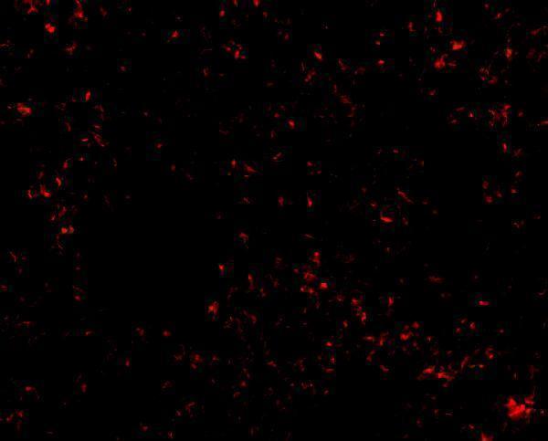
Two anti-SKIL antibodies have IHC images from human carcinoma or tonsil paraffin sections and mouse lung tissue, plus IF images from A431 or mouse lung cells (catalog image captions).

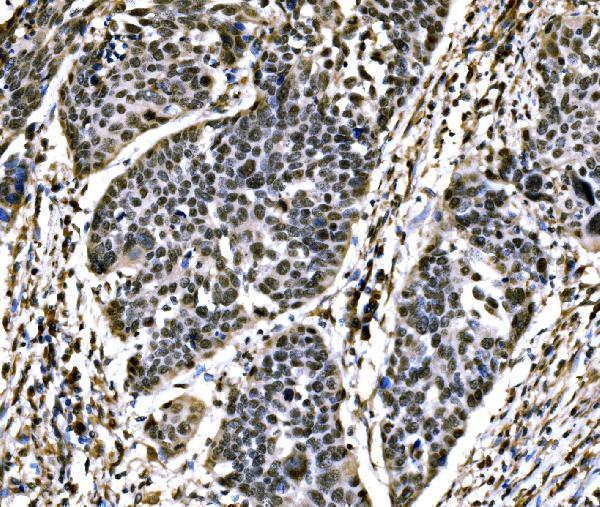
Real IHC data IHC analysis of SnoN/SNO/SKIL using anti-SnoN/SNO/SKIL antibody (A04131-2). SnoN/SNO/SKIL was detected in a paraffin-embedded section of human esophageal squamous carcinoma tissue. Heat mediated antigen retrieval was performed in EDTA buffer (pH 8.0, epitope retrieval solution). The tissue section was blocked with 10% goat serum. The tissue section was then incubated with 2 μg/ml rabbit anti-SnoN/SNO/SKIL Antibody (A04131-2) overnight at 4°C. Biotinylated goat anti-rabbit IgG was used as secondary antibody and incubated for 30 minutes at 37°C. The tissue section was developed using Strepavidin-Biotin-Complex (SABC) (Catalog # SA1022) with DAB as the chromogen.
Anti-SnoN/SNO/SKIL Antibody ®
Cat # A04131-2
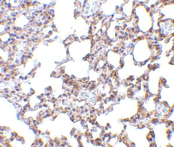
Real IHC data Immunohistochemistry of SnoN in mouse lung tissue with SnoN antibody at 5 μg/mL.
Anti-SnoN SKIL Antibody
Cat # A04131-1

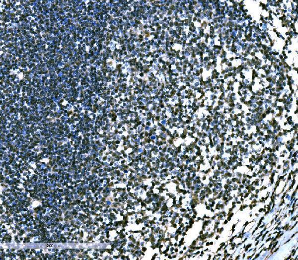
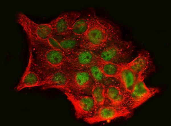
A04131-2 has IHC images from human esophageal squamous carcinoma and tonsil paraffin sections, plus an IF/ICC image from A431 cells (catalog image captions). A04131-1 has an IHC image from mouse lung tissue and an IF image from mouse lung cells (catalog image captions).

Which to pick: For human paraffin-section IHC, choose A04131-2 at 2–5 μg/mL; its IHC captions document paraffin sections and EDTA retrieval, but do not report the fixative (catalog IHC captions and dilution). For IF/ICC, A04131-2 has an A431-cell image at 5 μg/mL, while A04131-1 has a mouse lung-cell IF image at 20 μg/mL (catalog IF captions). Both list human, mouse and rat reactivity; for mouse tissue IHC, A04131-1 has a mouse lung image at 5 μg/mL, while rat tissue IHC has no image in the supplied catalog (catalog reactivity and IHC captions).

Each figure is that product's own IHC / IF validation image from its datasheet.

References

  1. UniProt Consortium. UniProt entry P12757 (SKIL_HUMAN, Ski-like protein).
  2. Human Protein Atlas. SKIL tissue IHC expression (reliability: Uncertain).
  3. Human Protein Atlas. SKIL subcellular location (ICC-IF): Mainly localized to the nucleoplasm. In addition localized to the cytosol..
  4. Human Protein Atlas. SKIL antibody validation summary (2 antibodies).
  5. Expression and clinical significance of the transforming growth factor-β signalling pathway in endometrial cancer. Histopathology 2011 — PMC5047385.
  6. Recurrent SKIL-activating rearrangements in ETS-negative prostate cancer. Oncotarget 2015 — PMC4467434.
  7. NSUN2 promotes colorectal cancer progression by enhancing SKIL mRNA stabilization. Clinical and translational medicine 2024 — PMC10928349.
  8. SKIL facilitates tumorigenesis and immune escape of NSCLC via upregulating TAZ/autophagy axis. Cell death & disease 2020 — PMC7710697.
  9. PubMed PMID:2762147 — UniProt-cited evidence.
  10. PubMed PMID:9207045 — UniProt-cited evidence.
  11. PubMed PMID:14702039 — UniProt-cited evidence.