This website uses cookies to ensure you get the best experience on our website.


- Table of Contents
Plan chromogenic SKP1 IHC on paraffin sections using breast glandular cells as a high-staining reference (HPA tissue IHC). Assess cytoplasmic and nuclear staining, and compare it with cell populations reported as low or undetected (HPA tissue IHC).

Expected localisation, validated protocols, controls and antibodies — the at-a-glance facts below, then the full design guide.
| Expected localisation | Cytoplasmic and nuclear staining in most tissues (HPA tissue IHC) | |
| Staining pattern | Cytoplasmic and nuclear signal across most tissues (HPA tissue IHC) | |
| Antigen retrieval | EDTA pH 8.0 HIER, heat-mediated (datasheet A00476-2) | |
| Positive control | Breast+4 more · see all | |
| Negative control | Appendix+3 more · see all |
| Fixation | Keep fixation consistent across paraffin sections (standard IHC practice; not target-specific) | |
| Caveat | Cell-type intensity varies across tissues (HPA tissue IHC) | |
| Regulation | No expression regulator specified (UniProt) | |
| Isoform / epitope | 2 isoforms; epitope coverage is unspecified (UniProt) |
The catalog antibody has an IHC-P protocol (datasheet A00476-2); the published SKP1 IHC procedures below cover lung cancer, pancreatic neuroendocrine neoplasms, mouse brain and rat brain (PMC4741501; PMC7728999; PMC3542602; PMC11814243).
| Sample | Paraffin-embedded human breast cancer tissue; fixative not specified (datasheet A00476-2) |
| Fixation | Image fixative and duration unreported (datasheet A00476-2); verify before use. |
| Sectioning | 4–5 µm sections on charged slides (standard) |
| Deparaffinisation | Xylene, graded ethanol series to water (standard) |
| Antigen retrieval | Heat retrieval: EDTA pH 8.0 (datasheet A00476-2); 20 min, 95–100 °C (standard) |
| Peroxidase block | 3% H2O2, 10 min, room temperature (standard) |
| Blocking | 10% goat serum (datasheet A00476-2) |
| Primary antibody | Rabbit anti-SKP1, 2-5μg/ml (datasheet A00476-2) |
| Primary incubation | Overnight at 4 °C (datasheet A00476-2) |
| Detection | HRP-conjugated secondary, DAB chromogen (datasheet A00476-2) |
| Counterstain | Hematoxylin, blue, dehydrate and mount (standard) |
| Expected result | SKP1-positive staining in glandular cells of breast (HPA tissue IHC: High). HPA tissue profile: Cytoplasmic and nuclear expression in most tissues. No signal in the no-primary control. |
SKP1 staining is expected in the cytoplasm and nuclei of cells in most tissues (HPA tissue IHC: Approved; medium consistency with RNA, pending external verification). High staining is reported in breast glandular cells, bronchial respiratory epithelial cells and placental trophoblastic cells (HPA tissue IHC: High). SKP1 has no transmembrane segment, and UniProt does not annotate its subcellular location (UniProt P63208 topology and subcellular record).
| Clear cytoplasmic and nuclear staining in breast glandular cells or bronchial respiratory epithelial cells (HPA tissue IHC: High). | This matches the reported tissue pattern and provides a useful positive reference (HPA tissue IHC: cytoplasmic and nuclear expression in most tissues; High in these cells). Compare cells within the same section; a positive reference supports detection but does not establish antibody specificity on its own (general IHC practice). |
| Staining is confined to a membrane rim or extracellular deposit, without a cytoplasmic or nuclear pattern (HPA tissue IHC: expected pattern). | That distribution conflicts with the reported IHC pattern and SKP1's lack of a transmembrane segment (HPA tissue IHC; UniProt P63208 topology). Review morphology and controls for artefact before scoring it as SKP1 (general IHC practice). |
| Strong signal appears in cardiomyocytes or skeletal myocytes while the expected positive cells stain weakly (HPA tissue IHC: Not detected in those myocytes). | Treat the unexpected signal as suspect; cross-reactivity or endogenous detection activity are possibilities, not established causes (general IHC practice). Compare a reagent control and an HPA High reference before assigning cell-specific positivity (HPA tissue IHC: High in breast glandular cells). |
| Brown colour covers stroma, empty spaces and many unrelated cell types, obscuring cellular boundaries (general IHC practice). | A diffuse pattern cannot reliably establish cytoplasmic or nuclear SKP1 localisation (HPA tissue IHC: expected pattern; general IHC practice). Background from detection reagents or insufficient blocking is possible; assess controls and morphology before scoring (general IHC practice). |
| No staining appears in breast glandular cells or placental trophoblastic cells (HPA tissue IHC: High in both). | This weakens confidence in a negative call for the run (general IHC practice). Check tissue preservation and detection controls, then review the catalog antibody's IHC-P conditions; HPA staining levels do not identify a fixation effect (HPA tissue IHC; general IHC practice). |
| Which tissue and cell type should anchor interpretation? (HPA tissue IHC) | Breast glandular cells, bronchial respiratory epithelial cells and placental trophoblastic cells are High; cardiomyocytes and skeletal myocytes are Not detected (HPA tissue IHC). Evaluate named cells within their tissue context rather than treating every cell in a section as an equivalent reference (general IHC practice). |
| How firm is the tissue-pattern evidence? (HPA tissue IHC) | HPA rates tissue IHC Approved, with medium consistency between staining and RNA and external verification pending (HPA tissue IHC). Its low tissue-specificity RNA classification does not mean every cell has equal protein staining (HPA tissue IHC: RNA specificity and cell-level staining). |
| What does protein topology contribute? (UniProt P63208) | SKP1 has no annotated transmembrane segment, signal peptide or propeptide; its annotated chain spans residues 2–163 (UniProt P63208 topology and processing). These features support scrutiny of a membrane-only pattern but do not predict antigen-retrieval conditions (UniProt P63208; general IHC practice). |
| Can isoforms or modifications explain an unusual slide? (UniProt P63208) | Two isoforms and a modified threonine at residue 131 are listed (UniProt P63208). The supplied record gives no antibody epitope or isoform-specific IHC pattern, so an unusual stain cannot be assigned to either feature (UniProt P63208; evidence limit). |
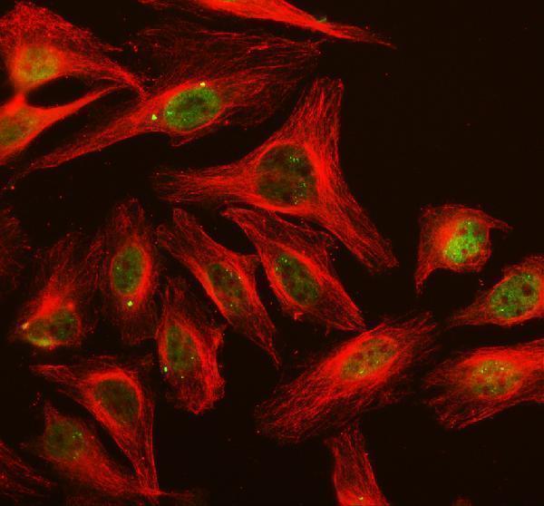
| Does IF/ICC show the same localisation? (HPA subcellular ICC-IF) | IF/ICC reports approved nucleoplasm and cytosol localisation, plus approved centrosome, cytokinetic bridge, primary cilium and basal body localisation (HPA subcellular ICC-IF). HPA cautions that these findings are based on antibodies targeting proteins from multiple genes; the extra structures are not an expected chromogenic tissue-IHC scoring criterion (HPA subcellular ICC-IF; HPA tissue IHC). |
| What does antibody validation establish? (HPA antibodies) | CAB012982 is Approved for IHC, while HPA053745 is Approved for ICC and has no listed IHC status (HPA antibodies). Those application statuses should be kept distinct when comparing a tissue section with IF/ICC images (HPA antibodies; general IHC practice). |
| Situation | Likely cause | Next action |
|---|---|---|
| Positive-reference cells show no chromogenic signal (HPA tissue IHC: High in breast glandular cells). | An unsuccessful staining or detection step is possible; the HPA tissue level alone does not establish why the run failed (general IHC practice; HPA tissue IHC). | Confirm tissue identity and morphology, inspect run controls, and repeat using the catalog antibody's IHC-P instructions if needed (general IHC practice). |
| Only membrane-edge staining is visible (HPA tissue IHC: cytoplasmic and nuclear pattern). | This conflicts with the reported pattern and absent transmembrane segment; edge artefact is possible (HPA tissue IHC; UniProt P63208 topology; general IHC practice). | Compare internal cells with section edges and check reagent controls before scoring the signal (general IHC practice). |
| A Not detected cell type stains strongly (HPA tissue IHC: cardiomyocytes and skeletal myocytes). | Cross-reactivity or endogenous detection activity is possible; neither is established by the HPA result alone (HPA tissue IHC; general IHC practice). | Compare a reagent control and an HPA High cell type on a suitable section; score the named cell populations separately (HPA tissue IHC; general IHC practice). |
| Diffuse colour makes nuclei and cytoplasm hard to distinguish (general IHC practice). | Background from detection reagents or blocking conditions is possible (general IHC practice). | Inspect negative controls and counterstain, then optimise blocking, detection and wash conditions as general IHC workflow variables (general IHC practice). |
| Weak staining occurs in a proposed low-signal reference (HPA tissue IHC: Low in alveolar cells or hepatocytes). | The selected cells may have low reported protein staining, so weak signal is not by itself a failed run (HPA tissue IHC). | Include an HPA High cell type for the run-level comparison and interpret the low-signal cells in context (HPA tissue IHC; general IHC practice). |
| Changing retrieval appears to alter the result (general IHC practice). | Target-specific fixation effects are not established by the supplied assay evidence. Verify with a matched IHC source before attributing a result to fixation. | Record the retrieval conditions and compare matched sections with the same positive reference and controls; avoid attributing the change to an established SKP1 fixation effect (general IHC practice). |
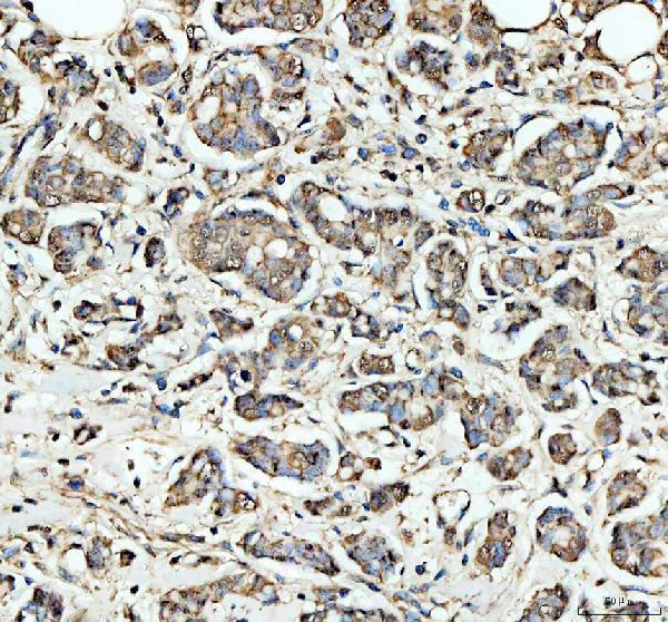
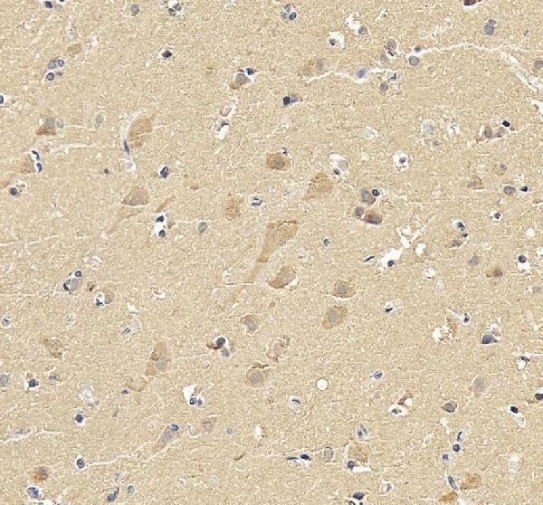
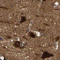
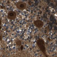


Comprehensive Human Protein Atlas IHC scoring per tissue (reliability: Approved — Medium consistency between antibody staining and RNA expression data. Pending external verification.). Rows are taken directly from the HPA tissue chart — click any row's HPA link to view the source.
| Tissue | Cell type | Level | Evidence | Source |
|---|---|---|---|---|
| Breast | Glandular cells | High | Protein (IHC) | HPA → |
| Bronchus | Respiratory epithelial cells | High | Protein (IHC) | HPA → |
| Caudate | Glial cells | High | Protein (IHC) | HPA → |
| Cerebellum | Cells in granular layer | High | Protein (IHC) | HPA → |
| Cerebral cortex | Glial cells | High | Protein (IHC) | HPA → |
| Tissue | Cell type | Level | Evidence | Source |
|---|---|---|---|---|
| Appendix | Glandular cells | Not detected | Protein (IHC) | HPA → |
| Heart muscle | Cardiomyocytes | Not detected | Protein (IHC) | HPA → |
| Skeletal muscle | Myocytes | Not detected | Protein (IHC) | HPA → |
| Smooth muscle | Smooth muscle cells | Not detected | Protein (IHC) | HPA → |
Troubleshoot SKP1 staining in paraffin sections by controlling retrieval and detection, then assessing signal by cell type and compartment.
A00476-2 has IHC images from human paraffin sections and an IF image from HeLa cells; both listed antibodies report human, mouse and rat reactivity (catalog image captions; catalog reactivity).
A00476-2 is listed for IHC and IF, with images from human paraffin sections of breast cancer and brain tissue and IF staining of HeLa cells (catalog applications; catalog image captions). M00476 is listed for IF/ICC, with human, mouse and rat reactivity, but no IHC or IF image is supplied (catalog applications; catalog reactivity; catalog image captions).
Which to pick: Choose A00476-2 for tissue IHC: its own images show human paraffin sections after EDTA pH 8.0 heat retrieval, with the fixative unreported (A00476-2 IHC captions). For IF/ICC, A00476-2 has a HeLa IF image; M00476 is a monoclonal option listed for IF/ICC without a supplied image (A00476-2 IF caption; M00476 clone and applications). Both list human, mouse and rat reactivity, while the supplied IHC and IF images show human samples only (catalog reactivity; catalog image captions).