This website uses cookies to ensure you get the best experience on our website.


- Table of Contents
Plan chromogenic SLAMF9 IHC with the catalog antibody starting at 2.5 μg/mL (datasheet). Assess the reported cytoplasmic tissue staining (HPA tissue IHC) alongside the expected membrane location (UniProt), and interpret staining cautiously given its poor concordance with RNA expression (HPA tissue IHC).

Expected localisation, validated protocols, controls and antibodies — the at-a-glance facts below, then the full design guide.
| Expected localisation | Cytoplasmic tissue staining (HPA tissue IHC); membrane expected (UniProt) | |
| Staining pattern | General cytoplasmic staining in tissue cells (HPA tissue IHC) | |
| Antigen retrieval | Citrate pH 6.0 HIER, 95–98 °C, 20 min (rule: cytoplasmic / membrane antigen) | |
| Positive control | Adrenal gland+4 more · see all | |
| Negative control | Adipose tissue+4 more · see all |
| Fixation | Keep fixation consistent across paraffin sections. (standard IHC practice; not target-specific) | |
| Caveat | Very low consistency between staining and RNA expression (HPA tissue IHC) | |
| Regulation | Predominantly hematopoietic expression (UniProt) | |
| Isoform / epitope | 2 isoforms; map the epitope to the extracellular or cytoplasmic region (UniProt) |
The catalog antibody IHC-P protocol is followed by 4 published SLAMF9 IHC protocols from target-verified articles (PMC8688141; PMC6145869; PMC11297030; PMC12885906).
| Sample | Tissue sections; selected-image fixative not specified (standard IHC workflow) |
| Fixation | Image fixative and duration unreported (datasheet A13838); verify before use. |
| Sectioning | 4–5 µm sections on charged slides (standard) |
| Deparaffinisation | Xylene, graded ethanol series to water (standard) |
| Antigen retrieval | Heat-induced epitope retrieval in citrate buffer, pH 6.0, 20 min at 95–98 °C (standard rule: cytoplasmic / membrane antigen) |
| Peroxidase block | 3% H2O2, 10 min, room temperature (standard) |
| Blocking | 10% normal serum of the secondary host, 30 min, room temperature (standard) |
| Primary antibody | Rabbit anti-SLAMF9, 2.5 μg/mL (datasheet A13838) |
| Primary incubation | Overnight at 4 °C (standard) |
| Detection | HRP-polymer secondary, DAB chromogen 5–10 min (standard) |
| Counterstain | Hematoxylin, blue, dehydrate and mount (standard) |
| Expected result | SLAMF9-positive staining in glandular cells of adrenal gland (HPA tissue IHC: Medium). HPA tissue profile: General cytoplasmic expression. No signal in the no-primary control. |
SLAMF9 is a membrane protein with an extracellular region at residues 19–236 and a cytoplasmic tail at 258–289 (UniProt Q96A28: topology). Immune cells, including monocytes and dendritic cells, are plausible cells to examine (UniProt Q96A28: tissue specificity). Treat any IHC pattern as provisional: HPA reports general cytoplasmic staining, RNA as not detected, and tissue IHC reliability as uncertain (HPA: tissue IHC).
| Immune cells show a distinct cell-edge signal in a chromogenic paraffin section. | This is compatible with membrane localisation (UniProt Q96A28: topology). Record which cells stain and compare with local controls; morphology alone does not establish SLAMF9 identity (general IHC practice). HPA has not established a reliable membrane pattern by tissue IHC (HPA: Uncertain). |
| Staining is predominantly cytoplasmic, with little visible cell-edge enrichment. | HPA describes general cytoplasmic expression, but rates its tissue IHC uncertain because antibody staining and RNA expression have very low consistency (HPA: tissue IHC). Since UniProt places SLAMF9 at the membrane, treat cytoplasmic staining as unresolved and check another validated approach before assigning localisation (UniProt Q96A28: subcellular location; general IHC practice). |
| Strong nuclear staining appears without a corresponding cell-edge signal. | A nuclear-only pattern conflicts with the annotated membrane location and transmembrane segment at 237–257 (UniProt Q96A28: topology). Consider nonspecific staining or a detection artefact; assess negative controls and cell morphology before calling the section positive (general IHC practice). |
| Adipocytes or other unexpected cells stain as strongly as the cells under study. | HPA reports SLAMF9 as not detected in adipocytes, although its tissue IHC reliability is uncertain (HPA: adipose tissue; HPA: Uncertain). Such staining warrants checks for cross-reactivity and endogenous detection activity; it does not, by itself, prove either cause (general IHC practice). |
| Color is spread across tissue, empty spaces, or the section edge. | A diffuse deposit that does not follow cell boundaries is difficult to interpret as membrane staining (UniProt Q96A28: membrane; general IHC practice). Compare a negative reagent control and inspect background across the whole section before scoring individual cells (general IHC practice). |
| Cell compartment and epitope | SLAMF9 has an extracellular region at 19–236, a membrane span at 237–257, and a cytoplasmic tail at 258–289 (UniProt Q96A28: topology). The supplied sources do not locate the catalog antibody's epitope, so they cannot predict which processing condition exposes it (UniProt Q96A28: topology; supplied record). |
| Strength of tissue evidence | HPA rates tissue IHC Uncertain, citing very low consistency between antibody staining and RNA expression, and lists RNA specificity as not detected (HPA: tissue IHC). Its reported positive cells are observations to investigate, rather than validated positive controls (HPA: tissue IHC). |
| Choice of candidate tissue and cell | HPA reports low staining in lung macrophages and not detected staining in lymph-node germinal-center cells (HPA: tissue IHC). UniProt describes expression in several immune-cell classes but reports no expression in peripheral blood leukocytes (UniProt Q96A28: tissue specificity). Specify the cell population when interpreting a section. |
| Antibody validation and target variation | The listed HPA antibody, HPA035153, has Uncertain IHC status and no reported ICC status (HPA: antibodies). UniProt lists 2 isoforms (UniProt Q96A28: isoforms); the supplied sources do not establish whether an antibody detects both. Interpret a negative result within that validation limit. |
| Situation | Likely cause | Next action |
|---|---|---|
| No signal appears in a section that previously stained with the same assay. | A failed run or a change in specimen or reagent handling is possible (general IHC practice). HPA supplies no dependable strongly positive SLAMF9 tissue control (HPA: Uncertain; HPA: tissue IHC). | Run the previously confirmed control alongside the sample; verify detection reagents, retrieval conditions, and antibody dilution against the assay record before interpreting absence of staining (general IHC practice). |
| Only diffuse cytoplasmic staining appears. | HPA reports a general cytoplasmic profile, but its IHC evidence is uncertain (HPA: tissue IHC); UniProt assigns SLAMF9 to the membrane (UniProt Q96A28: subcellular location). | Record the pattern as unresolved, inspect cell edges at higher magnification, and compare negative controls or an independently validated antibody if available (general IHC practice). |
| Unexpected cell populations stain strongly. | Cross-reactivity or endogenous detection activity may contribute (general IHC practice). HPA tissue calls alone cannot validate the identity of stained cells here (HPA: Uncertain). | Confirm cell identity by morphology, include an appropriate negative reagent control, and investigate endogenous activity relevant to the chosen chromogenic detection system (general IHC practice). |
| Background obscures cell boundaries. | Nonspecific binding or incomplete control of endogenous detection activity can produce background in chromogenic IHC (general IHC practice). | Check the negative control, blocking and wash steps, and detection-system controls; optimise conditions against a reproducible reference section before scoring localisation (general IHC practice). |
| The signal changes when antigen retrieval conditions change. | Retrieval conditions can affect staining in paraffin IHC generally (general IHC practice). Target-specific fixation sensitivity or an optimal SLAMF9 retrieval condition is not reported in the supplied sources (UniProt Q96A28; HPA: tissue IHC). | Compare conditions on matched sections with the same controls and record both signal and background; do not infer SLAMF9-specific epitope masking from this comparison alone (general IHC practice). |
| Can IF/ICC confirm the IHC result? | HPA summarises SLAMF9 as membrane-localised but provides no ICC-IF image cell lines; its listed antibody has no ICC status (HPA: subcellular; HPA: antibodies). | Treat IF/ICC as a separate validation question and consult the dedicated IF/ICC guide; do not present the HPA summary as image-based confirmation of this paraffin IHC pattern (HPA: subcellular). |


Comprehensive Human Protein Atlas IHC scoring per tissue (reliability: Uncertain — Very low consistency between antibody staining and RNA expression data.). Rows are taken directly from the HPA tissue chart — click any row's HPA link to view the source.
| Tissue | Cell type | Level | Evidence | Source |
|---|---|---|---|---|
| Adrenal gland | Glandular cells | Medium | Protein (IHC) | HPA → |
| Appendix | Glandular cells | Medium | Protein (IHC) | HPA → |
| Breast | Glandular cells | Medium | Protein (IHC) | HPA → |
| Bronchus | Respiratory epithelial cells | Medium | Protein (IHC) | HPA → |
| Caudate | Neuronal cells | Medium | Protein (IHC) | HPA → |
| Tissue | Cell type | Level | Evidence | Source |
|---|---|---|---|---|
| Adipose tissue | Adipocytes | Not detected | Protein (IHC) | HPA → |
| Epididymis | Glandular cells | Not detected | Protein (IHC) | HPA → |
| Fallopian tube | Glandular cells | Not detected | Protein (IHC) | HPA → |
| Heart muscle | Cardiomyocytes | Not detected | Protein (IHC) | HPA → |
| Lymph node | Germinal center cells | Not detected | Protein (IHC) | HPA → |
Troubleshoot SLAMF9 chromogenic IHC by checking retrieval, compartment, cell identity and control staining before interpreting signal intensity.
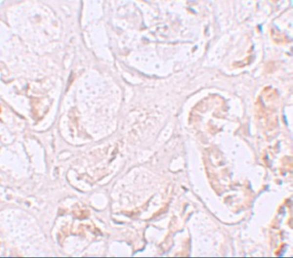
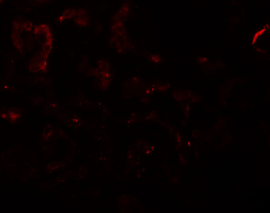
A13838 has IHC and IF images from human kidney tissue (catalog image captions). Its listed reactivity includes human, mouse and rat (catalog reactivity).
A13838 is listed for IHC-P and shows SLAMF9 staining in human kidney tissue at 2.5 μg/mL (catalog applications; IHC image caption). A13838 is also listed for IF and shows human kidney staining at 20 μg/mL (catalog applications; IF image caption).
Which to pick: Choose A13838 for paraffin-section IHC: IHC-P is listed, and its own IHC caption documents human kidney staining; the fixative is unreported (catalog applications; IHC image caption). For fluorescence, A13838 has a human kidney IF image, but ICC validation is unreported (catalog applications; IF image caption). A13838 lists human, mouse and rat reactivity; its supplied IHC and IF images document human tissue only (catalog reactivity; image captions).