This website uses cookies to ensure you get the best experience on our website.


- Table of Contents
This guide plans chromogenic paraffin IHC with antibody A02622 at 1:50–1:200 (datasheet A02622). Use salivary gland glandular cells as a high-staining reference and adipocytes as a negative comparison while assessing cytoplasmic and membranous signal (HPA tissue IHC).

Expected localisation, validated protocols, controls and antibodies — the at-a-glance facts below, then the full design guide.
| Expected localisation | Variable cytoplasmic and membranous tissue staining (HPA tissue IHC) | |
| Staining pattern | Glandular and other cells: variable cytoplasmic/membranous stain (HPA tissue IHC) | |
| Antigen retrieval | Tris pH 9.0 HIER, heat-mediated (datasheet A02622) | |
| Positive control | Salivary gland+4 more · see all | |
| Negative control | Adipose tissue+4 more · see all |
| Fixation | Keep formalin fixation consistent across sections. (standard IHC practice; not target-specific) | |
| Caveat | Staining has low concordance with RNA expression (HPA tissue IHC) | |
| Regulation | Iron/cobalt-linked neuronal degradation (UniProt) | |
| Isoform / epitope | Five isoforms; verify epitope conservation and membrane side (UniProt) |
The catalog antibody’s IHC-P protocol is complemented by published SLC11A2 tissue-staining methods with usable parameters (PMC13576891; PMC12845770).
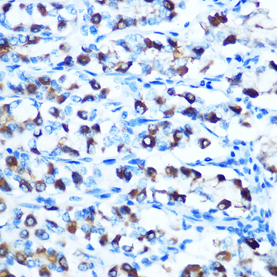
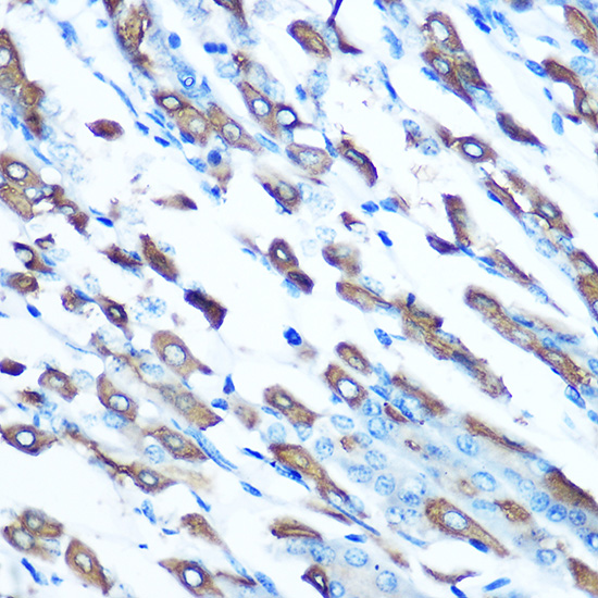
| Sample | Paraffin-embedded rat stomach tissue; fixative not specified (datasheet A02622) |
| Fixation | Image fixative and duration unreported (datasheet A02622); verify before use. |
| Sectioning | 4–5 µm sections on charged slides (standard) |
| Deparaffinisation | Xylene, graded ethanol series to water (standard) |
| Antigen retrieval | Heat retrieval: Tris pH 9.0 (datasheet A02622); 20 min, 95–100 °C (standard) |
| Peroxidase block | 3% H2O2, 10 min, room temperature (standard) |
| Blocking | 10% normal serum of the secondary host, 30 min, room temperature (standard) |
| Primary antibody | Rabbit anti-SLC11A2, 1:50-1:200 (datasheet A02622) |
| Primary incubation | Overnight at 4 °C (standard) |
| Detection | HRP-polymer secondary, DAB chromogen 5–10 min (standard) |
| Counterstain | Hematoxylin, blue, dehydrate and mount (standard) |
| Expected result | SLC11A2-positive staining in glandular cells of salivary gland (HPA tissue IHC: High). HPA tissue profile: Cytoplasmic and membranous expression at variable levels in several different cell types. No signal in the no-primary control. |
SLC11A2 is a 12-pass membrane protein found predominantly in early endosomes beneath the apical membrane of polarized epithelia, with other membrane locations annotated (UniProt P49281 topology and subcellular location). In paraffin sections, expect variable cytoplasmic and membranous staining; salivary glandular cells provide the strongest listed example (HPA tissue IHC: High). Interpret any pattern cautiously because tissue IHC reliability is Uncertain (HPA tissue IHC).
| Salivary glandular cells show distinct cytoplasmic and membranous chromogenic staining. | This fits the strongest listed tissue example, rated High, and HPA’s overall cytoplasmic and membranous profile (HPA tissue IHC). An apical or punctate intracellular emphasis is biologically plausible from early-endosome localization beneath the apical membrane; routine chromogenic IHC alone cannot identify individual organelles (UniProt P49281 subcellular location; standard IHC practice). |
| Glandular cells or respiratory epithelial cells stain less strongly than the salivary example. | Medium staining is reported in adrenal, breast, cervix and colon glandular cells and bronchial respiratory epithelial cells; duodenal and small-intestinal glandular cells are listed as Low (HPA tissue IHC). Compare the actual cell population with its corresponding HPA entry. Variable intensity across tissues is consistent with the HPA profile, but staining strength alone cannot establish specificity (HPA tissue IHC: Uncertain). |
| Signal is concentrated in nuclei or appears as a uniform nuclear stain. | A predominantly nuclear pattern conflicts with the annotated membrane locations and 12 transmembrane segments (UniProt P49281 topology and subcellular location). Treat it as suspect: inspect morphology and staining controls, then reassess antibody specificity and detection background (standard IHC practice). Some intracellular signal is expected, but a nuclear-only result does not match the supplied localization evidence (UniProt P49281; HPA tissue IHC). |
| Strong staining appears in a cell population HPA lists as Not detected. | Examples include adipocytes, lymph-node germinal-center cells and smooth-muscle cells (HPA tissue IHC: Not detected). Check cell identity and compare a no-primary control: cross-reactivity or endogenous detection activity can mimic target staining (standard IHC practice). A discrepant field is a reason to investigate, not proof of absence or artifact, because HPA rates tissue IHC reliability Uncertain (HPA tissue IHC). |
| The section shows diffuse haze, or salivary glandular cells show no discernible signal. | Diffuse staining obscures the cytoplasmic and membranous pattern reported by HPA; examine no-primary background and staining distribution (HPA tissue IHC; standard IHC practice). Absent signal in the listed High salivary glandular population prompts a review of section quality, retrieval and detection controls (HPA tissue IHC: High; standard IHC practice). One negative section cannot establish target absence (standard IHC practice). |
| Cell population and tissue context | HPA reports variable cytoplasmic and membranous staining, with High salivary glandular staining, several Medium populations and several Not detected populations (HPA tissue IHC). Score named cell populations rather than treating an entire tissue as uniformly positive or negative; HPA’s tissue IHC reliability is Uncertain (HPA tissue IHC; standard IHC practice). |
| Membrane topology and antibody epitope | SLC11A2 has 12 transmembrane segments and both cytoplasmic and extracellular regions (UniProt P49281 topology). The supplied record does not identify the antibody epitope, so topology cannot predict which antigen-retrieval condition will work. Compare retrieval conditions using appropriate controls as a general IHC optimization step, without assuming SLC11A2-specific fixation sensitivity (standard IHC practice). |
| Isoform coverage | UniProt lists 5 isoforms (UniProt P49281 isoforms). The supplied evidence does not map the catalog antibody’s epitope to those isoforms, so a stained section cannot establish which isoform contributed the signal. Report staining as SLC11A2-associated unless separate antibody characterization supports a narrower interpretation (UniProt P49281 isoforms; standard IHC practice). |
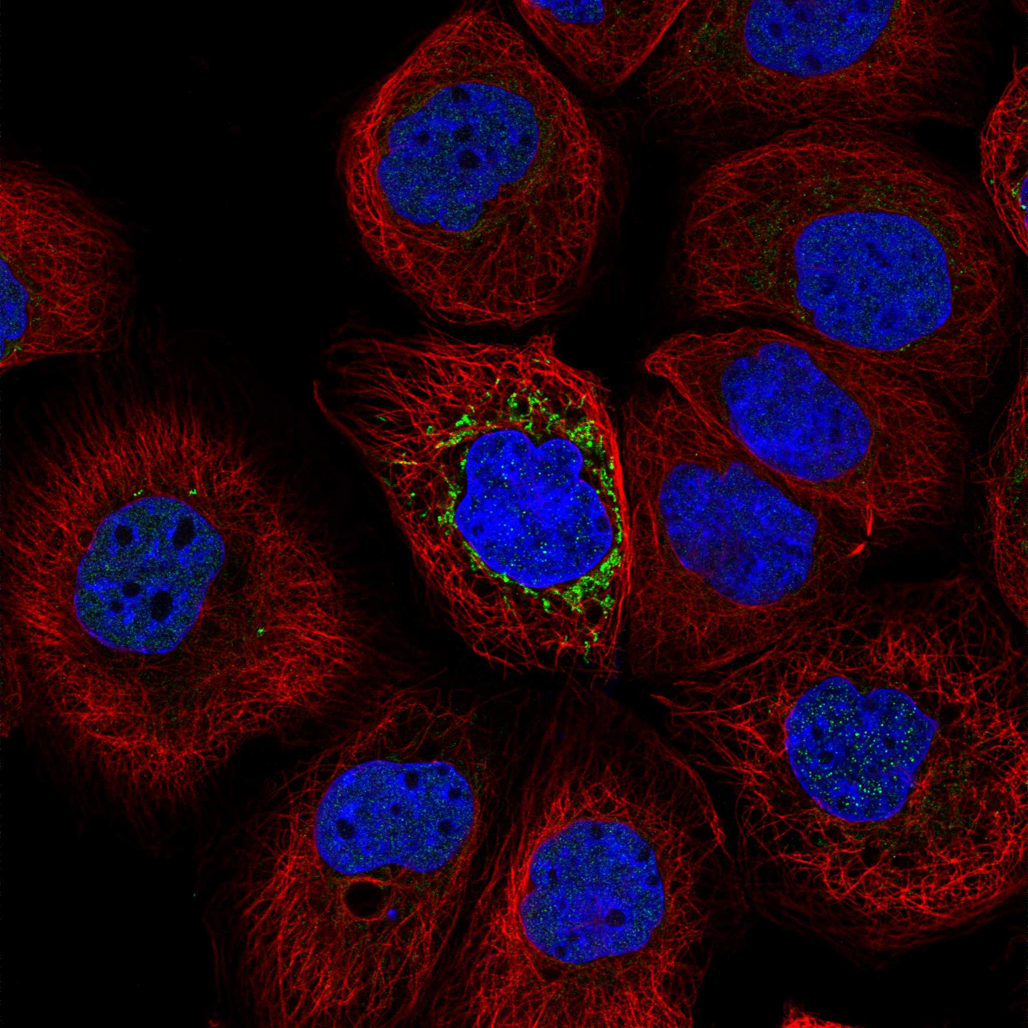
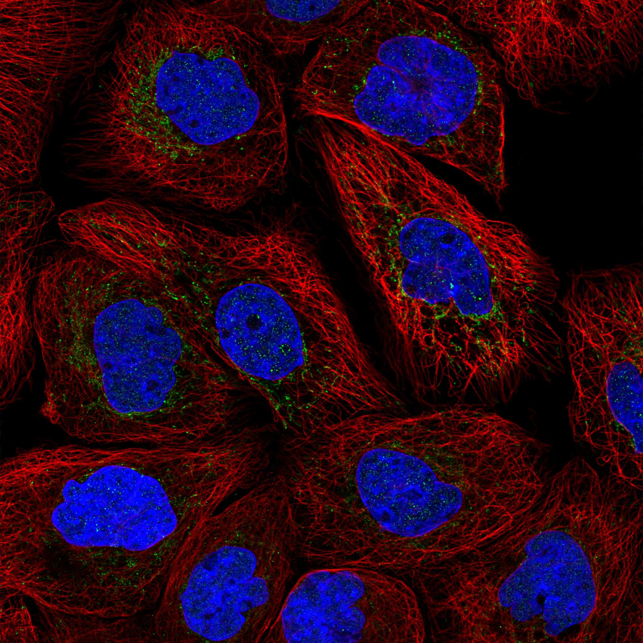
| IF/ICC Q&A: should the IHC pattern match fluorescence images? | HPA ICC-IF reports a supported mitochondrial localization and lists A-431, U-251MG and U2OS images (HPA subcellular ICC-IF). UniProt also annotates the mitochondrial outer membrane, alongside endosomal and other membranes (UniProt P49281 subcellular location). These observations can inform interpretation, but fluorescence localization does not establish an IHC-P protocol or validate every chromogenic tissue signal (HPA subcellular ICC-IF; standard IHC practice). |
| Situation | Likely cause | Next action |
|---|---|---|
| No staining in salivary glandular cells. | A technical failure is possible even though this population is listed as High; HPA tissue IHC reliability remains Uncertain (HPA tissue IHC; standard IHC practice). | Check section integrity and the run’s positive and detection controls; then review retrieval and antibody conditions using the catalog IHC-P instructions (standard IHC practice). |
| Only nuclear staining is visible. | Nuclear-only staining disagrees with the supplied membrane localization and topology (UniProt P49281 topology and subcellular location). | Inspect morphology and no-primary background, then compare with an independently validated antibody if available; do not score nuclear signal as the expected pattern (standard IHC practice; UniProt P49281). |
| Diffuse brown background hides cell boundaries. | Nonspecific reagent binding or detection background may obscure the variable cytoplasmic and membranous pattern (standard IHC practice; HPA tissue IHC). | Review no-primary control and wash and blocking conditions; score only signal that remains distinguishable in the relevant cells (standard IHC practice). |
| Strong signal appears in HPA Not detected cells. | Cell misidentification, antibody cross-reactivity or endogenous detection activity may explain the discrepancy; an HPA Not detected entry is not an absolute exclusion (HPA tissue IHC: Uncertain; standard IHC practice). | Confirm the cell population and check a no-primary control. For chromogenic detection, assess endogenous enzyme background with an appropriate control (standard IHC practice). |
| Duodenal or small-intestinal glandular staining is faint. | Both populations are listed as Low, so a weak result can agree with the tissue profile (HPA tissue IHC: Low). | Compare them with a stronger listed population and the same run’s controls before changing retrieval or detection conditions (HPA tissue IHC: High in salivary glandular cells; standard IHC practice). |
| Punctate signal and apparent surface staining differ between cells. | SLC11A2 has early-endosome and apical membrane annotations, while HPA reports variable cytoplasmic and membranous tissue staining (UniProt P49281 subcellular location; HPA tissue IHC). | Record compartment, cell type and intensity separately. Seek additional localization evidence before assigning individual chromogenic puncta to a named organelle (standard IHC practice). |


Comprehensive Human Protein Atlas IHC scoring per tissue (reliability: Uncertain — Low consistency between antibody staining and RNA expression data.). Rows are taken directly from the HPA tissue chart — click any row's HPA link to view the source.
| Tissue | Cell type | Level | Evidence | Source |
|---|---|---|---|---|
| Salivary gland | Glandular cells | High | Protein (IHC) | HPA → |
| Adrenal gland | Glandular cells | Medium | Protein (IHC) | HPA → |
| Breast | Glandular cells | Medium | Protein (IHC) | HPA → |
| Bronchus | Respiratory epithelial cells | Medium | Protein (IHC) | HPA → |
| Caudate | Neuronal cells | Medium | Protein (IHC) | HPA → |
| Tissue | Cell type | Level | Evidence | Source |
|---|---|---|---|---|
| Adipose tissue | Adipocytes | Not detected | Protein (IHC) | HPA → |
| Cerebellum | Cells in granular layer | Not detected | Protein (IHC) | HPA → |
| Hippocampus | Glial cells | Not detected | Protein (IHC) | HPA → |
| Lymph node | Germinal center cells | Not detected | Protein (IHC) | HPA → |
| Oral mucosa | Squamous epithelial cells | Not detected | Protein (IHC) | HPA → |
Troubleshoot SLC11A2 staining in paraffin sections by checking retrieval, compartment pattern and tissue controls before comparing chromogenic signal between samples.
The catalog lists human-, mouse-, and rat-reactive SLC11A2 antibodies for IHC, with paraffin-section images from rat and mouse stomach; IF is listed for one antibody without an image (catalog applications; A02622 image captions).
A02622 is listed for IHC in human, mouse, and rat, with paraffin-section images from rat and mouse stomach (catalog applications and reactivity; A02622 image captions). A02622-2 is listed for IHC and IF in all three species, but has no supplied IHC or IF image (catalog applications, reactivity, and image alts).
Which to pick: For paraffin-section IHC, choose A02622: its own captions document staining in rat and mouse stomach; the fixative is unreported (A02622 image captions). For IF, choose A02622-2 because IF is listed at 1:50; ICC is unlisted and no IF image is supplied (A02622-2 catalog applications, dilution, and image alts). Both are rabbit polyclonal antibodies listed as reactive with human, mouse, and rat, so species coverage alone does not distinguish them (catalog host, dilution_raw, and reactivity).