This website uses cookies to ensure you get the best experience on our website.


- Table of Contents
Plan paraffin IHC for SLC14A1 with the catalog antibody at 2–5 μg/ml (datasheet A04926-2). Use erythrocyte staining as a reference (HPA tissue IHC) and assess its cytoplasmic pattern alongside the expected membrane location (UniProt).

Expected localisation, validated protocols, controls and antibodies — the at-a-glance facts below, then the full design guide.
| Expected localisation | Cytoplasmic stain (HPA tissue IHC); membrane expected (UniProt) | |
| Staining pattern | Cytoplasmic staining, most abundant in erythrocytes (HPA tissue IHC) | |
| Antigen retrieval | EDTA pH 8.0 HIER, heat-mediated (datasheet A04926-2) | |
| Positive control | Cerebellum+4 more · see all | |
| Negative control | Adipose tissue+4 more · see all |
| Fixation | Keep fixation consistent across sections (standard IHC practice; not target-specific) | |
| Caveat | Antibody staining has low concordance with RNA (HPA tissue IHC) | |
| Regulation | Regulation not established (UniProt) | |
| Isoform / epitope | Two isoforms; map extracellular vs cytoplasmic epitopes (UniProt) |
The catalog antibody's IHC-P protocol (datasheet A04926-2) is accompanied by published SLC14A1 IHC methods for urothelial, colorectal and prostate specimens (PMC7546005; PMC11282742; PMC9579171).
| Sample | Paraffin-embedded human melanoma tissue; fixative not specified (datasheet A04926-2) |
| Fixation | Image fixative and duration unreported (datasheet A04926-2); verify before use. |
| Sectioning | 4–5 µm sections on charged slides (standard) |
| Deparaffinisation | Xylene, graded ethanol series to water (standard) |
| Antigen retrieval | Heat retrieval: EDTA pH 8.0 (datasheet A04926-2); 20 min, 95–100 °C (standard) |
| Peroxidase block | 3% H2O2, 10 min, room temperature (standard) |
| Blocking | 10% goat serum (datasheet A04926-2) |
| Primary antibody | Rabbit anti-SLC14A1, 2-5 μg/ml (datasheet A04926-2) |
| Primary incubation | Overnight at 4 °C (datasheet A04926-2) |
| Detection | HRP-conjugated secondary, DAB chromogen (datasheet A04926-2) |
| Counterstain | Hematoxylin, blue, dehydrate and mount (standard) |
| Expected result | SLC14A1-positive staining in processes in granular layer of cerebellum (HPA tissue IHC: High). HPA tissue profile: Cytoplasmic expression in several tissues, most abundant in erythrocytes. No signal in the no-primary control. |
SLC14A1 is a nine-pass membrane transporter; UniProt places it at the cell membrane and restricts it to the basolateral membrane in portions of urothelium (UniProt Q13336 topology and subcellular location). On IHC, expect conspicuous erythrocyte staining and assess cell-specific patterns such as colon enterocyte microvilli and placental endothelium (HPA tissue IHC). HPA rates the tissue IHC antibody Approved but reports low consistency between staining and RNA expression (HPA tissue IHC).
| Distinct staining outlines erythrocytes or follows the microvilli of colon enterocytes. | This fits the erythrocyte-rich tissue profile and High colon microvillar signal (HPA tissue IHC). Interpret a discrete surface pattern alongside tissue anatomy; UniProt places SLC14A1 at membranes but does not establish which IHC signal in a mixed section is specific (UniProt Q13336 subcellular location; HPA tissue IHC: low RNA–staining consistency). |
| Staining is predominantly nuclear in the cells being scored. | Nuclear dominance conflicts with the cell-membrane assignment and supported plasma-membrane ICC-IF localization (UniProt Q13336 subcellular location; HPA subcellular ICC-IF). Treat it as an unexpected pattern and review controls before calling the cells positive; HPA also describes cytoplasmic tissue staining, so a cytoplasmic component alone is not proof of artefact (HPA tissue IHC). |
| Strong staining appears in adipocytes or bone-marrow hematopoietic cells. | Those cell populations were Not detected in the supplied HPA tissue IHC observations (HPA tissue IHC). Consider cross-reactivity or endogenous chromogen activity, then compare a reagent-omission control and a known-positive section (general IHC practice). An HPA negative is an observed result under its conditions, not proof that every specimen must be negative (HPA tissue IHC). |
| Color spreads across cells and extracellular areas without a defined cellular pattern. | Diffuse background prevents reliable compartment or cell-type assignment. Excess primary or detection reagent, incomplete blocking, and inadequate washes are general IHC possibilities (general IHC practice). Do not score background as SLC14A1 merely because the tissue contains a listed positive cell population (HPA tissue IHC). |
| No signal appears in colon enterocyte microvilli or placental endothelium. | Both are listed as High in HPA tissue IHC (HPA tissue IHC). If an appropriate positive section fails in the same run, first assess section integrity, retrieval, antibody dilution, and detection controls (general IHC practice). A failed positive control makes a negative study section uninterpretable; it does not establish absent SLC14A1 expression (general IHC practice). |
| Membrane topology and compartment | SLC14A1 has 9 annotated transmembrane segments and a cell-membrane assignment (UniProt Q13336 topology and subcellular location). Use cell outlines and polarized epithelial surfaces as localization clues; the record does not identify the antibody epitope, so topology alone cannot prescribe permeabilisation or retrieval (UniProt Q13336 topology; general IHC practice). |
| Cell-specific tissue evidence | HPA lists High colon enterocyte microvilli, placental endothelial cells, and cerebellar granular-layer processes; its overall profile says staining is most abundant in erythrocytes (HPA tissue IHC). Choose and score a defined structure rather than treating every cell in one tissue as an expected positive (HPA tissue IHC; general IHC practice). |
| Agreement between staining and RNA | The HPA tissue IHC reliability label is Approved, with low consistency between antibody staining and RNA expression and staining in unannotated cells or structures (HPA tissue IHC). That caveat calls for cautious interpretation of an unexpected cell population, even when the slide has a visually strong signal (HPA tissue IHC). |
| Glycosylation and isoforms | UniProt lists one glycosylation site at residue 211 and 2 isoforms (UniProt Q13336). These annotations do not show whether either feature changes this antibody’s staining, epitope access, or performance in paraffin sections; antibody-specific evidence would be needed for that interpretation (UniProt Q13336; general IHC practice). |
| IF/ICC Q: What localization can be expected? | A: HPA reports supported plasma-membrane localization in ICC-IF images from RT-4 using an ICC-Supported antibody; its IHC-Approved antibody is a different listed reagent (HPA subcellular ICC-IF; HPA antibodies). Use that result as compartment context for IHC, without assuming the two antibody validations are interchangeable (HPA antibodies). |
| Situation | Likely cause | Next action |
|---|---|---|
| The known-positive section has no visible signal. | A run-level failure is possible when a structure listed as High, such as colon enterocyte microvilli, is also blank (HPA tissue IHC; general IHC practice). | Check tissue preservation and the detection run, then review the catalog antibody’s IHC-P instructions for the specified retrieval and dilution; change one general IHC variable at a time (general IHC practice). |
| Study tissue is negative, but the positive control stains as expected. | Expression may be absent or below the assay’s detection limit in the scored cells; HPA staining varies by cell type and tissue (HPA tissue IHC; general IHC practice). | Report the scored cell population and control result. Avoid inferring a tissue-wide negative from one compartment, and compare the relevant HPA cell-level observation when available (HPA tissue IHC; general IHC practice). |
| Adipocytes or marrow hematopoietic cells stain strongly. | HPA lists these populations as Not detected; nonspecific binding or endogenous detection activity is possible (HPA tissue IHC; general IHC practice). | Run a primary-antibody omission control, inspect whether color persists, and review blocking and detection reagents before assigning target positivity (general IHC practice). |
| Nuclear color dominates while cell surfaces are indistinct. | The pattern conflicts with UniProt’s membrane location and HPA’s supported plasma-membrane ICC-IF localization (UniProt Q13336 subcellular location; HPA subcellular ICC-IF). | Check a positive tissue structure and reagent-omission control, then review antibody dilution and detection background (HPA tissue IHC; general IHC practice). Do not use nuclear intensity alone as the positive score (UniProt Q13336 subcellular location). |
| Diffuse chromogen obscures enterocyte microvilli or endothelial borders. | Excess reagent, incomplete blocking, or insufficient washing can obscure cellular detail (general IHC practice). | Compare the reagent-omission control, review blocking and washes, and adjust detection or antibody concentration within the validated IHC workflow (general IHC practice). Score only a pattern that resolves the relevant cells or structures (HPA tissue IHC). |
| A strong tissue pattern conflicts with the expected cell population. | HPA explicitly reports low consistency between antibody staining and RNA expression and notes staining in unannotated cells or structures (HPA tissue IHC). | Document the exact cells and compartment, check controls, and avoid treating intensity alone as validation (HPA tissue IHC; general IHC practice). If testing another antibody, review its application-specific validation independently (HPA antibodies). |
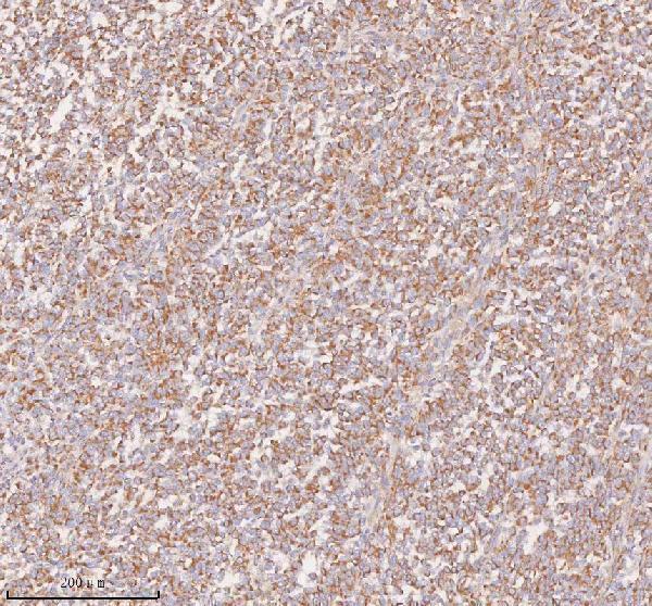
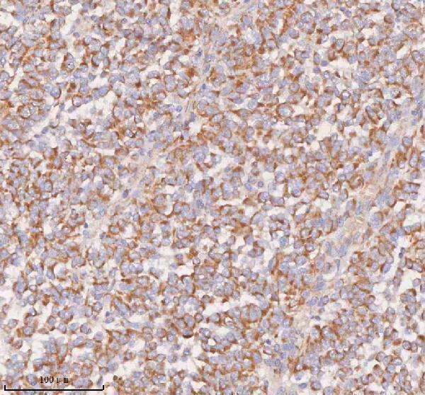


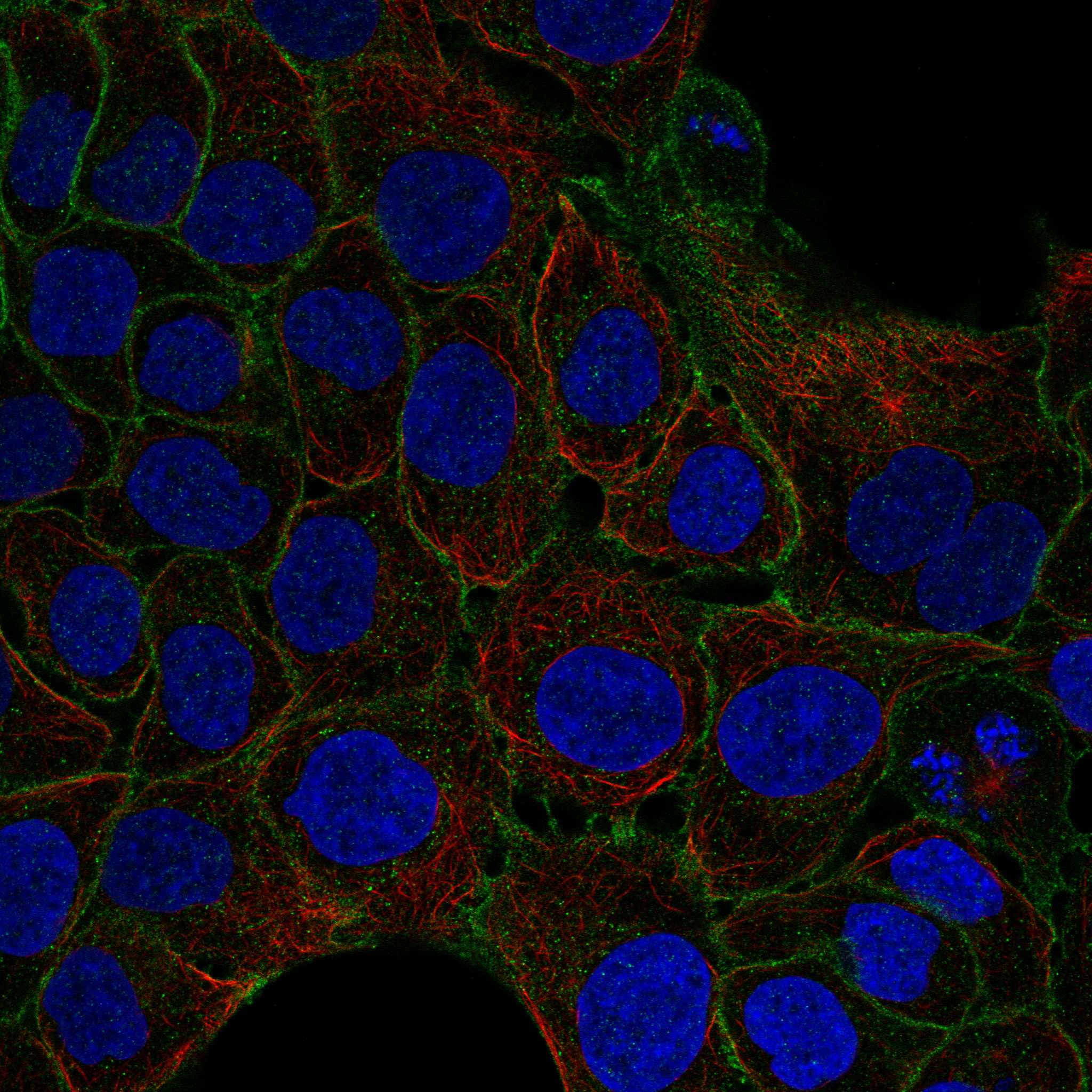
Comprehensive Human Protein Atlas IHC scoring per tissue (reliability: Approved — Low consistency between antibody staining and RNA expression data. Antibody staining in cells/structures not annotated, view images.). Rows are taken directly from the HPA tissue chart — click any row's HPA link to view the source.
| Tissue | Cell type | Level | Evidence | Source |
|---|---|---|---|---|
| Cerebellum | Processes in granular layer | High | Protein (IHC) | HPA → |
| Colon | Enterocytes - Microvilli | High | Protein (IHC) | HPA → |
| Placenta | Endothelial cells | High | Protein (IHC) | HPA → |
| Appendix | Enterocytes | Medium | Protein (IHC) | HPA → |
| Duodenum | Goblet cells | Medium | Protein (IHC) | HPA → |
| Tissue | Cell type | Level | Evidence | Source |
|---|---|---|---|---|
| Adipose tissue | Adipocytes | Not detected | Protein (IHC) | HPA → |
| Bone marrow | Hematopoietic cells | Not detected | Protein (IHC) | HPA → |
| Breast | Adipocytes | Not detected | Protein (IHC) | HPA → |
| Caudate | Glial cells | Not detected | Protein (IHC) | HPA → |
| Endometrium | Cells in endometrial stroma | Not detected | Protein (IHC) | HPA → |
Troubleshoot SLC14A1 chromogenic IHC in paraffin sections using the catalog antibody’s tissue image, protein annotations, and tissue staining data.
A04926-2 has real IHC data from paraffin-embedded human melanoma and pancreatic cancer sections (catalog image captions). Its listed reactivity covers human, mouse and rat (catalog reactivity).
A04926-2 is listed for IHC and for human, mouse and rat reactivity (catalog applications/reactivity). Its IHC images show paraffin-embedded human melanoma and pancreatic cancer sections (A04926-2 image captions).
Which to pick: For tissue IHC, choose A04926-2 at 2–5 μg/ml (catalog IHC dilution); its image caption documents EDTA retrieval at pH 8.0 and 2 μg/ml primary antibody on a paraffin-embedded human section, with the fixative unreported (A04926-2 image caption). No IF/ICC-validated SKU is supplied: A04926-2 has no IF application, image or dilution listed (catalog applications/IF fields). For cross-species IHC planning, A04926-2 lists human, mouse and rat reactivity, while its IHC dilution listing specifies human and rat and its shown IHC sections are human (catalog reactivity/dilution; A04926-2 image captions).