SLC16A1 / Monocarboxylate transporter 1 · IHC design guide

Design Immunohistochemistry for SLC16A1

Plan chromogenic IHC for SLC16A1 in paraffin sections using its cytoplasmic and membranous tissue pattern (HPA tissue IHC). Use high-staining colon glandular cells as a positive reference and assess membrane staining in light of BSG/EMB-dependent surface expression (HPA tissue IHC; UniProt).

Evidence assembled Oct 2026 · For research use; verify linked source records and product datasheet before use
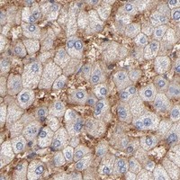
Immunohistochemistry protocol sheet for SLC16A1 (IHC for SLC16A1): expected localisation Cytoplasmic and membranous staining in most tissues (HPA tissue IHC), antibody A02240-1, validated IHC image, and IHC protocol steps
Printable SLC16A1 IHC protocol sheet — expected localisation Cytoplasmic and membranous staining in most tissues (HPA tissue IHC), antibody A02240-1, controls and protocol steps. Open the full SLC16A1 IHC guide →

SLC16A1 Immunohistochemistry Experimental Design Guide

Expected localisation, validated protocols, controls and antibodies — the at-a-glance facts below, then the full design guide.

Must know before staining
Expected localisation Cytoplasmic and membranous staining in most tissues (HPA tissue IHC)
Staining pattern Colon glandular cells: high cytoplasmic and membranous staining (HPA tissue IHC)
Antigen retrieval EDTA pH 8.0 HIER, heat-mediated (datasheet A02240-1)
Positive control ⓘ Appendix+4 more · see all
Negative control ⓘ Adipose tissue+4 more · see all
Important caveats
Reasons your staining may differ from the expected pattern.
Fixation Keep fixation consistent across sections (standard IHC practice; not target-specific); Matched tissue-IHC evidence does not establish the fixation claim. Validate the specimen-specific method before use. (selected-SKU IHC image A02240-1)
Caveat Staining has medium consistency with RNA expression (HPA tissue IHC)
Regulation Surface display depends on BSG/EMB (UniProt)
Isoform / epitope 2 isoforms; check epitope location and membrane side (UniProt)
Section 1

Recommended SLC16A1 IHC & IF Protocols

The catalog antibody uses EDTA pH 8.0 retrieval (datasheet A02240-1). The published IHC methods below cover pancreatic cancer, colon and liver biopsies, and mucosal sections (PMC11094506; PMC11237055; PMC12740759).

Recommended immunohistochemistry (IHC-P) protocol parameters
SampleParaffin-embedded mouse kidney tissue; fixative not specified (datasheet A02240-1)
FixationImage fixative and duration unreported (datasheet A02240-1); verify before use.
Sectioning4–5 µm sections on charged slides (standard)
DeparaffinisationXylene, graded ethanol series to water (standard)
Antigen retrievalHeat retrieval: EDTA pH 8.0 (datasheet A02240-1); 20 min, 95–100 °C (standard)
Peroxidase block3% H2O2, 10 min, room temperature (standard)
Blocking10% goat serum (datasheet A02240-1)
Primary antibodyRabbit anti-SLC16A1, 1:50 recommended; image 1:100 (datasheet A02240-1)
Primary incubationOvernight at 4 °C (datasheet A02240-1)
DetectionHRP-conjugated secondary, DAB chromogen (datasheet A02240-1)
CounterstainHematoxylin, blue, dehydrate and mount (standard)
Expected resultSLC16A1-positive staining in glandular cells of appendix (HPA tissue IHC: High). HPA tissue profile: Cytoplasmic and membranous expression in most tissues. No signal in the no-primary control.
💡Decision noteStart with heat-mediated EDTA pH 8.0 retrieval (datasheet A02240-1); citrate for 60 minutes is a published alternative for colon and liver biopsies (PMC11237055).
Section 2

What Is the Expected SLC16A1 Staining Pattern?

SLC16A1 (MCT1) is a 12-pass membrane transporter expected at the cell surface, including apical or basolateral membranes (UniProt P53985 topology and subcellular location). On paraffin IHC, expect membranous and cytoplasmic staining in many tissues, with strong staining in specified glandular cells and testicular Sertoli cells (HPA tissue IHC). HPA rates tissue staining reliability “Enhanced,” while noting medium consistency with RNA expression (HPA tissue IHC).

What am I looking at on my slide?
Distinct membrane staining in colon or duodenal glandular cells, possibly with cytoplasmic signal.This fits a positive tissue pattern: HPA reports High staining in those cells and describes expression as cytoplasmic and membranous across most tissues (HPA tissue IHC). Record the membrane component and the stained cell population separately; cytoplasmic signal alone does not establish cell-surface localisation (UniProt P53985 subcellular location).
Strong nuclear-only staining, with no convincing membrane outline.Nuclear-only signal conflicts with the documented membrane localisation and HPA’s cytoplasmic and membranous tissue profile (UniProt P53985 subcellular location; HPA tissue IHC). Treat it as a suspected artefact and review controls and morphology before assigning it to SLC16A1. Cytoplasmic staining by itself is not automatically erroneous (HPA tissue IHC).
Prominent staining in adipocytes or pancreatic exocrine glandular cells.HPA lists those particular cell populations as Not detected, so a strong result there warrants scrutiny for cross-reactivity or endogenous chromogenic activity (HPA tissue IHC; general IHC practice). This comparison applies to the named cells, not every cell in adipose tissue or pancreas; check tissue anatomy and controls before calling it nonspecific.
Uniform colour over tissue, extracellular spaces, or the whole slide, without cell boundaries.A diffuse deposit that does not follow the documented cellular pattern is less convincing than defined glandular or membrane-associated staining (HPA tissue IHC; UniProt P53985 subcellular location). In chromogenic IHC, assess secondary-reagent background, residual endogenous detection activity, and wash quality with appropriate controls (general IHC practice).
No staining in colon glandular cells or testicular Sertoli cells.Absence of signal in a well-preserved positive-control population is a run-level warning: HPA reports High staining in both populations (HPA tissue IHC). First verify that the expected cells are present; then review antibody suitability, detection reagents, and the control slide before interpreting a study specimen as negative (general IHC practice).
💡Expected SLC16A1 appearanceA convincing positive has defined membrane-associated staining, sometimes with cytoplasmic signal, in High-staining glandular cells or Sertoli cells; nuclear-only or diffuse cell-independent colour is suspect (UniProt P53985 subcellular location; HPA tissue IHC; general IHC practice).
How each factor affects the staining
Membrane topology and traffickingSLC16A1 has 12 transmembrane segments, and cell-surface expression requires BSG or EMB association (UniProt P53985 topology and subunit data). Interpret the visible compartment as an observation; staining intensity alone cannot show whether transporter reached the surface.
Cell population chosen for comparisonHPA reports High staining in appendix, colon, duodenum, endometrium, epididymis, prostate, and stomach glandular cells, plus testicular Sertoli cells (HPA tissue IHC). Its negative calls name specific populations, including adipocytes and pancreatic exocrine glandular cells (HPA tissue IHC).
Strength of the reference patternHPA assigns tissue IHC reliability “Enhanced” but describes only medium consistency between antibody staining and RNA expression (HPA tissue IHC). Use the listed cell-level patterns as references, while checking morphology and assay controls when a specimen differs.
Antibody validationHPA003324 has “Enhanced” IHC validation; CAB079035 has “Supported” IHC validation (HPA antibodies). These grades describe the listed antibodies, not every catalog antibody. HPA071055 and CAB017489 have no IHC grade in the supplied record (HPA antibodies).
IF/ICC: where should fluorescence appear?Mainly at the plasma membrane and cell junctions; HPA marks both locations “enhanced” in ICC-IF (HPA subcellular). This is an IF/ICC localisation reference, not an additional paraffin IHC protocol or a promise that every tissue cell will show junctional staining.
Why is my staining missing, weak or wrong?
SituationLikely causeNext action
Positive-control glandular cells show no chromogenic signal.The expected population may be absent from the section, or an assay step may have failed; HPA reports High staining in colon glandular cells (HPA tissue IHC).Confirm cell identity and section integrity, then inspect the IHC-validated antibody, retrieval conditions, detection reagents, and a known-positive control together (general IHC practice). No SLC16A1-specific retrieval condition is established here.
Staining is nuclear-only.The compartment conflicts with the documented membrane location and tissue pattern (UniProt P53985 subcellular location; HPA tissue IHC).Compare with a positive-control section and a no-primary control; assess whether colour follows nuclei or tissue pigment before accepting the signal (general IHC practice).
Adipocytes or pancreatic exocrine cells stain strongly.Those exact populations are listed as Not detected by HPA; cross-reactivity or endogenous activity may explain the discrepancy (HPA tissue IHC; general IHC practice).Recheck cell identity, examine the no-primary control, and compare staining with a documented positive population on a matched run (general IHC practice).
Diffuse background obscures gland boundaries.Background deposition or insufficient blocking or washing can obscure cellular localisation in chromogenic IHC (general IHC practice).Inspect no-primary and detection-only controls, review blocking and washes, and score only signal that resolves to identifiable cells (general IHC practice).
Cytoplasmic staining is present but membrane outlines are weak.HPA describes both cytoplasmic and membranous tissue expression; weak outlines therefore do not, by themselves, prove a false result (HPA tissue IHC).Document cytoplasmic and membrane components separately and compare the cell population with HPA’s tissue-level reference; avoid claiming confirmed surface localisation from diffuse cytoplasm (HPA tissue IHC; UniProt P53985 subcellular location).
A purported negative-control section has scattered stained cells.HPA’s “Not detected” entries refer to named cell types, and SLC16A1 is widely expressed across tissues (HPA tissue IHC; UniProt P53985 tissue specificity).Identify the stained cells before judging the control; compare the same cell type with its HPA entry, and use a no-primary control to assess assay background (HPA tissue IHC; general IHC practice).

Sample controls for SLC16A1 IHC & IF

🧪Run colon first: its glandular cells must show staining for a successful positive control (HPA: High in colon glandular cells; UniProt P53985: cell membrane). Run cervix glandular cells as the negative tissue (HPA: Not detected in cervix glandular cells); on the colon slide, stromal cells without membranous staining can serve as local background comparators, but their biological negative status is unverified (HPA: colon row specifies glandular cells; standard IHC practice).
Positive control tissue: Appendix (Glandular cells, HPA High)
Negative control tissue: Adipose tissue (HPA Not detected)
ICC-IF cell lines (HPA subcellular resource): HPA ICC-IF images show SLC16A1 in A-431, U-251MG, U2OS, HaCaT, U2OS, siRNA 2 (10x), U2OS, scrambled (10x), with annotated localisation: Plasma membrane (enhanced), Cell Junctions (enhanced) (HPA subcellular).
Technical controls: Include a no-primary, secondary-only control, a rabbit IgG isotype control matched to the primary's class and clonality where known, and validated SLC16A1 knockout material if available (selected A02240-1 caption: rabbit primary; standard IHC practice). Quench endogenous peroxidase for chromogenic detection and check colon luminal contents for nonspecific DAB signal (selected A02240-1 caption: HRP/DAB detection; standard IHC practice).
⚠️Feasibility: No target-specific fixation window or fixation effect is reported, and the selected A02240-1 paraffin-section caption does not state a fixative (selected A02240-1 caption). That caption uses heat-mediated retrieval in EDTA at pH 8.0; this is a starting condition to evaluate, not proof that retrieval is required for colon (selected A02240-1 caption). Whether frozen sections or IF are easier is unreported; HPA shows plasma membrane and cell junction IF localization in its imaged cell lines, while colon luminal contents can complicate chromogenic scoring (HPA subcellular; standard IHC practice).

HPA tissue IHC evidence for SLC16A1

Comprehensive Human Protein Atlas IHC scoring per tissue (reliability: Enhanced — Medium consistency between antibody staining and RNA expression data.). Rows are taken directly from the HPA tissue chart — click any row's HPA link to view the source.

Positive expression · recommended positive controls

TissueCell typeLevelEvidenceSource
Appendix Glandular cells High Protein (IHC) HPA →
Colon Glandular cells High Protein (IHC) HPA →
Duodenum Glandular cells High Protein (IHC) HPA →
Endometrium Glandular cells High Protein (IHC) HPA →
Epididymis Glandular cells High Protein (IHC) HPA →

Undetected expression · recommended negative controls

TissueCell typeLevelEvidenceSource
Adipose tissue Adipocytes Not detected Protein (IHC) HPA →
Caudate Glial cells Not detected Protein (IHC) HPA →
Cerebellum Cells in granular layer Not detected Protein (IHC) HPA →
Cervix Glandular cells Not detected Protein (IHC) HPA →
Hippocampus Glial cells Not detected Protein (IHC) HPA →
Section 3

Advanced SLC16A1 IHC Tips

Troubleshoot SLC16A1 staining by checking retrieval, membrane localisation, controls and scoring in paraffin-section chromogenic IHC (datasheet A02240-1; UniProt P53985).

What retrieval should I try first when SLC16A1 staining is weak?
Start with heat-mediated EDTA at pH 8.0 for paraffin-section IHC (datasheet A02240-1). The selected mouse kidney image used this retrieval before 1:100 primary incubation overnight at 4°C (datasheet A02240-1). If signal is weak, compare heating durations on serial sections while keeping antibody and detection conditions constant (standard IHC practice). SLC16A1 has 12 transmembrane segments, so epitope position may affect accessibility after retrieval; the catalog antibody's epitope is unspecified here (UniProt P53985 topology; datasheet A02240-1). Judge any gain in signal alongside tissue preservation, no-primary controls and the expected glandular-cell pattern (standard IHC practice; HPA tissue IHC).
Could fixation explain weak or uneven SLC16A1 staining?
The selected paraffin-section caption does not state a fixative, so target-specific fixation sensitivity is unknown (datasheet A02240-1). Record the fixative, fixation duration, tissue thickness and processing history for each specimen before comparing staining (standard IHC practice). On serial sections, hold retrieval at EDTA pH 8.0 and the primary at 1:100 overnight at 4°C while assessing processing-related variation (datasheet A02240-1; standard IHC practice). Check whether staining changes track fixation batches or occur alongside poor morphology, and review no-primary controls before attributing differences to SLC16A1 abundance (standard IHC practice). HPA tissue patterns and protein topology do not establish this antibody's fixation tolerance (HPA tissue IHC; UniProt P53985 topology).
Should I accept diffuse cytoplasmic staining as SLC16A1?
Look first for plasma-membrane staining, which may be apical or basolateral depending on the cell, and note any junctional accentuation (UniProt P53985 subcellular; HPA subcellular). HPA tissue IHC also reports cytoplasmic and membranous expression in most tissues, so cytoplasmic signal alone needs careful control-based assessment (HPA tissue IHC). Compare the same cell compartment across serial sections and examine boundaries at high magnification before scoring membrane positivity (standard IHC practice). Cell-surface expression requires BSG or EMB, making reduced membrane localisation biologically plausible without proving that a cytoplasmic stain is specific (UniProt P53985 subcellular). Record membranous and cytoplasmic staining separately, with a no-primary control for diffuse chromogen (standard IHC practice).
How do isoforms and epitope position affect this stain?
SLC16A1 has 2 annotated isoforms, but this payload does not map the catalog antibody's epitope or establish isoform recognition (UniProt P53985 isoforms; datasheet A02240-1). Its 12 transmembrane segments separate short extracellular loops from cytoplasmic regions, including residues 195–261 and 444–500 (UniProt P53985 topology). Phosphorylation is annotated at residues including 210, 213, 231, 461 and 466, but no effect on this antibody's staining is supplied (UniProt P53985 modified residues; datasheet A02240-1). Check the antibody's documented immunogen or epitope before assigning an isoform-specific interpretation (standard IHC practice). If that information remains unavailable, report staining as SLC16A1 immunoreactivity and use tissue and no-primary controls to assess its distribution (standard IHC practice).
How can IF help assess an ambiguous IHC pattern?
Use IF/ICC as a separate localisation check, with its own validated conditions rather than treating the paraffin-section caption as an IF protocol (datasheet A02240-1; standard IF practice). Multiplex SLC16A1 with a marker for the expected cell population, such as glandular cells in an HPA-positive tissue, and inspect whether membrane signal belongs to those cells (HPA tissue IHC; UniProt P53985 subcellular). Choose fluorophores after checking tissue autofluorescence and include single-label controls to assess channel bleed-through (standard IF practice). Adjust permeabilisation to the documented epitope: a cytoplasmic epitope may require access, whereas excessive permeabilisation can complicate membrane-boundary assessment (UniProt P53985 topology; standard IF practice). The antibody's epitope and IF fixation conditions are not supplied here, so verify both before interpreting discordance with IHC (datasheet A02240-1).
What should I check when DAB background obscures membrane staining?
Inspect a no-primary control first to separate detection-system background from primary-antibody staining (standard IHC practice). The selected caption used 10% goat serum block, 1:100 primary overnight at 4°C, a peroxidase-conjugated secondary and DAB development (datasheet A02240-1). Apply an endogenous peroxidase block and control DAB development as general chromogenic IHC steps, especially when diffuse colour hides cell borders (standard IHC practice). If background remains high, compare primary dilutions and wash conditions on serial sections while retaining the validated EDTA pH 8.0 retrieval as the reference (datasheet A02240-1; standard IHC practice). Require spatially resolved cell staining before calling broad brown deposits SLC16A1 positive (standard IHC practice).
How should I score SLC16A1 in paraffin sections? ⚠ ANSWER MARKED FOR VERIFICATION
Define the cell population and compartment before scoring, separating membrane from cytoplasmic signal because both patterns are reported in tissue IHC (HPA tissue IHC; standard IHC practice). For a defined cell population, record percent positive cells and intensity, then calculate an H-score if graded intensity is reproducible (standard IHC practice). When cells are sparse, report positive-cell density per mm² of viable tissue or a defined region of interest (standard IHC practice). Normalise comparisons to the same cell type, viable area, section thickness, staining run and threshold, with controls reviewed for each run (standard IHC practice). Report membrane and cytoplasmic scores separately so a compartment shift does not masquerade as a change in total expression (UniProt P53985 subcellular; standard IHC practice).
When is SLC16A1 staining convincing rather than artefactual?
A convincing result has cell-associated staining with a plausible membrane distribution and agrees with control behaviour; HPA also reports cytoplasmic tissue staining (UniProt P53985 subcellular; HPA tissue IHC; standard IHC practice). Compare candidate positive cells with documented high-staining glandular cells or Sertoli cells, while respecting the specimen's actual cell composition (HPA tissue IHC; standard IHC practice). Treat nuclear-only staining, cut-edge accentuation, necrotic deposits and colour persisting in a no-primary control as reasons to investigate artefact (UniProt P53985 subcellular; standard IHC practice). Endogenous peroxidase can mimic DAB positivity, so examine the peroxidase control when staining follows that pattern (standard IHC practice). Interpret a negative area cautiously because HPA reports variable cell-type staining and only medium staining–RNA consistency (HPA tissue IHC).
Boster reagents

Best SLC16A1 / Monocarboxylate transporter 1 IHC Antibodies

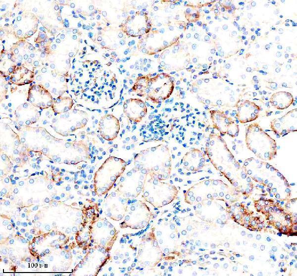
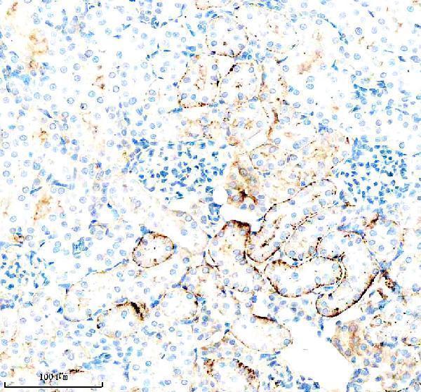
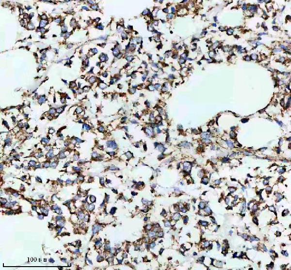
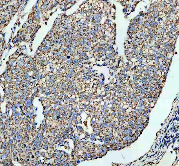
Two anti-SLC16A1 antibodies have paraffin-section IHC images: A02240-1 in mouse and rat kidney, and M02240 in human breast and cervical cancer tissue (catalog IHC image captions).

Real IHC data IHC analysis of MCT1/SLC16A1 using anti-MCT1/SLC16A1 antibody (A02240-1). MCT1/SLC16A1 was detected in a paraffin-embedded section of mouse kidney tissue. Heat mediated antigen retrieval was performed in EDTA buffer (pH 8.0, epitope retrieval solution). The tissue section was blocked with 10% goat serum. The tissue section was then incubated with 1:100 rabbit anti-MCT1/SLC16A1 Antibody (A02240-1) overnight at 4°C. Peroxidase Conjugated Goat Anti-rabbit IgG was used as secondary antibody and incubated for 30 minutes at 37°C. The tissue section was developed using HRP Conjugated Rabbit IgG Super Vision Assay Kit (Catalog # SV0002) with DAB as the chromogen.
Anti-MCT1/SLC16A1 Antibody
Cat # A02240-1
Real IHC data IHC analysis of MCT1 using anti-MCT1 antibody (M02240). MCT1 was detected in a paraffin-embedded section of human breast cancer tissue. Heat mediated antigen retrieval was performed in EDTA buffer (pH 8.0, epitope retrieval solution). The tissue section was blocked with 10% goat serum. The tissue section was then incubated with 1:50 rabbit anti-MCT1 Antibody (M02240) overnight at 4°C. Peroxidase Conjugated Goat Anti-rabbit IgG was used as secondary antibody and incubated for 30 minutes at 37°C. The tissue section was developed using HRP Conjugated Rabbit IgG Super Vision Assay Kit (Catalog # SV0002) with DAB as the chromogen.
Anti-MCT1 Rabbit Monoclonal Antibody
Cat # M02240

A02240-1 lists IHC reactivity with human, mouse and rat, with paraffin-section IHC images from mouse and rat kidney (catalog applications, reactivity and IHC image captions). M02240 lists human IHC reactivity, with paraffin-section IHC images from human breast and cervical cancer tissue (catalog applications, reactivity and IHC image captions).

Which to pick: For human tissue IHC, choose monoclonal M02240 if its human paraffin-section examples match your needs; its image captions do not report a fixative (catalog clone and IHC image captions). For cross-species IHC, choose polyclonal A02240-1, which lists human, mouse and rat reactivity and has mouse and rat paraffin-section images; its image captions also do not report a fixative (catalog dilution_raw, reactivity and IHC image captions). Neither SKU lists IF/ICC validation, so there is no evidence-based IF/ICC pick here (catalog applications and IF image alts).

Each figure is that product's own IHC / IF validation image from its datasheet.

References

  1. UniProt Consortium. UniProt entry P53985 (MOT1_HUMAN, Monocarboxylate transporter 1).
  2. Human Protein Atlas. SLC16A1 tissue IHC expression (reliability: Enhanced).
  3. Human Protein Atlas. SLC16A1 subcellular location (ICC-IF): Mainly localized to the plasma membrane and cell junctions..
  4. Human Protein Atlas. SLC16A1 antibody validation summary (4 antibodies).
  5. Solute carrier transporter superfamily member SLC16A1 is a potential prognostic biomarker and associated with immune infiltration in skin cutaneous melanoma. Channels (Austin, Tex.) 2021 — PMC8279094.
  6. Solute carrier family 16 member 1 as a potential prognostic factor for pancreatic ductal adenocarcinoma and its influence on tumor immunity. Journal of gastrointestinal oncology 2024 — PMC11094506.
  7. Exploring the butyrate metabolism-related shared genes in metabolic associated steatohepatitis and ulcerative colitis. Scientific reports 2024 — PMC11237055.
  8. Identification and validation of biomarkers related to centrosome replication in ulcerative colitis based on bulk transcriptome, single-cell RNA sequencing and experiments. Frontiers in immunology 2025 — PMC12740759.
  9. PubMed PMID:7835905 — UniProt-cited evidence.
  10. PubMed PMID:11944921 — UniProt-cited evidence.
  11. PubMed PMID:17974005 — UniProt-cited evidence.