This website uses cookies to ensure you get the best experience on our website.


- Table of Contents
Plan chromogenic IHC for SLC20A1 using the catalog antibody’s paraffin-section conditions (datasheet A03537-1). Compare the observed cytoplasmic tissue pattern with the protein’s annotated membrane location, using high-staining gut glandular cells as a reference (HPA tissue IHC; UniProt).

Expected localisation, validated protocols, controls and antibodies — the at-a-glance facts below, then the full design guide.
| Expected localisation | Cytoplasmic tissue staining (HPA tissue IHC); cell membrane localization (UniProt) | |
| Staining pattern | Cytoplasmic staining in gut glandular cells (HPA tissue IHC) | |
| Antigen retrieval | EDTA pH 8.0 HIER, heat-mediated (datasheet A03537-1) | |
| Positive control | Appendix+4 more · see all | |
| Negative control | Adipose tissue |
| Fixation | Keep fixation consistent across sections (standard IHC practice; not target-specific); Matched tissue-IHC evidence does not establish the fixation claim. Validate the specimen-specific method before use. (selected-SKU IHC image A03537-1) | |
| Caveat | Staining and RNA expression show medium consistency (HPA tissue IHC) | |
| Regulation | No specific expression regulator annotated (UniProt) | |
| Isoform / epitope | No isoforms annotated; epitope side matters across 10 membrane spans (UniProt) |
The catalog antibody has an IHC-P protocol (datasheet A03537-1). The published protocols below report additional SLC20A1 staining conditions (PMC12612327; PMC11024302; PMC3537628; PMC7426641).
| Sample | Paraffin-embedded human breast cancer tissue; fixative not specified (datasheet A03537-1) |
| Fixation | Image fixative and duration unreported (datasheet A03537-1); verify before use. |
| Sectioning | 4–5 µm sections on charged slides (standard) |
| Deparaffinisation | Xylene, graded ethanol series to water (standard) |
| Antigen retrieval | Heat retrieval: EDTA pH 8.0 (datasheet A03537-1); 20 min, 95–100 °C (standard) |
| Peroxidase block | 3% H2O2, 10 min, room temperature (standard) |
| Blocking | 10% goat serum (datasheet A03537-1) |
| Primary antibody | Rabbit anti-SLC20A1, 2-5 μg/ml (datasheet A03537-1) |
| Primary incubation | Overnight at 4 °C (datasheet A03537-1) |
| Detection | HRP-conjugated secondary, DAB chromogen (datasheet A03537-1) |
| Counterstain | Hematoxylin, blue, dehydrate and mount (standard) |
| Expected result | SLC20A1-positive staining in glandular cells of appendix (HPA tissue IHC: High). HPA tissue profile: Cytoplasmic expression in most tissues. No signal in the no-primary control. |
SLC20A1 is a cell membrane protein with 10 transmembrane segments (UniProt Q8WUM9 topology). In tissue IHC, expect staining in many cell types, including glandular cells of the appendix, colon, duodenum, rectum, and stomach, and hepatocytes in liver (HPA: tissue IHC). HPA describes the observed tissue pattern as predominantly cytoplasmic; its IHC reliability is Approved, with medium consistency between staining and RNA expression (HPA: tissue IHC).
| Glandular cells stain strongly in colon or duodenum (HPA: High in glandular cells). | This matches an observed IHC pattern. Assess the signal in the named cells; HPA reports cytoplasmic expression in most tissues and supported plasma membrane localisation by ICC-IF (HPA: tissue IHC; HPA: subcellular). |
| The dominant signal is nuclear, with little staining in the expected cells. | A nuclear-dominant pattern is unsupported by the supplied localisation records (UniProt Q8WUM9: cell membrane; HPA: tissue IHC; HPA: subcellular). Consider nonspecific staining or a detection artefact before assigning it to SLC20A1 (general IHC practice). |
| Adipocytes stain strongly in adipose tissue (HPA: Not detected in adipocytes). | This conflicts with the HPA tissue observation and warrants a specificity check; cross-reactivity or endogenous detection activity are possibilities, not established causes (HPA: tissue IHC; general IHC practice). |
| Colour spreads across stroma or blank areas without a clear cell pattern. | Diffuse background cannot establish SLC20A1 localisation. Review the no-primary control, blocking, washes, and chromogen development to locate nonspecific or detection-related signal (general IHC practice). |
| No signal appears in glandular cells of appendix or hepatocytes of liver. | Both are reported as High in HPA tissue IHC, so a blank result raises a technical or reagent concern. Check tissue integrity and run the same antibody with appropriate controls before interpreting absence of expression (HPA: tissue IHC; general IHC practice). |
| Readout and compartment | UniProt places SLC20A1 at the cell membrane, and HPA supports plasma membrane localisation in ICC-IF; HPA tissue IHC nevertheless describes cytoplasmic expression in most tissues. Interpret paraffin IHC against its observed tissue pattern rather than requiring a crisp membrane outline (UniProt Q8WUM9; HPA: subcellular; HPA: tissue IHC). |
| Choice of tissue and cell type | HPA reports High staining in glandular cells of appendix, colon, duodenum, rectum, and stomach, and in liver hepatocytes and placenta decidual cells. It reports adipocytes as Not detected; use those observations as comparisons, with tissue context retained (HPA: tissue IHC). |
| Expression breadth | UniProt describes SLC20A1 as ubiquitously expressed, while HPA assigns low tissue RNA specificity and reports variable IHC levels across named cells. A broadly positive slide therefore needs cell-specific interpretation; expression breadth alone does not validate staining in every cell (UniProt Q8WUM9: tissue specificity; HPA: tissue IHC). |
| Antibody evidence | Two listed antibodies have IHC status Approved (HPA035834 and CAB019279), whereas HPA064386 has ICC status Approved and no listed IHC status. These labels identify the supplied validation context; they do not prove that any staining pattern is specific in an individual run (HPA: antibodies). |
| Topology and unknown epitope | The protein has 10 annotated transmembrane segments, but the supplied record does not map an antibody epitope or document target-specific retrieval sensitivity. Choose and assess retrieval conditions as a general paraffin IHC workflow variable, without predicting an SLC20A1-specific effect (UniProt Q8WUM9 topology; general IHC practice). |
| Situation | Likely cause | Next action |
|---|---|---|
| Expected high-staining cells are blank (HPA: High in colon glandular cells or liver hepatocytes). | The run may have weak detection, unsuitable antibody working conditions, or compromised tissue; the blank slide alone cannot identify which (general IHC practice). | Check section quality and a same-run positive tissue control; review the IHC-validated antibody's documented retrieval and dilution instructions, then verify detection reagents (general IHC practice). |
| Signal is mainly nuclear. | This compartment disagrees with the supplied membrane and tissue IHC localisation evidence; nonspecific staining or detection artefact is possible (UniProt Q8WUM9; HPA: tissue IHC; general IHC practice). | Compare no-primary and positive tissue controls, then reassess antibody concentration and detection conditions before scoring the nuclear signal (general IHC practice). |
| Adipocytes show strong staining. | HPA reports adipocytes as Not detected; antibody cross-reactivity or endogenous activity could explain the discrepancy, but neither is established by that observation (HPA: tissue IHC; general IHC practice). | Inspect a no-primary control and the expected positive cells in the same run; investigate endogenous enzyme activity if the chromogenic control is positive (general IHC practice). |
| The whole section has diffuse colour. | Background may reflect insufficient blocking or washing, excessive antibody or detection reagent, or prolonged chromogen development (general IHC practice). | Use a no-primary control to localise the source; adjust one general workflow variable at a time and preserve the named positive-cell pattern when judging improvement (general IHC practice; HPA: tissue IHC). |
| Only a faint membrane rim appears, with little cytoplasmic tissue signal. | A membrane rim is compatible with protein localisation, but HPA describes predominantly cytoplasmic expression in tissue IHC; staining intensity may also vary by cell type (UniProt Q8WUM9; HPA: subcellular; HPA: tissue IHC). | Score the specified cells against a same-run positive tissue and controls. Do not require ICC-IF membrane sharpness as the sole criterion for paraffin IHC (HPA: tissue IHC; HPA: subcellular; general IHC practice). |
| An ICC-IF image appears membrane-positive while paraffin IHC looks cytoplasmic. | The two HPA readouts describe different observed appearances: supported plasma membrane localisation by ICC-IF and cytoplasmic expression in most tissue IHC samples (HPA: subcellular; HPA: tissue IHC). | Interpret each image within its assay and controls. Use the HPA tissue IHC pattern for this paraffin IHC decision; consult the separate IF/ICC guide for IF workflow (HPA: tissue IHC; HPA: subcellular; general IHC practice). |
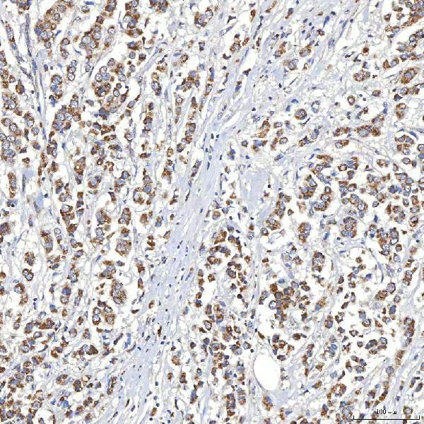
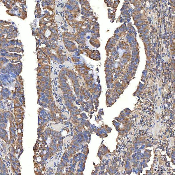
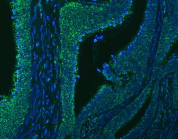


Comprehensive Human Protein Atlas IHC scoring per tissue (reliability: Approved — Medium consistency between antibody staining and RNA expression data.). Rows are taken directly from the HPA tissue chart — click any row's HPA link to view the source.
| Tissue | Cell type | Level | Evidence | Source |
|---|---|---|---|---|
| Appendix | Glandular cells | High | Protein (IHC) | HPA → |
| Colon | Glandular cells | High | Protein (IHC) | HPA → |
| Duodenum | Glandular cells | High | Protein (IHC) | HPA → |
| Liver | Hepatocytes | High | Protein (IHC) | HPA → |
| Placenta | Decidual cells | High | Protein (IHC) | HPA → |
| Tissue | Cell type | Level | Evidence | Source |
|---|---|---|---|---|
| Adipose tissue | Adipocytes | Not detected | Protein (IHC) | HPA → |
Troubleshoot SLC20A1 staining in paraffin sections by checking retrieval, cellular localisation, controls and scoring before interpreting differences in signal.
A03537-1 has IHC images from human paraffin sections and an IF image from a human paraffin section (catalog image captions); IHC reactivity is listed for human, mouse and rat (catalog applications).
A03537-1 is listed for IHC in human, mouse and rat and IF in human (catalog applications). Its images show IHC in human breast, colon, larynx and liver cancers, plus IF in human rectal cancer, all on paraffin sections (catalog image captions).
Which to pick: Choose A03537-1 for tissue IHC: its own images show chromogenic staining of human paraffin sections (IHC image captions), with a listed starting concentration of 2–5 μg/ml (catalog dilution). For IF, A03537-1 has a human paraffin-section image at 5 μg/ml (IF image caption); ICC validation is unreported (catalog applications and images). The catalog lists human, mouse and rat IHC reactivity (catalog applications), although the pictured IHC sections are human (IHC image captions); the fixative is unreported (catalog image captions).