This website uses cookies to ensure you get the best experience on our website.


- Table of Contents
Plan chromogenic IHC on paraffin kidney sections with the IHC-validated antibody A02515-2, starting at the listed 1:50 dilution (datasheet A02515-2). Assess proximal-tubule microvillar membrane staining and interpret unexpected staining cautiously because presumed off-target binding was observed (HPA tissue IHC).

Expected localisation, validated protocols, controls and antibodies — the at-a-glance facts below, then the full design guide.
| Expected localisation | Proximal-tubule microvillar membrane (HPA tissue IHC) | |
| Staining pattern | Membranous staining in renal proximal-tubule microvilli (HPA tissue IHC) | |
| Antigen retrieval | EDTA pH 8.0 HIER, heat-mediated (datasheet A02515-2) | |
| Positive control | Kidney | |
| Negative control | Adipose tissue+4 more · see all |
| Fixation | Keep fixation consistent across sections. (standard IHC practice; not target-specific) | |
| Caveat | Presumed off-target binding may mimic positive staining (HPA tissue IHC) | |
| Regulation | No specific regulator reported (UniProt) | |
| Isoform / epitope | 4 isoforms; assess epitope position and loop orientation (UniProt) |
The catalog antibody’s IHC-P protocol (datasheet: A02515-2) is accompanied by published renal tissue IHC protocols (PMC8262335; PMC7771643).
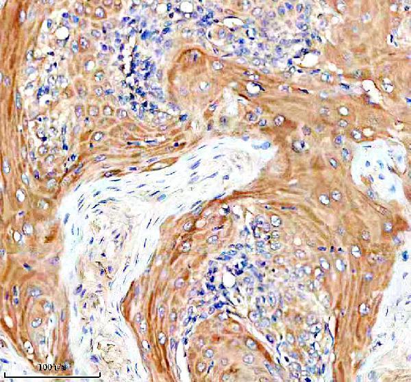
| Sample | Paraffin-embedded human skin cancer tissue; fixative not specified (datasheet A02515-2) |
| Fixation | Image fixative and duration unreported (datasheet A02515-2); verify before use. |
| Sectioning | 4–5 µm sections on charged slides (standard) |
| Deparaffinisation | Xylene, graded ethanol series to water (standard) |
| Antigen retrieval | Heat retrieval: EDTA pH 8.0 (datasheet A02515-2); 20 min, 95–100 °C (standard) |
| Peroxidase block | 3% H2O2, 10 min, room temperature (standard) |
| Blocking | 10% goat serum (datasheet A02515-2) |
| Primary antibody | Rabbit anti-SLC22A12, 1:50 recommended; image 1:100 (datasheet A02515-2) |
| Primary incubation | Overnight at 4 °C (datasheet A02515-2) |
| Detection | HRP-conjugated secondary, DAB chromogen (datasheet A02515-2) |
| Counterstain | Hematoxylin, blue, dehydrate and mount (standard) |
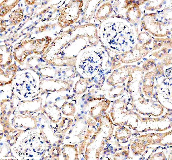
| Expected result | SLC22A12-positive staining in proximal tubules (microvilli) of kidney (HPA tissue IHC: Medium). HPA tissue profile: Distinct membranous expression in renal proximal tubules. No signal in the no-primary control. |
SLC22A12 is an apical membrane transporter with 12 transmembrane segments (UniProt Q96S37 topology). In kidney sections, expect membranous staining of proximal tubule microvilli; HPA reports a medium staining level and rates its tissue IHC evidence Enhanced (HPA tissue IHC). UniProt also places the protein in renal cortical proximal tubule epithelial cells (UniProt Q96S37 tissue specificity).
| A defined luminal rim outlines proximal tubules, with medium staining (HPA tissue IHC). | This fits the reported microvillar pattern and apical membrane location (HPA tissue IHC; UniProt Q96S37 subcellular location). Judge localisation as well as intensity: a brown tubular profile alone does not establish that the signal lies at the luminal surface (general IHC practice). |
| Staining chiefly fills proximal tubule cytoplasm or outlines the basolateral edge. | That compartment differs from the expected apical membrane pattern (UniProt Q96S37 subcellular location; HPA tissue IHC). Treat it as unconfirmed until the luminal border, counterstained morphology, and background controls are reviewed (general IHC practice). |
| Strong staining appears in cells outside proximal tubules or in an HPA listed negative tissue. | HPA reports proximal tubule microvilli as positive and, for example, adrenal glandular cells as not detected (HPA tissue IHC). Consider cross-reactivity or endogenous detection activity; check a reagent omission control before assigning biological expression (general IHC practice). |
| Colour spreads across the section without a clear luminal rim. | Diffuse background cannot support the reported distinct membranous pattern (HPA tissue IHC). Compare a no primary antibody control, inspect washes and detection reagents, and score only signal that remains anatomically resolved (general IHC practice). |
| No convincing signal appears in identifiable renal proximal tubules. | The expected positive compartment is absent (HPA tissue IHC). First verify tubule identity and the IHC staining run; then review antigen retrieval, antibody dilution, and detection conditions using the antibody's own instructions (general IHC practice). |
| Membrane topology | SLC22A12 has 12 transmembrane segments and an apical membrane location (UniProt Q96S37 topology and subcellular location). Interpret the chromogenic result against the luminal border of intact proximal tubules, where HPA describes microvillar staining (HPA tissue IHC). |
| Tissue evidence and antibody confidence | Kidney proximal tubule microvilli show medium staining; HPA rates tissue IHC Enhanced but notes medium agreement with RNA and presumed off target binding that was disregarded (HPA tissue IHC). Use that caveat when judging unexpected staining. |
| Isoforms and modifications | UniProt lists 4 isoforms and glycosylation sites at residues 56 and 102 (UniProt Q96S37). The supplied evidence does not map the antibody epitope to an isoform or establish how these features affect this IHC stain. |
| IF/ICC Q&A: what location should an IF image show? | Expect membrane localisation from the HPA subcellular summary and apical localisation from UniProt (HPA subcellular; UniProt Q96S37). HPA supplies no ICC-IF images or main location for this record, so this evidence does not validate an IF staining pattern (HPA subcellular). |
| Situation | Likely cause | Next action |
|---|---|---|
| The known positive kidney section has no proximal tubule rim. | An IHC run or staining condition may have failed; HPA reports proximal tubule microvilli as positive (HPA tissue IHC). | Confirm proximal tubules on the counterstain, then check the antibody instructions, retrieval run, dilution, and detection controls (general IHC practice). Do not infer SLC22A12 specific fixation sensitivity from this result. |
| Brown signal is present but sits mainly in cytoplasm. | Its location conflicts with the expected apical membrane pattern (UniProt Q96S37; HPA tissue IHC). | Examine an intact luminal edge at higher magnification and compare the no primary antibody control (general IHC practice). Record the stain as unconfirmed if a distinct membrane rim cannot be resolved. |
| Negative tissue or an unexpected cell type stains. | The signal may reflect cross-reactivity or endogenous detection activity (general IHC practice); HPA notes disregarded presumed off target binding (HPA tissue IHC). | Compare the reagent omission control and the expected kidney pattern in the same run (general IHC practice). Limit any negative tissue comparison to cells actually listed as not detected by HPA (HPA tissue IHC). |
| The entire section has diffuse chromogen. | Background or detection activity may obscure the distinct membranous pattern (general IHC practice; HPA tissue IHC). | Review blocking, washes, chromogen development, and a no primary antibody control (general IHC practice). Reassess only after individual tubule borders can be seen. |
| A faint rim is difficult to score. | HPA reports a medium level in proximal tubule microvilli, so intensity alone is an incomplete criterion (HPA tissue IHC). | Score the presence and continuity of the luminal rim alongside intensity, using comparable tubules and the same staining run (general IHC practice). |


Comprehensive Human Protein Atlas IHC scoring per tissue (reliability: Enhanced — Medium consistency between antibody staining and RNA expression data. Presumed off target binding observed and disregarded.). Rows are taken directly from the HPA tissue chart — click any row's HPA link to view the source.
| Tissue | Cell type | Level | Evidence | Source |
|---|---|---|---|---|
| Kidney | Proximal tubules (microvilli) | Medium | Protein (IHC) | HPA → |
| Tissue | Cell type | Level | Evidence | Source |
|---|---|---|---|---|
| Adipose tissue | Adipocytes | Not detected | Protein (IHC) | HPA → |
| Adrenal gland | Glandular cells | Not detected | Protein (IHC) | HPA → |
| Appendix | Glandular cells | Not detected | Protein (IHC) | HPA → |
| Bone marrow | Hematopoietic cells | Not detected | Protein (IHC) | HPA → |
| Breast | Adipocytes | Not detected | Protein (IHC) | HPA → |
Use renal proximal tubules and their apical staining pattern as the main reference when troubleshooting SLC22A12 chromogenic IHC (HPA tissue IHC; UniProt Q96S37).
A02515-2 has real IHC images from paraffin sections of human skin cancer and mouse and rat kidney (A02515-2 IHC captions). No IF data are supplied (catalog if_image_alts).
A02515-2 is listed for IHC and for human, mouse and rat reactivity (catalog applications/reactivity). Its IHC images show paraffin sections of human skin cancer and mouse and rat kidney (A02515-2 IHC captions).
Which to pick: Choose A02515-2 for paraffin-section tissue IHC: it is a rabbit polyclonal antibody with IHC listed and images from those tissues (catalog host/dilution_raw/applications; A02515-2 IHC captions). It is also the cross-species choice here, with human, mouse and rat reactivity listed and IHC images for each species (catalog reactivity; A02515-2 IHC captions). No IF/ICC antibody can be recommended from this payload because A02515-2 has neither a listed IF/ICC application nor an IF image; the fixative for its paraffin sections is unreported (catalog applications/if_image_alts; A02515-2 IHC captions).