SLC22A2 / Solute carrier family 22 member 2 · IHC design guide

Design Immunohistochemistry for SLC22A2

Plan SLC22A2 chromogenic IHC in paraffin sections using kidney tubules as positive tissue and the catalog antibody at 1:25 (HPA tissue IHC; datasheet M01612). Assess membranous and cytoplasmic staining while accounting for segment-specific membrane polarity (HPA tissue IHC; UniProt).

Evidence assembled Oct 2026 · For research use; verify linked source records and product datasheet before use
Immunohistochemistry protocol sheet for SLC22A2 (IHC for SLC22A2): expected localisation Renal tubules: membranous and cytoplasmic (HPA tissue IHC), antibody M01612, validated IHC image, and IHC protocol steps
Printable SLC22A2 IHC protocol sheet — expected localisation Renal tubules: membranous and cytoplasmic (HPA tissue IHC), antibody M01612, controls and protocol steps. Open the full SLC22A2 IHC guide →

SLC22A2 Immunohistochemistry Experimental Design Guide

Expected localisation, validated protocols, controls and antibodies — the at-a-glance facts below, then the full design guide.

Must know before staining
Expected localisation Renal tubules: membranous and cytoplasmic (HPA tissue IHC)
Staining pattern Tubule cells show distinct membranous and cytoplasmic staining (HPA tissue IHC)
Antigen retrieval Citrate pH 6 HIER, heat-mediated (datasheet M01612)
Positive control ⓘ Kidney
Negative control ⓘ Adipose tissue+4 more · see all
Important caveats
Reasons your staining may differ from the expected pattern.
Fixation Formaldehyde-fixed paraffin sections (selected-SKU IHC image M01612)
Caveat Proximal basolateral vs distal apical polarity (UniProt)
Regulation Kidney-enriched expression (HPA tissue IHC)
Isoform / epitope 3 isoforms; epitope map unknown; extracellular vs cytoplasmic matters (UniProt)
Section 1

Recommended SLC22A2 IHC & IF Protocols

The catalog antibody protocol is accompanied by one published SLC22A2 IHC protocol using paraffin-embedded tumor sections (PMC8301305).

Recommended immunohistochemistry (IHC-P) protocol parameters
SampleFormaldehyde-fixed, paraffin-embedded human kidney tissue (datasheet M01612)
FixationImage formalin-fixed; duration unreported (datasheet M01612); verify before use.
Sectioning4–5 µm sections on charged slides (standard)
DeparaffinisationXylene, graded ethanol series to water (standard)
Antigen retrievalHeat retrieval: Citrate pH 6 (datasheet M01612); 20 min, 95–100 °C (standard)
Peroxidase block3% H2O2, 10 min, room temperature (standard)
Blocking10% normal serum of the secondary host, 30 min, room temperature (standard)
Primary antibodyRabbit anti-SLC22A2, 1:25 (datasheet M01612)
Primary incubation1 hours at 37°C (datasheet M01612)
DetectionHRP-polymer secondary, DAB chromogen 5–10 min (standard)
CounterstainHematoxylin, blue, dehydrate and mount (standard)
Expected resultSLC22A2-positive staining in cells in tubules of kidney (HPA tissue IHC: Medium). HPA tissue profile: Distinct membranous and cytoplasmic expression in renal tubules. No signal in the no-primary control.
💡Decision noteStart with heat-mediated citrate pH 6 retrieval (datasheet M01612); the published tumor protocol reports antigen retrieval without specifying its method (PMC8301305).
Section 2

What Is the Expected SLC22A2 Staining Pattern?

SLC22A2 is a 12-pass membrane transporter (UniProt O15244 topology). In paraffin-section IHC, expect staining in renal tubule cells: HPA reports medium staining with distinct membranous and cytoplasmic expression (HPA tissue IHC). UniProt places SLC22A2 at the basolateral membrane of proximal tubules and the luminal membrane of distal tubules (UniProt O15244). HPA rates its tissue IHC evidence Enhanced, with medium consistency between staining and RNA data (HPA tissue IHC).

What am I looking at on my slide?
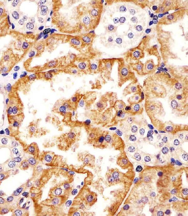
Renal tubule cells show distinct membrane staining with some cytoplasmic signal.This matches the observed kidney pattern (HPA tissue IHC). Judge membrane orientation by tubule type: UniProt reports proximal basolateral and distal luminal localization (UniProt O15244).
Strong nuclear staining dominates, with little recognizable tubule membrane signal.Nuclear localization is outside the reported membrane pattern (UniProt O15244; HPA tissue IHC). Treat it as suspect staining and check controls before scoring it as SLC22A2 (general IHC practice).
Prominent staining appears in cells outside the expected renal tubule pattern.HPA identifies renal tubule cells as its positive tissue-IHC population (HPA tissue IHC). Cross-reactivity or endogenous detection activity is possible; neither can be diagnosed from appearance alone (general IHC practice).
Color is diffuse across the section or present in cell-free areas.An indistinct background pattern cannot establish tubule localization (general IHC practice). Compare a matched negative control and assess blocking, washing, and detection conditions (general IHC practice).
No signal appears in a kidney section expected to contain tubules.HPA reports medium staining in renal tubule cells, so an absent result warrants a technical check (HPA tissue IHC). Confirm tubules are present, then review antibody and detection conditions (general IHC practice).
💡Expected SLC22A2 appearanceCall a positive result when renal tubule cells show distinct membrane-associated staining, potentially with cytoplasmic signal, at an approximately medium level (HPA tissue IHC); isolated strong nuclear or diffuse section-wide color is suspect (UniProt O15244; general IHC practice).
How each factor affects the staining
Tubule identity and membrane sideProximal tubules are reported as basolateral and distal tubules as luminal/apical (UniProt O15244). Assess orientation only where tubule morphology permits it (general IHC practice).
Evidence strengthHPA calls kidney tubule staining medium and IHC reliability Enhanced, while describing only medium agreement with RNA data (HPA tissue IHC). Use controls for each staining run (general IHC practice).
Antibody choiceHPA lists HPA008549 as IHC Enhanced and CAB068236/CAB068237 as IHC Approved (HPA antibodies). These ratings do not specify a working dilution for this experiment (HPA antibodies).
Other tissues as controlsHPA reports no detectable bronchial respiratory epithelial staining, although UniProt reports bronchial ciliated epithelial expression (HPA tissue IHC; UniProt O15244). Kidney is the better documented positive tissue here (HPA tissue IHC).
Isoforms and epitopeUniProt lists three isoforms and 12 membrane-spanning segments (UniProt O15244). The supplied sources do not identify which isoforms or epitopes these antibodies recognize; check antibody documentation before interpreting a variant-specific result.
IF/ICC?HPA summarizes the subcellular location as membrane but supplies no ICC-IF image-bearing cell lines or ICC validation for the listed antibodies (HPA subcellular; HPA antibodies). Use the separate IF/ICC guide for assay design.
Why is my staining missing, weak or wrong?
SituationLikely causeNext action
Kidney tubules are negative while the run control develops color.The kidney result conflicts with HPA's medium tubule staining (HPA tissue IHC); the cause is unresolved.Confirm tubules are in the section, then check antibody use, retrieval settings, incubation, and detection against the reagent instructions (general IHC practice).
Most of the section has a uniform chromogenic haze.Diffuse color obscures the distinct tubule pattern reported by HPA (HPA tissue IHC). Background or endogenous detection activity may contribute (general IHC practice).Review the matched negative control; adjust blocking, washing, or endogenous-activity suppression as appropriate for the detection system (general IHC practice).
Nuclei stain strongly but renal tubule membranes do not.This conflicts with reported membrane localization and the observed tubule pattern (UniProt O15244; HPA tissue IHC).Check the negative control and antibody specificity; score nuclear-only staining as unconfirmed (general IHC practice).
An unexpected cell population stains more strongly than renal tubules.The reported HPA positive population is renal tubule cells (HPA tissue IHC); cross-reactivity or endogenous detection activity remains possible (general IHC practice).Verify cell identity on the counterstained section and compare negative controls before attributing the signal to SLC22A2 (general IHC practice).
Only one side of some tubules stains.Sided staining can be compatible with proximal basolateral or distal luminal localization (UniProt O15244). Tubule identity cannot be inferred from stain position alone (general IHC practice).Review tubule morphology and section orientation; retain the sided pattern as a description if segment identity is uncertain (general IHC practice).
A bronchus section is negative despite an expected airway signal.UniProt reports bronchial ciliated epithelial expression, but HPA tissue IHC reports bronchial respiratory epithelial staining as not detected (UniProt O15244; HPA tissue IHC).Use kidney tubules to assess whether this IHC run detects the HPA-supported pattern; do not use bronchus negativity alone to call the run a failure (HPA tissue IHC).

Sample controls for SLC22A2 IHC & IF

🧪Run kidney first: tubular cells should stain at the HPA-reported Medium level (HPA: kidney, cells in tubules, Medium). Use adipose tissue as a negative tissue (HPA: adipocytes, Not detected); on the kidney slide, assess cells outside stained tubules as an internal background reference rather than assuming they are confirmed SLC22A2-negative (HPA: kidney, cells in tubules, Medium).
Positive control tissue: Kidney (Cells in tubules, HPA Medium)
Negative control tissue: Adipose tissue (HPA Not detected)
ICC-IF cell lines (HPA subcellular resource): HPA carries no ICC-IF cell line for SLC22A2; derive a cell-line control from the positive tissue's cell type (Cells in tubules) and confirm it by RNA or western blot first.
Technical controls: Include a no-primary, secondary-only control and an isotype control matched to the primary antibody’s host species and subclass; use SLC22A2-knockout material as a biological negative where available (standard IHC practice). For chromogenic kidney sections, check endogenous peroxidase and endogenous biotin background, particularly with the caption’s biotinylated secondary antibody (selected-SKU IHC caption: biotinylated secondary; standard IHC practice).
⚠️Feasibility: A target-specific fixation window or fixation effect is unreported, and the selected-SKU kidney IHC caption does not establish a fixative (selected-SKU IHC caption: fixative unreported). The caption uses heat retrieval in citrate buffer at pH 6, but retrieval dependence has not been established (selected-SKU IHC caption: heat retrieval, citrate pH 6). There is no supplied matched evidence that frozen sections or IF are easier; kidney background from endogenous biotin or peroxidase should be checked during chromogenic detection (HPA: kidney, cells in tubules, Medium; selected-SKU IHC caption: biotinylated secondary; standard IHC practice).

HPA tissue IHC evidence for SLC22A2

Comprehensive Human Protein Atlas IHC scoring per tissue (reliability: Enhanced — Medium consistency between antibody staining and RNA expression data.). Rows are taken directly from the HPA tissue chart — click any row's HPA link to view the source.

Positive expression · recommended positive controls

TissueCell typeLevelEvidenceSource
Kidney Cells in tubules Medium Protein (IHC) HPA →

Undetected expression · recommended negative controls

TissueCell typeLevelEvidenceSource
Adipose tissue Adipocytes Not detected Protein (IHC) HPA →
Adrenal gland Glandular cells Not detected Protein (IHC) HPA →
Appendix Glandular cells Not detected Protein (IHC) HPA →
Bone marrow Hematopoietic cells Not detected Protein (IHC) HPA →
Breast Adipocytes Not detected Protein (IHC) HPA →
Section 3

Advanced SLC22A2 IHC Tips

Use renal tubule distribution and membrane orientation to evaluate SLC22A2 staining, while checking each technical step against the available IHC evidence.

What retrieval should I use for SLC22A2 in paraffin kidney sections?
Start with heat-mediated citrate buffer at pH 6 for SLC22A2 paraffin IHC (datasheet M01612). Keep retrieval conditions consistent across the study, because differences in heating can change epitope exposure and tissue morphology (standard IHC practice). The M01612 kidney image used this retrieval, followed by 3% BSA blocking for 0.5 hour and primary antibody at 1/25 for 1 hour at 37°C (M01612 IHC caption). If staining remains weak, adjust heating duration on matched sections before considering another retrieval buffer; compare membrane detail and background alongside signal intensity (standard IHC practice).
How should I assess whether fixation is masking SLC22A2 staining?
Target-specific fixation sensitivity is unknown from the supplied evidence, so treat fixation as a variable to evaluate rather than an established cause of weak SLC22A2 staining. Compare sections processed with documented, consistent fixation and section handling, using the same citrate pH 6 retrieval and detection conditions (datasheet M01612; standard IHC practice). Check whether renal tubule morphology remains intact while staining changes between preparations; tissue damage can make an apparent loss of membrane signal difficult to interpret (standard IHC practice). Include a matched positive kidney section in each run, and record fixation conditions before changing retrieval or antibody concentration (HPA tissue IHC; standard IHC practice).
Which renal staining pattern fits SLC22A2, and when is cytoplasmic signal plausible?
Expect staining in renal tubule cells, with membranous and cytoplasmic expression reported in tissue IHC (HPA tissue IHC). Assess membrane orientation by tubule type: SLC22A2 is reported at the basolateral membrane of proximal tubules and the luminal or apical membrane of distal tubules (UniProt O15244 localisation). Do not classify every apical profile as artefact without identifying the tubule, or every diffuse cytoplasmic profile as specific solely because HPA reports cytoplasmic staining (HPA tissue IHC; standard IHC practice). Compare serial sections and the same anatomical regions across runs, giving greatest weight to reproducible cellular patterns with preserved morphology (standard IHC practice).
How can epitope position and isoforms affect SLC22A2 IHC interpretation?
SLC22A2 has 12 predicted transmembrane segments and 3 listed isoforms, so epitope assignment matters when comparing antibodies or staining patterns (UniProt O15244 topology and isoforms). Its extracellular region spans residues 44–150, while residues 516–555 are cytoplasmic; those regions can differ in accessibility after processing (UniProt O15244 topology; standard IHC practice). Check the catalog antibody’s stated immunogen or epitope against the isoform sequences before claiming that a negative section excludes all isoforms (standard IHC practice). A glycosylation site is annotated at residue 72, but the supplied evidence does not establish its effect on this antibody’s IHC staining (UniProt O15244 glycosylation).
How should IF clarify a disputed chromogenic SLC22A2 pattern?
Use IF as a follow-up to localize an IHC signal within renal tubules, pairing SLC22A2 with a validated marker of the expected tubule cell type (HPA tissue IHC; standard IF practice). Choose a far-red fluorophore where kidney autofluorescence compromises shorter wavelengths, and inspect unstained and single-label controls before calling overlap (standard IF practice). Match permeabilisation to the antibody’s documented epitope: an extracellular epitope may be accessible without detergent, whereas a cytoplasmic epitope generally requires access across the membrane (UniProt O15244 topology; standard IF practice). The supplied HPA subcellular record lists no ICC/IF images, so establish IF specificity independently of the IHC pattern (HPA subcellular record).
What should I change when SLC22A2 kidney sections stain diffusely?
First compare diffuse colour with a no-primary control and inspect whether it follows tissue edges, damaged areas, or the expected tubule cells (standard IHC practice; HPA tissue IHC). The M01612 kidney image used 3% BSA for 0.5 hour before primary antibody at 1/25, providing documented conditions for a controlled repeat (M01612 IHC caption). With biotin-based detection, assess endogenous biotin and secondary-reagent background; with peroxidase and DAB detection, include a peroxidase block as a general workflow step (standard IHC practice). Adjust washing and detection exposure one factor at a time while retaining a matched positive kidney section (standard IHC practice).
How should I score SLC22A2 staining across kidney sections? ⚠ ANSWER MARKED FOR VERIFICATION
Define the renal tubule compartment and scoring rule before reviewing treatment groups, because SLC22A2 expression is reported in tubule cells (HPA tissue IHC; standard IHC practice). Report the percentage of positive tubule cells and an H-score calculated from intensity grades 0–3, or measure positive membrane length per tubule area when membrane distribution is the question (standard IHC practice). Normalise cell-based scores to evaluable tubule cells, and area-based measurements to intact tubule area, excluding folds and necrotic regions by a preset rule (standard IHC practice). Score comparable cortical or medullary regions separately when sampling differs, and retain identical staining and image thresholds across sections (UniProt O15244 tissue specificity; standard IHC practice).
What distinguishes genuine SLC22A2 staining from an IHC artefact?
A credible positive pattern should be reproducible in renal tubule cells and compatible with the reported membranous and cytoplasmic distribution (HPA tissue IHC). Check membrane orientation against anatomy: proximal tubules have reported basolateral SLC22A2, while distal tubules have reported luminal or apical localisation (UniProt O15244 localisation). Treat staining confined to section edges, folds, necrotic tissue, or no-primary controls as suspect; investigate endogenous enzyme signal when using peroxidase and DAB (standard IHC practice). HPA rates its tissue evidence Enhanced but describes only medium agreement with RNA expression, so avoid interpreting staining intensity alone as a transport activity measurement (HPA tissue IHC; UniProt O15244 function).
Boster reagents

Best SLC22A2 / Solute carrier family 22 member 2 IHC Antibodies

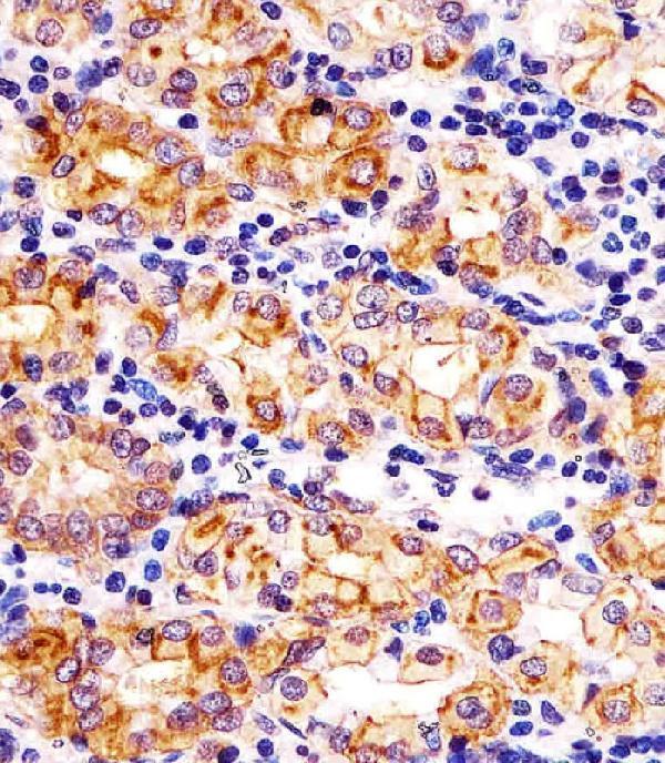
Anti-SLC22A2 images show IHC in human kidney and stomach sections (M01612 image captions) and IF/ICC in HeLa cells (PB9394 image caption); PB9394 lists human, mouse and rat reactivity (catalog).

Real IHC data M01612 staining SLC22A2 in human kidney tissue sections by Immunohistochemistry (IHC-P -paraformaldehyde-fixed, paraffin-embedded sections). Tissue was fixed with formaldehyde and blocked with 3% BSA for 0. 5 hour at room temperature; antigen retrieval was by heat mediation with a citrate buffer (pH6). Samples were incubated with primary antibody (1/25) for 1 hours at 37°C. A undiluted biotinylated goat polyvalent antibody was used as the secondary antibody.
Anti-SLC22A2 Antibody (N-term)
Cat # M01612

M01612 has IHC-P images from human kidney and stomach paraffin sections (M01612 image captions). PB9394 has a HeLa IF/ICC image (PB9394 image caption) and lists IHC with human, mouse and rat reactivity (catalog applications and reactivity).

Which to pick: Choose M01612 for tissue IHC: its rabbit polyclonal antibody is listed for IHC-P (catalog), and its images show formaldehyde-fixed human kidney and stomach paraffin sections (M01612 image captions). Choose PB9394 for IF/ICC because it has a HeLa fluorescence image, although IF is absent from its application list; the image caption does not report the fixative (PB9394 image caption; catalog applications). For cross-species tissue IHC, PB9394 lists human, mouse and rat reactivity and paraffin-section IHC, but its supplied image documents HeLa IF/ICC rather than tissue IHC (PB9394 catalog reactivity and dilution listing; PB9394 image caption).

Each figure is that product's own IHC / IF validation image from its datasheet.

References

  1. UniProt Consortium. UniProt entry O15244 (S22A2_HUMAN, Solute carrier family 22 member 2).
  2. Human Protein Atlas. SLC22A2 tissue IHC expression (reliability: Enhanced).
  3. Human Protein Atlas. SLC22A2 subcellular location (ICC-IF): Membrane.
  4. Human Protein Atlas. SLC22A2 antibody validation summary (3 antibodies).
  5. Effective Detection and Monitoring of Glioma Using [(18)F]FPIA PET Imaging. Biomedicines 2021 — PMC8301305.
  6. DNA methylation is associated with downregulation of the organic cation transporter OCT1 (SLC22A1) in human hepatocellular carcinoma. Genome medicine 2011 — PMC3334547.
  7. PubMed PMID:9260930 — UniProt-cited evidence.
  8. PubMed PMID:12089365 — UniProt-cited evidence.
  9. PubMed PMID:14702039 — UniProt-cited evidence.