SLC22A8 / Organic anion transporter 3 · IHC design guide

Design Immunohistochemistry for SLC22A8

Plan paraffin-section SLC22A8 IHC around kidney proximal tubules and their observed cytoplasmic staining (HPA tissue IHC). This guide relates that pattern to the expected basolateral location (UniProt) and the catalog antibody’s IHC conditions (datasheet A04087-2).

Evidence assembled Oct 2026 · For research use; verify linked source records and product datasheet before use
Immunohistochemistry protocol sheet for SLC22A8 (IHC for SLC22A8): expected localisation Proximal tubule cytoplasm (HPA tissue IHC), antibody A04087-2, validated IHC image, and IHC protocol steps
Printable SLC22A8 IHC protocol sheet — expected localisation Proximal tubule cytoplasm (HPA tissue IHC), antibody A04087-2, controls and protocol steps. Open the full SLC22A8 IHC guide →

SLC22A8 Immunohistochemistry Experimental Design Guide

Expected localisation, validated protocols, controls and antibodies — the at-a-glance facts below, then the full design guide.

Must know before staining
Expected localisation Proximal tubule cytoplasm (HPA tissue IHC)
Staining pattern Selective cytoplasmic staining in proximal renal tubules (HPA tissue IHC)
Antigen retrieval EDTA pH 8.0 HIER, heat-mediated (datasheet A04087-2)
Positive control ⓘ Kidney
Negative control ⓘ Adipose tissue+4 more · see all
Important caveats
Reasons your staining may differ from the expected pattern.
Fixation Keep fixation consistent across sections. (standard IHC practice; not target-specific)
Caveat Cytoplasmic staining may obscure basolateral enrichment (HPA tissue IHC; UniProt)
Regulation No specific expression regulator reported (UniProt)
Isoform / epitope 5 isoforms; epitope differences unreported; topology spans both membrane sides (UniProt)
Section 1

Recommended SLC22A8 IHC & IF Protocols

Compare the catalog antibody’s IHC-P protocol (datasheet A04087-2) with the published HCC tissue microarray protocol (PMC9402792).

Recommended immunohistochemistry (IHC-P) protocol parameters
SampleParaffin-embedded human breast cancer tissue; fixative not specified (datasheet A04087-2)
FixationImage fixative and duration unreported (datasheet A04087-2); verify before use.
Sectioning4–5 µm sections on charged slides (standard)
DeparaffinisationXylene, graded ethanol series to water (standard)
Antigen retrievalHeat retrieval: EDTA pH 8.0 (datasheet A04087-2); 20 min, 95–100 °C (standard)
Peroxidase block3% H2O2, 10 min, room temperature (standard)
Blocking10% goat serum (datasheet A04087-2)
Primary antibodyRabbit anti-SLC22A8, 2-5 μg/ml (datasheet A04087-2)
Primary incubationOvernight at 4 °C (datasheet A04087-2)
DetectionHRP-conjugated secondary, DAB chromogen (datasheet A04087-2)
CounterstainHematoxylin, blue, dehydrate and mount (standard)
Expected resultSLC22A8-positive staining in proximal tubules (cell body) of kidney (HPA tissue IHC: Medium). HPA tissue profile: Selective cytoplasmic expression in proximal renal tubules. No signal in the no-primary control.
💡Decision noteStart with EDTA pH 8.0 heat retrieval for the catalog antibody (datasheet A04087-2). The published protocol used 0.08% citrate pressure cooking for 20 min (PMC9402792).
Section 2

What Is the Expected SLC22A8 Staining Pattern?

SLC22A8 is a multi-pass transporter assigned to the basolateral membrane of proximal tubular cells (UniProt Q8TCC7 topology and localisation). In paraffin-section IHC, expect selective staining of proximal renal tubule cell bodies at medium intensity; HPA describes the observed signal as cytoplasmic (HPA tissue IHC: Enhanced; proximal tubules, Medium). Read the observed cell-body pattern alongside the membrane assignment rather than requiring a sharply outlined membrane.

What am I looking at on my slide?
Selective, medium-intensity staining in proximal renal tubule cell bodies (HPA tissue IHC).This matches the reported kidney IHC pattern and is the clearest positive reference in these sources (HPA tissue IHC: Enhanced). A visible cytoplasmic or cell-body signal can agree with the HPA result even when a basolateral membrane edge is unresolved (HPA tissue IHC; UniProt Q8TCC7 localisation).
A strong, exclusively apical rim or predominantly nuclear signal in renal tubules.That distribution does not match the reported proximal-tubule cell-body IHC pattern or the basolateral assignment for these cells (HPA tissue IHC; UniProt Q8TCC7 localisation). Treat it as a localisation discrepancy: review morphology and controls before calling it SLC22A8 (general IHC practice).
Prominent staining of unrelated cells, including adrenal glandular cells.HPA reports adrenal glandular cells as not detected by tissue IHC, although UniProt reports adrenal expression (HPA tissue IHC; UniProt Q8TCC7 tissue specificity). Consider cross-reactivity or detection-system activity and check controls; the two sources do not justify assigning that stain to SLC22A8.
Diffuse color across tubules, interstitium, and the section background.A widespread signal obscures the selective proximal-tubule pattern reported by HPA (HPA tissue IHC). Assess background with a no-primary control and review blocking, washes, and chromogen development (general IHC practice); diffuse color alone cannot establish target localisation.
No convincing staining in proximal renal tubules on a kidney section.This conflicts with HPA's medium-intensity positive reference (HPA tissue IHC). First verify that identifiable proximal tubules and an interpretable section are present, then inspect the antibody and detection controls (general IHC practice); absence of color alone cannot distinguish biology from assay failure.
💡Expected SLC22A8 appearanceCall a section positive when identifiable proximal renal tubule cell bodies show selective, approximately medium-intensity staining (HPA tissue IHC); strong nuclear, exclusively apical, or broad unrelated-cell staining warrants a false-positive check against the reported pattern (HPA tissue IHC; UniProt Q8TCC7 localisation).
How each factor affects the staining
Tissue and cell selectionKidney proximal tubules provide the observed medium-staining reference; HPA lists several other sampled cell types as not detected (HPA tissue IHC). Its sampled negatives are controls for interpretation, not proof that every cell in those organs lacks the protein.
Subcellular resolutionUniProt assigns SLC22A8 to the proximal-tubule basolateral membrane and lists 11 transmembrane segments (UniProt Q8TCC7 topology and localisation). HPA describes paraffin-section staining as cytoplasmic in proximal tubules; that observation does not resolve the precise membrane face (HPA tissue IHC).
Antibody evidenceHPA rates tissue IHC reliability Enhanced and lists antibody HPA044174 as IHC Enhanced (HPA tissue IHC; HPA antibodies). This supports the reported tissue pattern, but does not validate every antibody or establish a required chromogenic staining intensity under other assay conditions.
Molecular annotationsUniProt lists 5 isoforms and glycosylation sites at residues 86 and 102 (UniProt Q8TCC7). Without an epitope map or isoform-specific IHC evidence here, these annotations cannot predict which variants an antibody detects or explain a staining difference.
IF/ICC evidenceHPA's subcellular summary says membrane, but supplies no main location or cell-line ICC-IF images; the listed antibody has no ICC validation entry (HPA subcellular; HPA antibodies). That record does not establish an expected IF image or justify an IF/ICC protocol here.
Why is my staining missing, weak or wrong?
SituationLikely causeNext action
Kidney positive control has no proximal-tubule signal.The result is inconsistent with HPA's medium proximal-tubule staining; these sources cannot identify a specific failed step (HPA tissue IHC).Confirm proximal-tubule morphology and section integrity, then check antibody identity, detection reagents, and a working assay control (general IHC practice). If considering retrieval changes, optimize them empirically; no SLC22A8-specific retrieval condition is supplied.
The entire section develops diffuse chromogen.Nonspecific binding, incomplete washing, or detection-system activity can create broad background (general IHC practice); it is unlike HPA's selective pattern (HPA tissue IHC).Run a no-primary control, review blocking and washes, and limit development to a readable signal (general IHC practice). Reassess localisation only after the background is controlled.
Adrenal glandular cells or another HPA-negative cell type stain strongly.That conflicts with the sampled HPA IHC result; UniProt's adrenal-expression annotation does not validate the observed glandular-cell stain (HPA tissue IHC; UniProt Q8TCC7 tissue specificity).Compare with a kidney positive section and no-primary control, then assess antibody specificity and detection-system activity (general IHC practice). Record the discrepancy rather than scoring those cells as confirmed positive.
Signal appears only on the apical edge of proximal tubules.An exclusively apical pattern differs from UniProt's proximal-tubule basolateral localisation; HPA's cell-body IHC report cannot resolve a membrane face (UniProt Q8TCC7 localisation; HPA tissue IHC).Check cell orientation and adjacent morphology, and compare independent sections or controls (general IHC practice). Do not infer basolateral localisation from an apical-looking chromogenic rim.
Color is concentrated in nuclei instead of tubule cell bodies.Nuclear localisation is not the supplied proximal-tubule IHC pattern or UniProt membrane assignment (HPA tissue IHC; UniProt Q8TCC7 localisation).Check the counterstain and no-primary control, then reassess whether the chromogen truly occupies nuclei (general IHC practice). Treat persistent nuclear-only staining as unconfirmed for SLC22A8.
Can an IF/ICC image confirm the membrane pattern?HPA summarizes a membrane location but provides no cell-line ICC-IF images or ICC validation for the listed antibody (HPA subcellular; HPA antibodies).Use the separate IF/ICC guide for that application. Here, report the IHC cell-body observation and UniProt's basolateral assignment separately; do not present the HPA summary as image-level IF confirmation.

Sample controls for SLC22A8 IHC & IF

🧪Run kidney first: proximal tubule cell bodies should stain at the medium level (HPA: kidney proximal tubules, Medium). Run adipose tissue as a negative, where adipocytes are not detected (HPA: adipose tissue adipocytes, Not detected); on the kidney slide, use cells outside the proximal tubules to assess background without assuming they are target negative.
Positive control tissue: Kidney (Proximal tubules (cell body), HPA Medium)
Negative control tissue: Adipose tissue (HPA Not detected)
ICC-IF cell lines (HPA subcellular resource): HPA carries no ICC-IF cell line for SLC22A8; derive a cell-line control from the positive tissue's cell type (Proximal tubules (cell body)) and confirm it by RNA or western blot first.
Technical controls: Include a no-primary, secondary-only control; a concentration-matched rabbit IgG isotype control (selected-SKU tissue-IHC caption: rabbit primary); and a SLC22A8 knockout control or peptide competition if the immunizing peptide is available. For chromogenic kidney sections, block endogenous peroxidase and assess residual tubular background alongside the controls (standard IHC practice).
⚠️Feasibility: The selected-SKU paraffin-section caption reports heat retrieval in EDTA, pH 8.0, but its fixative is unreported; no target-specific fixation window or fixation effect is reported in the supplied evidence (selected-SKU tissue-IHC caption). Test retrieval conditions when establishing the assay, since that caption documents one successful condition but does not establish a universal requirement (selected-SKU tissue-IHC caption). The supplied evidence does not establish whether frozen sections or IF are easier; kidney interpretation should emphasize proximal tubule localization against tubular background (HPA: kidney proximal tubules, Medium; UniProt Q8TCC7: proximal tubular basolateral membrane).

HPA tissue IHC evidence for SLC22A8

Comprehensive Human Protein Atlas IHC scoring per tissue (reliability: Enhanced — High consistency between antibody staining and RNA expression data.). Rows are taken directly from the HPA tissue chart — click any row's HPA link to view the source.

Positive expression · recommended positive controls

TissueCell typeLevelEvidenceSource
Kidney Proximal tubules (cell body) Medium Protein (IHC) HPA →

Undetected expression · recommended negative controls

TissueCell typeLevelEvidenceSource
Adipose tissue Adipocytes Not detected Protein (IHC) HPA →
Adrenal gland Glandular cells Not detected Protein (IHC) HPA →
Appendix Glandular cells Not detected Protein (IHC) HPA →
Bone marrow Hematopoietic cells Not detected Protein (IHC) HPA →
Breast Adipocytes Not detected Protein (IHC) HPA →
Section 3

Advanced SLC22A8 IHC Tips

Troubleshoot SLC22A8 staining in paraffin sections by checking retrieval, tissue controls and cellular localisation before comparing chromogenic signal across samples.

Which retrieval condition should I use when SLC22A8 staining is weak?
Start with heat-mediated retrieval in EDTA pH 8.0 for paraffin-section chromogenic IHC (datasheet A04087-2). The catalog antibody’s tissue image used this condition, followed by 2 μg/ml primary antibody overnight at 4°C; its caption does not isolate retrieval as the cause of staining (datasheet A04087-2). If signal is weak, check that sections remain attached and that the retrieval bath reaches and maintains its intended temperature before changing the antibody concentration (standard IHC practice). Compare any retrieval adjustments on adjacent sections with the same detection and exposure conditions, using proximal renal tubules as a reference (HPA: Medium staining in proximal tubules).
Can fixation explain inconsistent SLC22A8 staining between paraffin blocks?
Target-specific fixation sensitivity is unknown: the catalog image describes a paraffin-embedded section but does not state its fixative (datasheet A04087-2). Record each block’s fixative and fixation duration, then compare matched sections using the same EDTA pH 8.0 retrieval and 2 μg/ml primary concentration (datasheet A04087-2; standard IHC practice). If one block stains poorly, check tissue preservation, section adhesion and the detection controls before attributing the difference to fixation (standard IHC practice). Use a consistently processed kidney control for comparison, focusing on proximal tubules rather than whole-section color intensity (HPA: Medium staining in proximal tubules; standard IHC practice).
Should renal SLC22A8 appear cytoplasmic or confined to a membrane?
Expect the strongest interpretable renal pattern in proximal tubules, where SLC22A8 is assigned to the basolateral membrane (UniProt Q8TCC7 localisation). HPA instead describes selective cytoplasmic staining in proximal renal tubules, so assess the observed pattern against both references rather than assuming that diffuse color resolves a membrane boundary (HPA: tissue IHC profile; UniProt Q8TCC7 localisation). Examine well-preserved tubules at higher magnification and compare staining along the basal and lateral borders with any luminal or diffuse signal (standard IHC practice). Score cell type and compartment separately; a brown deposit outside intact proximal tubules needs control-based scrutiny before assignment to SLC22A8 (HPA: proximal tubule profile; standard IHC practice).
How can an unknown antibody epitope affect SLC22A8 IHC interpretation?
SLC22A8 has 5 listed isoforms and 11 transmembrane segments, but the supplied antibody caption does not identify its epitope (UniProt Q8TCC7 isoforms and topology; datasheet A04087-2). Therefore, do not infer which isoforms the catalog antibody detects or whether retrieval exposes a cytoplasmic or extracellular sequence (UniProt Q8TCC7 topology; datasheet A04087-2). The record places glycosylation sites at residues 86 and 102 in an extracellular region, without establishing their effect on this antibody’s staining (UniProt Q8TCC7 topology and glycosylation). If isoform specificity matters, obtain the immunogen sequence and compare it with the isoforms before interpreting a negative section (standard antibody validation practice).
How should I assess SLC22A8 in a multiplex IF experiment?
For the separate IF/ICC workflow, pair SLC22A8 with an independently validated proximal tubule marker and assess whether signals occur in the same cells (HPA: proximal tubule IHC profile; standard IF practice). Choose fluorophores after inspecting unstained tissue for autofluorescence, and reserve a spectrally distinct channel for the weaker signal (standard IF practice). Because the supplied caption does not identify the antibody epitope, test permeabilised and minimally permeabilised conditions before deciding whether access to a cytoplasmic or extracellular face is required (datasheet A04087-2; UniProt Q8TCC7 topology; standard IF practice). Include single-label and secondary-only controls to distinguish bleed-through and nonspecific fluorescence from the expected cellular pattern (standard IF practice).
What should I check when DAB staining is widespread?
First compare a section without primary antibody with the full stain to identify signal from the secondary reagent, endogenous peroxidase or DAB deposition (standard chromogenic IHC practice). The catalog caption used peroxidase-linked detection and DAB after 10% goat-serum blocking, so keep those steps consistent while troubleshooting (datasheet A04087-2). Check peroxidase blocking, washing and section drying, then titrate the 2 μg/ml primary concentration if the control is clean but the test section remains diffuse (datasheet A04087-2; standard IHC practice). Judge improvement in proximal tubules against surrounding tissue rather than by overall darkness, since HPA reports selective proximal tubule staining (HPA: tissue IHC profile).
How should I quantify SLC22A8 staining across kidney sections? ⚠ ANSWER MARKED FOR VERIFICATION
Define proximal tubules before scoring, because the reference IHC pattern is selective in that cell population (HPA: tissue IHC profile). Report the percentage of positive proximal tubule cells and staining intensity, or calculate an H-score from intensity categories 0–3 for a 0–300 range (standard IHC scoring practice). Normalise counts to the number of evaluable proximal tubule cells, or report positive tubules per mm² of evaluable cortex when cell boundaries cannot be scored reliably (standard histology quantification practice). Apply identical retrieval, DAB development, counterstaining and scoring thresholds across sections, and record excluded damaged or edge regions (standard IHC practice).
How do I distinguish genuine SLC22A8 signal from staining artefact?
A convincing renal result localises to intact proximal tubules and is compatible with basolateral localisation, while HPA’s tissue IHC profile describes selective cytoplasmic staining in those cells (UniProt Q8TCC7 localisation; HPA: tissue IHC profile). Examine unexpected luminal, nuclear or widespread staining alongside a no-primary control before assigning it to SLC22A8 (UniProt Q8TCC7 localisation; standard IHC practice). Discount signal concentrated at section edges, in necrotic areas or where endogenous peroxidase persists, and confirm that adjacent intact tissue shows the same cellular pattern (standard IHC practice). The breast cancer paraffin image documents staining with this antibody but does not by itself establish cell-specific expression or specificity there (datasheet A04087-2).
Boster reagents

Best SLC22A8 / Organic anion transporter 3 IHC Antibodies

A04087-2 has human paraffin-section IHC data and human paraffin-section IF data (catalog image captions); its listed reactivity is human (catalog: reactivity).

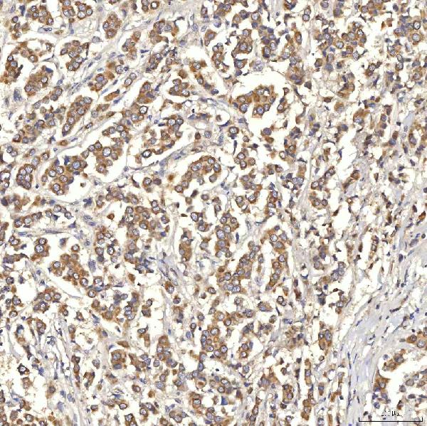
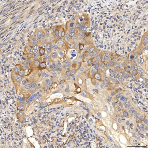
Real IHC data IHC analysis of OAT3/SLC22A8 using anti-OAT3/SLC22A8 antibody (A04087-2). OAT3/SLC22A8 was detected in a paraffin-embedded section of human breast cancer tissue. Heat mediated antigen retrieval was performed in EDTA buffer (pH 8.0, epitope retrieval solution). The tissue section was blocked with 10% goat serum. The tissue section was then incubated with 2 μg/ml rabbit anti-OAT3/SLC22A8 Antibody (A04087-2) overnight at 4°C. Peroxidase Conjugated Goat Anti-rabbit IgG was used as secondary antibody and incubated for 30 minutes at 37°C. The tissue section was developed using HRP Conjugated Rabbit IgG Super Vision Assay Kit (Catalog # SV0002) with DAB as the chromogen.
Anti-OAT3/SLC22A8 Antibody ®
Cat # A04087-2

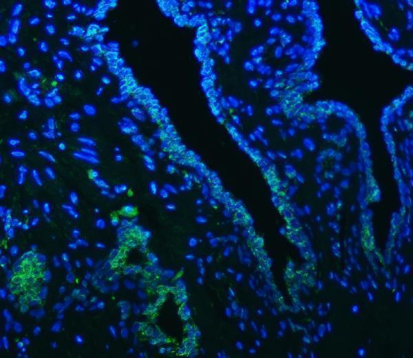
A04087-2 was tested for IHC on a paraffin-embedded human breast cancer section (catalog IHC image caption). A04087-2 also has IF data from a paraffin-embedded human prostate cancer section (catalog IF image caption).

Which to pick: Choose A04087-2 for human tissue IHC; its breast cancer image documents paraffin sections, while the fixative is unreported (catalog IHC image caption). For IF, A04087-2 has a human tissue image and a listed starting concentration of 5 μg/ml (catalog IF image caption; datasheet: IF dilution); ICC validation is unreported (catalog: applications). No cross-species option is documented because A04087-2 lists human reactivity only (catalog: reactivity).

Each figure is that product's own IHC / IF validation image from its datasheet.

References

  1. UniProt Consortium. UniProt entry Q8TCC7 (S22A8_HUMAN, Organic anion transporter 3).
  2. Human Protein Atlas. SLC22A8 tissue IHC expression (reliability: Enhanced).
  3. Human Protein Atlas. SLC22A8 subcellular location (ICC-IF): Membrane.
  4. Human Protein Atlas. SLC22A8 antibody validation summary (1 antibodies).
  5. The mRNA-miRNA-lncRNA Regulatory Network and Factors Associated with Prognosis Prediction of Hepatocellular Carcinoma. Genomics, proteomics & bioinformatics 2021 — PMC9402792.
  6. Expression of six drug transporters in vaginal, cervical, and colorectal tissues: Implications for drug disposition in HIV prevention. Journal of clinical pharmacology 2014 — PMC4061289.
  7. PubMed PMID:10049739 — UniProt-cited evidence.
  8. PubMed PMID:14702039 — UniProt-cited evidence.
  9. PubMed PMID:16554811 — UniProt-cited evidence.