This website uses cookies to ensure you get the best experience on our website.


- Table of Contents
Plan SLC25A10 staining in paraffin sections using the catalog antibody’s IHC conditions. Compare granular cytoplasmic staining with HPA tissue observations, and account for the HPA antibody’s stated cross-reactivity caution (HPA tissue IHC).

Expected localisation, validated protocols, controls and antibodies — the at-a-glance facts below, then the full design guide.
| Expected localisation | Granular cytoplasm (HPA tissue IHC); mitochondrial inner membrane (UniProt) | |
| Staining pattern | Granular cytoplasmic staining in hepatocytes and other cells (HPA tissue IHC) | |
| Antigen retrieval | EDTA pH 8.0 HIER, heat-mediated (datasheet A10727-3) | |
| Positive control | Appendix+4 more · see all | |
| Negative control | Caudate+3 more · see all |
| Fixation | Keep fixation consistent across sections. (standard IHC practice; not target-specific) | |
| Caveat | The HPA antibody may detect proteins from more than one gene (HPA tissue IHC) | |
| Regulation | No specific expression regulator reported (UniProt) | |
| Isoform / epitope | 2 isoforms; epitope coverage across them is unknown (UniProt) |
The catalog antibody’s IHC-P protocol (datasheet A10727-3) is paired with three published SLC25A10 IHC protocols (PMC7405602; PMC11371862; PMC12092617).
| Sample | Paraffin-embedded mouse kidney tissue; fixative not specified (datasheet A10727-3) |
| Fixation | Image fixative and duration unreported (datasheet A10727-3); verify before use. |
| Sectioning | 4–5 µm sections on charged slides (standard) |
| Deparaffinisation | Xylene, graded ethanol series to water (standard) |
| Antigen retrieval | Heat retrieval: EDTA pH 8.0 (datasheet A10727-3); 20 min, 95–100 °C (standard) |
| Peroxidase block | 3% H2O2, 10 min, room temperature (standard) |
| Blocking | 10% goat serum (datasheet A10727-3) |
| Primary antibody | Rabbit anti-SLC25A10, 2-5 μg/ml (datasheet A10727-3) |
| Primary incubation | Overnight at 4 °C (datasheet A10727-3) |
| Detection | HRP-conjugated secondary, DAB chromogen (datasheet A10727-3) |
| Counterstain | Hematoxylin, blue, dehydrate and mount (standard) |
| Expected result | SLC25A10-positive staining in glandular cells of appendix (HPA tissue IHC: High). HPA tissue profile: General cytoplasmic expression with a granular pattern. No signal in the no-primary control. |
SLC25A10 is an inner mitochondrial membrane carrier with six transmembrane segments (UniProt Q9UBX3 topology). In paraffin sections, expect granular cytoplasmic staining, particularly in liver hepatocytes and kidney Bowman’s capsule cells (HPA tissue IHC: High; general cytoplasmic granular pattern). HPA rates tissue staining Supported, with medium consistency against RNA data, pending external verification, and cautions that the antibody targets proteins from more than one gene (HPA tissue IHC).
| Granular cytoplasmic staining in hepatocytes, with little nuclear emphasis. | This matches the reported tissue pattern and a high-staining cell population (HPA tissue IHC: general granular cytoplasmic pattern; liver hepatocytes High). Mitochondrial inner membrane location provides a plausible basis for the granularity (UniProt Q9UBX3 subcellular location). The pattern alone cannot prove antibody specificity because HPA flags targeting of proteins from more than one gene (HPA tissue IHC caution). |
| Predominantly nuclear or membrane-outline staining, without cytoplasmic granules. | Treat this as a compartment mismatch with the expected paraffin-section pattern (HPA tissue IHC: granular cytoplasmic expression; UniProt Q9UBX3: mitochondrial inner membrane). A separate ICC-IF record lists nucleoplasm as uncertain, so nuclear signal alone should not be accepted as the expected IHC result (HPA subcellular: nucleoplasm uncertain). |
| Strong staining in caudate glial cells or lymph-node germinal-center cells. | Those specific cell populations were not detected in the HPA tissue survey (HPA tissue IHC: caudate glial cells Not detected; lymph-node germinal-center cells Not detected). Check for cross-reactivity or endogenous detection activity before scoring such staining as SLC25A10; HPA explicitly cautions that its antibody targets proteins from more than one gene (HPA tissue IHC caution). |
| Uniform haze across cells and tissue spaces obscures granular detail. | Diffuse staining does not match HPA’s reported granular cytoplasmic pattern (HPA tissue IHC). In a chromogenic IHC run, assess reagent background with a no-primary control and review blocking, washes, and detection conditions (general IHC practice). Background that hides cell boundaries cannot support a cell-specific intensity call. |
| No staining in hepatocytes on a section expected to be positive. | HPA reports high hepatocyte staining, and UniProt reports high amounts in liver (HPA tissue IHC: liver hepatocytes High; UniProt Q9UBX3 tissue specificity). Review the run and tissue quality before interpreting absence biologically (general IHC practice). HPA’s Supported rating and pending external verification limit how conclusively one negative section can contradict the reference pattern (HPA tissue IHC reliability). |
| Cell population and tissue | Interpret staining by cell type: hepatocytes and Bowman’s capsule cells are High, while adipocytes are Low (HPA tissue IHC). A low result in adipocytes should not be judged against the hepatocyte benchmark; neither rating applies to every cell in its tissue. |
| Compartment and topology | Six transmembrane segments place SLC25A10 in the mitochondrial inner membrane (UniProt Q9UBX3 topology and subcellular location). HPA describes the tissue IHC appearance as granular cytoplasmic, so score that visible pattern rather than requiring individual mitochondria to be resolved (HPA tissue IHC). |
| Antibody evidence | The listed antibody is Supported for IHC, while HPA reports medium staining–RNA consistency, pending external verification, and targeting of proteins from more than one gene (HPA antibody validation; HPA tissue IHC reliability). These caveats matter most for unexpected compartments or cell populations. |
| IF/ICC Q&A | What should IF/ICC show? Mainly mitochondria (HPA subcellular: mitochondria supported). HPA also lists nucleoplasm as uncertain and warns that the antibodies target proteins from multiple genes (HPA subcellular). This observation should not override the granular cytoplasmic IHC benchmark (HPA tissue IHC). |
| Situation | Likely cause | Next action |
|---|---|---|
| Hepatocytes have no visible chromogen. | A failed IHC run or weak detection is possible; hepatocytes are reported High (HPA tissue IHC: liver hepatocytes High). | Check that the positive-control section, retrieval, primary incubation, detection reagents, and chromogen worked (general IHC practice). Interpret persistent absence cautiously because the HPA tissue rating awaits external verification (HPA tissue IHC reliability). |
| Granules are present, but the entire section is brown. | Diffuse reagent or chromogen background may be obscuring the reported granular cytoplasmic pattern (HPA tissue IHC; general IHC practice). | Compare a no-primary control, review blocking and washes, and adjust detection conditions according to the assay controls (general IHC practice). Score cells only where granular cytoplasmic staining remains distinguishable. |
| Nuclei dominate the staining. | A nuclear-dominant paraffin-section result conflicts with the expected granular cytoplasmic pattern (HPA tissue IHC). Nucleoplasm is an uncertain additional ICC-IF location (HPA subcellular). | Review the counterstain and no-primary control, then compare cytoplasmic granularity in hepatocytes (general IHC practice; HPA tissue IHC: liver hepatocytes High). Do not use the uncertain IF observation to validate nuclear-only IHC. |
| Strong staining appears in cells reported as undetected. | Cross-reactivity or endogenous detection activity is possible (general IHC practice); HPA cautions that the antibody targets proteins from more than one gene (HPA tissue IHC). | Confirm cell identity and compare no-primary controls before scoring (general IHC practice). Use the reported cell-level negatives as comparators, such as lymph-node germinal-center cells (HPA tissue IHC: Not detected). |
| Adipocytes stain faintly beside a strongly positive tissue. | Low adipocyte staining is within the reported range and need not indicate a failed run (HPA tissue IHC: adipocytes Low). | Judge the run with a high-staining cell population, such as hepatocytes, and evaluate adipocytes separately (HPA tissue IHC: liver hepatocytes High; adipocytes Low). Keep the same scoring threshold within each comparison. |
| A different antibody gives a discordant pattern. | HPA reports medium agreement with RNA data and cautions about proteins from more than one gene (HPA tissue IHC reliability); discordance alone does not identify which result is specific. | Compare cell-level patterns and assay controls, then use independent validation before making a new localization claim (general IHC practice). The HPA IHC entry is Supported, with external verification pending (HPA tissue IHC reliability). |


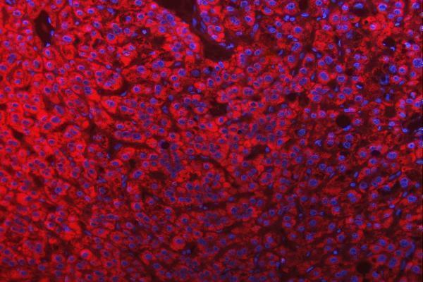


Comprehensive Human Protein Atlas IHC scoring per tissue (reliability: Supported — Medium consistency between antibody staining and RNA expression data. Pending external verification. Caution, targets protein from more than one gene.). Rows are taken directly from the HPA tissue chart — click any row's HPA link to view the source.
| Tissue | Cell type | Level | Evidence | Source |
|---|---|---|---|---|
| Appendix | Glandular cells | High | Protein (IHC) | HPA → |
| Colon | Endothelial cells | High | Protein (IHC) | HPA → |
| Gallbladder | Glandular cells | High | Protein (IHC) | HPA → |
| Kidney | Bowman's capsule | High | Protein (IHC) | HPA → |
| Liver | Hepatocytes | High | Protein (IHC) | HPA → |
| Tissue | Cell type | Level | Evidence | Source |
|---|---|---|---|---|
| Caudate | Glial cells | Not detected | Protein (IHC) | HPA → |
| Hippocampus | Glial cells | Not detected | Protein (IHC) | HPA → |
| Lymph node | Germinal center cells | Not detected | Protein (IHC) | HPA → |
| Spleen | Cells in red pulp | Not detected | Protein (IHC) | HPA → |
Troubleshoot SLC25A10 staining by checking retrieval, mitochondrial localisation, cell identity, and controls before interpreting chromogenic signal.
A10727-3 has IHC images from paraffin sections of human, mouse, and rat tissue, plus an IF image from a human paraffin section (catalog image captions).
A10727-3 has IHC images from mouse and rat kidney, human appendix adenocarcinoma, and human breast cancer paraffin sections (catalog IHC image captions). Its IF image shows a human liver cancer paraffin section (catalog IF image caption).
Which to pick: Choose A10727-3 for tissue IHC in paraffin sections: its own IHC captions document staining at 2 μg/ml, while the fixative is unreported (catalog IHC image captions). For IF, its own caption documents a human liver cancer paraffin section at 5 μg/ml; ICC is not listed among its applications (catalog IF image caption; catalog applications). It is the listed choice for work across human, mouse, and rat because those species are listed as reactive; the antibody is rabbit hosted and its clone is unreported (catalog reactivity; catalog host and clone fields).