This website uses cookies to ensure you get the best experience on our website.


- Table of Contents
Plan paraffin-section SLC25A6 IHC using the granular cytoplasmic tissue pattern (HPA tissue IHC). Compare cardiomyocytes with high staining against epididymal glandular cells with no detected staining (HPA tissue IHC), while accounting for possible detection of products from more than one gene (HPA tissue IHC).

Expected localisation, validated protocols, controls and antibodies — the at-a-glance facts below, then the full design guide.
| Expected localisation | Granular cytoplasm (HPA tissue IHC); inner mitochondrial membrane (UniProt) | |
| Staining pattern | Granular cytoplasmic staining in most tissues (HPA tissue IHC) | |
| Antigen retrieval | Citrate pH 6.0 HIER, 95–98 °C, 20 min (rule: cytoplasmic / membrane antigen) | |
| Positive control | Cerebellum+4 more · see all | |
| Negative control | Epididymis+3 more · see all |
| Fixation | Keep fixation consistent across sections (standard IHC practice; not target-specific); Matched tissue-IHC evidence does not establish the fixation claim. Validate the specimen-specific method before use. (selected-SKU IHC image A09457-1) | |
| Caveat | Antibody may detect products of more than one gene (HPA tissue IHC) | |
| Regulation | No specific expression regulator reported (UniProt) | |
| Isoform / epitope | No isoforms; N-terminal processing may affect an N-terminal epitope (UniProt) |
The catalog antibody’s IHC-P protocol is paired with one published SLC25A6 tissue-section protocol (datasheet; PMC13468794 methods).
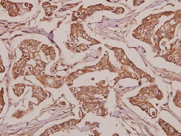
| Sample | Paraffin-embedded human breast carcinoma tissue; fixative not specified (datasheet A09457-1) |
| Fixation | Image fixative and duration unreported (datasheet A09457-1); verify before use. |
| Sectioning | 4–5 µm sections on charged slides (standard) |
| Deparaffinisation | Xylene, graded ethanol series to water (standard) |
| Antigen retrieval | Heat-induced epitope retrieval in citrate buffer, pH 6.0, 20 min at 95–98 °C (standard rule: cytoplasmic / membrane antigen) |
| Peroxidase block | 3% H2O2, 10 min, room temperature (standard) |
| Blocking | 10% normal serum of the secondary host, 30 min, room temperature (standard) |
| Primary antibody | Rabbit anti-SLC25A6, 1:50-1:200 (datasheet A09457-1) |
| Primary incubation | Overnight at 4 °C (standard) |
| Detection | HRP-polymer secondary, DAB chromogen 5–10 min (standard) |
| Counterstain | Hematoxylin, blue, dehydrate and mount (standard) |
| Expected result | SLC25A6-positive staining in purkinje cells of cerebellum (HPA tissue IHC: High). HPA tissue profile: Granular cytoplasmic expression in most tissues. No signal in the no-primary control. |
SLC25A6 is an inner mitochondrial membrane protein with six transmembrane segments (UniProt P12236 topology). In paraffin-section IHC, expect granular cytoplasmic staining in many tissues, with high staining reported in cardiomyocytes, Purkinje cells, and several glandular cell populations (HPA tissue IHC). Interpret that pattern cautiously: HPA rates its tissue IHC evidence Approved, with medium consistency between staining and RNA data, and warns that the antibodies target proteins from more than one gene (HPA tissue IHC).
| Granular cytoplasmic stain in cardiomyocytes or Purkinje cells, with little nuclear stain (HPA tissue IHC). | This fits the reported high staining in those cells and a mitochondrial location (HPA tissue IHC; UniProt P12236). Morphology alone cannot establish that every granule contains SLC25A6, given HPA's multi-gene antibody warning (HPA tissue IHC). |
| Predominantly nuclear or uniform membrane-edge stain in otherwise intact cells. | That distribution conflicts with the expected granular cytoplasmic pattern and inner mitochondrial membrane location (HPA tissue IHC; UniProt P12236). Review morphology and controls before assigning it to the target; the location evidence does not establish this pattern as SLC25A6 (HPA tissue IHC). |
| Strong stain in epididymal glandular cells or ovarian stromal cells. | HPA reports these specific cell populations as not detected (HPA tissue IHC). Consider cross-reactivity, endogenous chromogen-producing activity, or a specimen-specific result (HPA multi-gene antibody warning; general IHC practice). An HPA negative is a reference observation, not proof that every specimen must be negative (HPA tissue IHC). |
| Broad, diffuse color across cells and extracellular areas obscures cytoplasmic granules. | The slide cannot be scored confidently against HPA's granular cytoplasmic profile (HPA tissue IHC). Diffuse background can arise from nonspecific binding or detection chemistry; assess the reagent controls and staining distribution before changing the biological interpretation (general IHC practice). |
| No convincing stain in heart-muscle cardiomyocytes or cerebellar Purkinje cells. | Both are reported as high-staining reference cells (HPA tissue IHC). First suspect a staining-run or specimen issue and compare controls; absence in one section alone does not overturn the tissue profile, especially given its medium staining–RNA consistency (HPA tissue IHC; general IHC practice). |
| Cell and tissue selection | High staining is reported in heart-muscle cardiomyocytes, cerebellar Purkinje cells, rectal and stomach glandular cells, and testicular Leydig cells (HPA tissue IHC). HPA reports lower or undetected staining in other named cell populations, so score the cell type as well as the tissue (HPA tissue IHC). |
| Subcellular expectation | The protein has six transmembrane segments in the mitochondrial inner membrane (UniProt P12236 topology). HPA describes tissue staining as granular cytoplasmic and ICC-IF localization as mitochondrial (HPA tissue IHC; HPA subcellular). Neither source defines an antibody epitope or an antigen-retrieval setting. |
| Antibody evidence and specificity | HPA rates the tissue IHC profile Approved, with medium staining–RNA consistency, and cautions that antibodies target proteins from more than one gene (HPA tissue IHC). Its listed IHC antibody, HPA071684, is Approved; the listed ICC antibody, HPA046835, is Supported for ICC (HPA antibodies). |
| Processing and variants | UniProt lists full-length residues 1–298 and an N-terminally processed form, residues 2–298, with no annotated isoforms or glycosylation sites (UniProt P12236). These annotations do not predict a different tissue staining pattern or a retrieval response without epitope-specific evidence. |
| IF/ICC Q: Should fluorescence appear in mitochondria? | Yes. HPA reports approved mitochondrial localization in ICC-IF images from MCF-7, PC-3, and U2OS cells, while warning that the antibodies target proteins from multiple genes (HPA subcellular). This supports a localization expectation; it supplies no IF/ICC protocol for this IHC section. |
| Chromogenic background | Endogenous enzyme activity and nonspecific reagent binding can add color during chromogenic IHC (general IHC practice). Use appropriate detection controls to distinguish that color from the reported cell-specific granular cytoplasmic pattern (general IHC practice; HPA tissue IHC). |
| Situation | Likely cause | Next action |
|---|---|---|
| A high-staining reference tissue shows no signal. | The run may have failed, or the selected section may not show the relevant cells (general IHC practice; HPA tissue IHC). | Check that cardiomyocytes or Purkinje cells are present, then review the positive control, primary-antibody step, detection reagents, and counterstain before judging the tissue negative (HPA tissue IHC; general IHC practice). |
| Signal is diffuse or masks cell boundaries. | Nonspecific binding or detection background can obscure HPA's granular cytoplasmic profile (general IHC practice; HPA tissue IHC). | Inspect a no-primary control and the distribution of color; review blocking, washes, primary-antibody concentration, and detection conditions as general IHC troubleshooting steps (general IHC practice). |
| Nuclear stain dominates an otherwise positive section. | A nuclear-dominant pattern does not match the reported mitochondrial or granular cytoplasmic localization (UniProt P12236; HPA tissue IHC). | Compare the suspect cells with an expected positive cell population and the no-primary control; avoid calling the nuclear signal SLC25A6 solely from stain intensity (HPA tissue IHC; general IHC practice). |
| A reported undetected cell population stains strongly. | Cross-reactivity or detection background is plausible, particularly because HPA cautions that antibodies target proteins from more than one gene (HPA tissue IHC; general IHC practice). | Verify the cell identity, compare a high-staining reference on the same run, and inspect detection controls; record the discrepancy rather than treating HPA's undetected call as an absolute biological rule (HPA tissue IHC; general IHC practice). |
| Only an IF/ICC image is available for comparison. | HPA's mitochondrial ICC-IF localization and its tissue IHC profile come from different assay contexts, with separate antibody validation entries (HPA subcellular; HPA antibodies). | Use IF/ICC to assess the expected compartment, then judge the paraffin-section result against tissue cell types and granular cytoplasmic staining reported for IHC (HPA subcellular; HPA tissue IHC). |
| Changing antigen retrieval changes the apparent pattern. | The supplied UniProt topology and HPA staining profile do not establish SLC25A6-specific retrieval sensitivity (UniProt P12236; HPA tissue IHC). | Compare conditions using the same positive tissue and appropriate controls, and report the observed result as an assay finding rather than a documented target-specific retrieval effect (HPA tissue IHC; general IHC practice). |


Comprehensive Human Protein Atlas IHC scoring per tissue (reliability: Approved — Medium consistency between antibody staining and RNA expression data. Caution, targets protein from more than one gene. Pending external verification.). Rows are taken directly from the HPA tissue chart — click any row's HPA link to view the source.
| Tissue | Cell type | Level | Evidence | Source |
|---|---|---|---|---|
| Cerebellum | Purkinje cells | High | Protein (IHC) | HPA → |
| Heart muscle | Cardiomyocytes | High | Protein (IHC) | HPA → |
| Parathyroid gland | Glandular cells | High | Protein (IHC) | HPA → |
| Rectum | Glandular cells | High | Protein (IHC) | HPA → |
| Stomach | Glandular cells | High | Protein (IHC) | HPA → |
| Tissue | Cell type | Level | Evidence | Source |
|---|---|---|---|---|
| Epididymis | Glandular cells | Not detected | Protein (IHC) | HPA → |
| Ovary | Ovarian stroma cells | Not detected | Protein (IHC) | HPA → |
| Soft tissue | Chondrocytes | Not detected | Protein (IHC) | HPA → |
| Vagina | Squamous epithelial cells | Not detected | Protein (IHC) | HPA → |
Use compartment, cell type and matched controls to troubleshoot SLC25A6 staining in chromogenic IHC; the IF/ICC considerations appear in one entry.
A09457-1 has a real IHC image from paraffin-embedded human breast carcinoma (image caption). The catalog lists human, mouse and rat reactivity; IF/ICC is listed for A09457-2 without figure data (catalog).
The rendered A09457-1 card shows IHC in paraffin-embedded human breast carcinoma at 1:50 (image caption). A09457-2 is listed for IHC and IF/ICC in human and mouse, but has no IHC or IF figure in the payload (catalog: applications, reactivity, image alts).
Which to pick: Choose A09457-1 for tissue IHC because its own image documents a paraffin section; the caption does not report the fixative (image caption). Choose A09457-2 for IF/ICC based on its listed applications, while treating imaging conditions as unverified here because no IF figure is supplied (catalog: applications, image alts). Both are rabbit polyclonals; for rat samples, choose A09457-1 because only it lists rat reactivity (catalog: host, dilution_raw, reactivity).